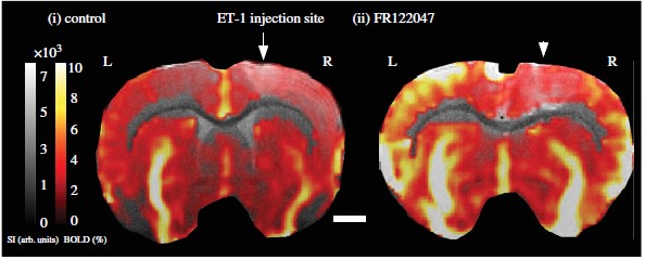
The latest theme issue of Philosophical Transactions B brings together cognitive and cellular neuroscientists to showcase the latest research on the use and interpretation of BOLD, an important tool used in studying brain function in humans.

What is BOLD and how does it work?
Blood Oxygenation Level Dependent (BOLD) imaging is a technique that is commonly used for measuring brain activity in humans using magnetic resonance imaging (MRI). Blood supplies oxygen to brain cells. When these cells are active, there is an increase in blood flow and blood oxygen in the surrounding area. BOLD imaging uses magnetic fields to measure this change in oxygenation. This signal is often used to infer the activity of brain cells and, thus, which brain areas are involved in a particular task.
What is BOLD best used for?
BOLD has an enormous range of uses, from a diagnostic tool in the hospital or clinic, to research into how the brain works, to investigating changes that occur in disease. The best and most reliable use of BOLD currently is in studying the mechanisms that underlie healthy brain function.

How is it applied to help our understanding of neuroscience?
BOLD revolutionised neuroscience by enabling scientists to record activity from human brains in a completely safe, non-invasive way. This allows us to investigate complex brain functions such as cognition and consciousness, which cannot be studied in animal models. It can also be used to study dysfunction of these processes during disease.
Another terrific thing about BOLD is that it measures from the whole brain more or less simultaneously. For example, BOLD can see if a region in the front of the brain is becoming less active at the same time that a region in the back is becoming more active. This provides incredible insight into not only the workings of each part of the brain, but also the complex interactions between different regions.
Are there limitations to interpreting BOLD?
Absolutely. The central limitation is that BOLD is not a direct readout of brain activity, but an indirect measurement that relies on the change in blood oxygen. Blood oxygenation changes on the order of seconds. This is very slow relative to the speed of brain activity, which can change every few milliseconds – that’s one thousandth of one second. Therefore, BOLD cannot detect rapid changes in neuronal activity, and there is debate amongst scientists about what speeds of imaging are required to extract meaningful information about the brain.
A second limitation is the resolution – our current techniques allow us to measure BOLD signals at a millimeter range, but there are thousands, maybe millions of brain cells in this small space. So BOLD can only measure the collective activity of a large number of neurons.
Finally, the process by which active brain regions cause the increase in blood flow is complex and involves multiple types of brain cells. This means that the BOLD signal may differ depending on the brain region and under conditions that can affect the blood supply such as diseases. This is an active area of research amongst scientists who study brain blood flow.
It is important to factor these limitations into our interpretation of BOLD signals, so we form the most accurate conclusions from our experiments. The Royal Society Theo Murphy meeting and the special issue of Philosophical Transactions B were important meetings of minds between the scientists who study the processes by which the brain generates BOLD signals and those who use BOLD signals in their research. We hope the exchange of ideas that this represents will help everyone to use the latest information and techniques to help us understand how the brain works.
What are you currently working on?
Anusha Mishra is currently working on how active neurons differently regulate the blood supply from large and small blood vessels. Clare Howarth is focusing on how astrocytes (a glial cell within the brain) are involved in regulating brain blood flow. Catherine Hall is researching how the relationship between brain activity and blood supply is affected in different states of alertness and in disease.
In the future, what do you think BOLD can reveal about the brain that it cannot tell us now? What advancements are helping improve the technique?
Computational methods are currently transforming the use of BOLD. This is happening in two main ways. First, computational models of information processing let us make extremely detailed predictions about patterns of brain activity. Second, big data sets can be analyzed with machine learning and lots of computing power.
In addition, as already mentioned, disease may alter the complex mechanisms underlying BOLD in unpredictable ways, limiting the use of BOLD in the clinic and in some research areas. Experiments using multiple approaches in parallel, from tiny recordings from single cells to recording the activity of the whole brain, are currently being conducted to help us understand exactly how brain cells signal to blood vessels and how brain activity and blood oxygen are related under different conditions. This will give us more power to interpret findings from BOLD studies in the future, especially in the context of disease or ageing.
To find out more, you can read the papers in our theme issue Interpreting BOLD. The Introduction to the issue is free to read.
