Guest editors Ricardo Henriques, Christophe Leterrier and Aubrey Weigel tell us about the background and content of this new special feature in Open Biology.
In this special feature of Open Biology ‘Advances in Quantitative Bioimaging’, we present breakthrough research demonstrating how innovative imaging modalities combined with new computational analysis methods are providing an intimate glimpse into up to now unseen molecular aspects of dynamic cellular processes.
Insight into Quantitative Bioimaging
Exploring life's intricate mechanisms calls for an up-close examination of biological structures from every vantage point, from molecules to cells to tissues. Recent progress in quantitative bioimaging technologies has empowered us to visualise complex molecular architectures, track individual proteins, compare cellular dynamics with nanoscale precision and single-molecule resolution. This leap forward holds significant potential for driving our understanding into uncharted territory by offering fresh mechanistic insights into living systems.
This feature explores innovative applications of quantitative bioimaging techniques in diverse biological domains. Key topics include the synergy of super-resolution microscopy (SRM) and single-particle analysis (SPA) in mapping molecular complexes, as discussed by Mendes et al. The review underscores SRM–SPA's efficacy in revealing nanoscale structures and complex arrangements within biological entities, with ongoing challenges being addressed by template-free algorithms and live-cell imaging correlation.
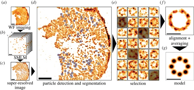
Mendes et al. DOI: 10.1098/rsob.220079
Hurley et al. correlate calcium signaling events with the structural organization of calcium channels in cardiac muscle cells, revealing insights into healthy and failing heart cells. Utilising image analysis and machine learning to characterise microglia behaviour in various conditions, the contribution from Martinez et al. offers a quantitative understanding of morphological alterations and behavioural shifts during neuroinflammation.
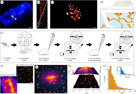
Hurley et al. DOI: 10.1098/rsob.230045
Riquelme-Guzmán et al. employ confocal Brillouin microscopy to assess tissue mechanical changes in axolotl development and regeneration, focusing on limb and digit cartilage. In a study by Ragaller et al., smart probes (Pro12A, NR12S, and NR12A) are introduced for assessing membrane properties. The probes demonstrate distinct sensitivities to lipid composition, saturation, headgroup changes, cholesterol content, and fluidity, providing a guide for selectively applying or combining these probes to quantitatively assess different aspects of membrane biophysics relevant to cellular processes and disease states.
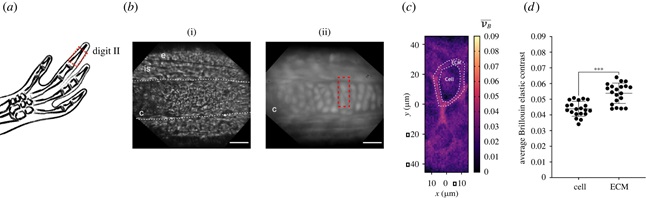
Riquelme-Guzmán et al. DOI: 10.1098/rsob.220078
The articles showcased cover various topics, including modern imaging methods that enable visualisation on a nanoscale, such as super-resolution microscopy and single-particle analysis.
What’s next?
The studies presented in the feature highlight scientific progress made possible by advanced quantitative bioimaging techniques and image analysis. These tools allow us to better understand the intricate layers of cellular functions and expand our perception of how living systems operate across different scales. Over time, these advancements will help bridge the gap between experimental observation and theoretical vision, progressively tackling the formidable complexity intrinsic to biology.
Keep up to date with the latest Open Biology content by signing up for article alerts, and browse previous special features on the journal website.
------------------------------------------------------
Image credit: 'Strange Fruit' - butterfly eggs. Coloured electron microscopy of butterfly eggs. ZEISS Sigma SEM Credit: ZEISS Microscopy. Source: Flickr.com CC-BY 2.0.