Links to external sources may no longer work as intended. The content may not represent the latest thinking in this area or the Society’s current position on the topic.
The transformative potential of data and image analysis for eye care

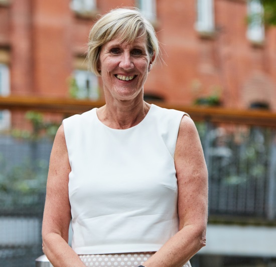
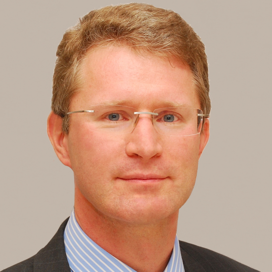
Science+ meeting organised by Professor Emanuele Trucco, Professor Caroline MacEwen, Professor Paul Foster and Professor Tunde Peto.
A Science+ interdisciplinary meeting that captured scientific and translational needs and opportunities for eye care research within data and image analysis, including harnessing the potential of the eye as a source of biomarkers for systemic conditions. The meeting brought together scientists and clinicians, including those from the Royal College of Ophthalmologists and the College of Optometrists.
More information on the speakers and programme is available below. Recorded audio of the presentations is also available below.
Attending the event
This meeting has taken place.
Enquiries: contact the Scientific Programmes team
Organisers
Schedule
| 09:00 - 09:05 | Welcome from the Royal Society | |
|---|---|---|
| 09:05 - 09:15 |
Introduction to the event from lead organiser Professor Emanuele Trucco, University of Dundee |
|
| 09:15 - 10:00 |
Gearing an entire country for Health Data Science
Healthcare is arguably the last major industry to be transformed by the information age. Deployments of information technology have only scratched the surface of possibilities for the potential influence of information and computer science on the quality and cost-effectiveness of healthcare. In this talk, the opportunities provided by computer science and "big data" to transform health care delivery models will be discussed. Examples will be given from nationwide research and development programmes that integrate electronic patient records with biologic and health system data. Two themes will be explored; specifically: 1) How the size of the UK (65M residents), allied to a relatively stable population and unified health care structures facilitate the application of health informatics to support nationwide quality-assured provision of diabetes care. 2) How population-based datasets and disease registries can be integrated with biologic information to facilitate (i) epidemiology; (ii) drug safety studies; (iii) enhanced efficiency of clinical trials through automated follow-up of clinical events and treatment response; and, (iv) the conduct of large-scale genetic, pharmacogenetics, and family-based studies essential for precision medicine. 
Professor Andrew Morris, UK Biomedical Informatics Research Institute, UK

Professor Andrew Morris, UK Biomedical Informatics Research Institute, UKSince August 2017 Andrew Morris has been the inaugural Director of Health Data Research UK, the multi-funder UK Institute for health and biomedical informatics research that will capitalise on the UK’s renowned data resources and research strengths to transform lives through health data science. He is seconded from his position as Professor of Medicine, and Vice Principal of Data Science at the University of Edinburgh, having taken up position in August 2014. Prior to this Andrew was Dean of Medicine at the University of Dundee. Andrew was Chief Scientist at the Scottish Government Health Directorate (2012-2017) and has served and chaired numerous national and international grant committees and Governmental bodies. |
|
| 10:00 - 10:45 |
How do new imaging technologies and their novel analysis contribute to the understanding of disease? Dr Srinivas Sadda
The last two decades, and in particular the last five years, have witnessed an explosion in the development of new imaging technologies. A significant advance is the availability of higher speed, higher resolution, and deeper penetrating swept source OCT technology which allows retinal disease to be studied in an en face plane in a depth-resolved fashion. Adaptive optics techniques have further advanced the axial resolution of our imaging technologies. Other major advances included enhances in contrast, facilitating molecular and functional imaging such as hyperspectral imaging, quantitative autofluroescence, fluorescence lifetime imaging ophthalmoscopy, and OCT angiography. Mutlidimensional analysis techniques are being developed to cope with these enormous amounts of data. Imaging technology has also benefited from the introduction of widefield devices with specialized mirrors and optics allowing the entire retina to be photographed in a single acquisition. Widefield imaging has provided new insights into various diseases including diabetic retinopathy. Recent studies have highlighted the prognostic importance of peripheral lesions in diabetic retinopathy, with eyes with predominantly peripheral disease having a four-fold higher risk of progression. Combining widefield imaging with artificial intelligence/deep learning approaches, it is now possible to automatically identify and classify diabetic retinopathy, which may have significant public health applications in diabetic retinopathy screening programs. |
|
| 10:45 - 11:15 | Tea break | |
| 11:15 - 12:00 |
From bench to clinical applications: opportunities in ophthalmic imaging from the lab to the individualised patient therapies - Professor Tunde Peto
Ophthalmology has one of the fastest growing imaging arsenal. With these new technologies nearly all components of the human eye can now be imaged. It is a major undertaking to make sure that all imaging modalities are scientifically evaluated for their validity and clinical utility. However, novel clinical imaging modalities raise new, often unexpected questions. These can only be answered by incorporating laboratory research into the evaluations which, in turn, lead to better understanding of the clinical phenotypes. Humans have an incredible ability to recognise patterns and with the new imaging modalities being high enough quality, new patterns are continuously emerging. Therefore, image analysis is still predominantly done manually. As such it is fraught with slow reporting and the need for ever increasing attention to detail. There is risk of not recognizing crucial features contained in the images leading to the recording of wrong phenotype. Therefore, if we are to cope with the amount of images we generate, there is a need for intelligent, automated, non-human based, though probably human supervised, image analysis. It is very likely that the development of new algorithms will lead to the development of new treatment paradigms to benefit the patients. 
Professor Tunde Peto, Queen's University Belfast, UK

Professor Tunde Peto, Queen's University Belfast, UKTunde Peto is Professor of Clinical Ophthalmology at Queen’s University Belfast where she is responsible for teaching both undergraduate and postgraduate students. Her remit also covers the development of International image analysis, training and educating in Countries throughout the world. In addition to her academic work, Professor Peto is also responsible for the Belfast Ophthalmic Image Reading Centre and a wide research portfolio. Her main interest has always been common blinding retinal diseases, with special emphasis on characterisation of diabetic retinopathy and its imaging modalities. Professor Peto has been involved in UK National Diabetic Retinopathy Screening programmes since 2002, She was clinical lead for London programmes and is currently the clinical lead for the Northern Irish Programme. Professor Tunde Peto trained in Hungary and Australia and spent 15 years at Moorfields Eye Hospital in London before taking up her current position in Belfast. |
|
| 12:00 - 12:45 |
Speaker to be confirmed
|
|
| 12:45 - 13:30 | Lunch |
Chair

Professor Emanuele Trucco, University of Dundee, UK

Professor Emanuele Trucco, University of Dundee, UK
Manuel has been active since 1984 in computer vision, and since 2002 in medical image analysis, publishing more than 250 refereed papers and 2 textbooks, and serving on the organizing or program committee of major international and UK conferences. Manuel is co-director of VAMPIRE (Vessel Assessment and Measurement Platform for Images of the Retina), an international research initiative led by the Universities of Dundee and Edinburgh (co-director Dr Tom MacGillivray), and part of the UK Biobank Eye andVision Consortium. VAMPIRE develops software tools for efficient data and image analysis with a focus on multi-modal retinal images. VAMPIRE has been used in UK and international biomarker studies on cardiovascular risk, stroke, dementia, diabetes and complications, cognitive performance, neurodegenerative diseases, and genetics. Industrial collaboratorss include Canon (ex Toshiba) Medical, OPTOS plc, NIDEK, and Epipole plc. The portfolio of recent VAMPIRE projects led or co-led by Manuel include a £7M NIHR grant Dundee-Chennai on precision medicine for diabetes, a £1.1M EPSRC grant on multi-modal biomarkers for vascular dementia, the 3M-euro ITN "REVAMMAD", and PhD studentships sponsored by OPTOS plc, SINAPSE and Toshiba.
| 13:30 - 14:30 |
The UK Biobank Eye & Vision Consortium as a model for synergy and collaboration in big data analysis - Professor Paul Foster
UK Biobank was initially conceived as a platform for studies of gene environment interaction in major chronic diseases of older age, such as cancer, stroke, MI and diabetes. Towards the end of the study, the UKBB steering committee advised the broadening of the scope of the study to include more detailed examination of participants, including assessment of physical fitness, brain and cardiac imaging, as well as an examination of eyes and vision. Moorfields Eye Hospital and the UCL Institute of Ophthalmology in London developed the eye and vision module. The core funding for the examination was provided by the Wellcome Trust, The Medical Research Council and The Department of Health. Additional support for training, implementation and quality control came from the NIHR Biomedical Research Centre at Moorfields Eye Hospital.
|
|
|---|---|---|
| 14:30 - 15:30 |
Multi-modal retinal imaging in the Northern Ireland Cohort for the Longitudinal Study of Aging (NICOLA)
The Northern Ireland Cohort for Longitudinal Aging Study is a comprehensive, long-term epidemiological study of adult development and ageing which started in February 2014 and consists of a random sample of men and women aged 50 years, representative of the Northern Ireland population. As part of the study approximately 3,600 participants have attended the Wellcome-Wolfson Clinical Research Facility (CRF) at Belfast City Hospital for a health assessment. The health assessment included anthropometry, respiratory, cardiovascular, cognitive and ophthalmology tests. The ophthalmic component involved visual acuity, auto refraction, corneal compensated intra-ocular pressure, stereo colour fundus photographs, infra-red and autofluorescent retinal images, wide field Optos colour images and Spectral Domain Optical Coherence Tomography (OCT) with Enhanced Depth Imaging (EDI) of the choroid. Macular pigment was also assessed using dual-wavelength autofluorescence. To date most epidemiological studies have only used colour fundus photographs for estimating ocular disease prevalence. The talk will discuss the opportunities and challenges of using multi-modal imaging and incorporating the data into a larger framework that also encompasses genomic, epigenomic, dietary and biochemical biomarkers.D 
Dr Ruth Hogg, Queen’s University Belfast, UK

Dr Ruth Hogg, Queen’s University Belfast, UKDr Hogg is an academic Optometrist and Epidemiologist based at the Centre for Public Health, Queen's University Belfast (QUB). After her professional training she completed a PhD at Queen's University Belfast supervised by Professor Usha Chakravarthy, followed by post-doctoral fellowships in Melbourne and Cambridge. In 2010 she returned to QUB to take up a Lectureship. Her research interests focus on the intersection between ocular aging and the development of age-related diseases such as Age-related macular degeneration (AMD), Diabetic Retinopathy and Glaucoma and also risk factors for progression of these diseases to their sight threatening forms. Her lab is focused on the following themes: 1. The epidemiology of age-related eye diseases such as Age-related macular degeneration (AMD), Diabetic Retinopathy and Glaucoma. She also leads the eye component of the Northern Ireland Cohort for the Longitudinal Study of Aging, an epidemiological study following 8,000 older adults in Northern Ireland. |
|
| 15:30 - 16:00 | Coffee | |
| 16:00 - 17:00 |
Facilitated open discussion
Facilitated open discussion: imaging, data and vision- priorities for collaboration, research and eye health. Led by Michael Bowen, College of Optometrists 
Michael Bowen, College of Optometrists, UK

Michael Bowen, College of Optometrists, UKMichael is the Director of Research at the College of Optometrists, where he is responsible for the strategic development of the College's Research activities. Michael has led various research projects within the sector, including: PrOVIDe - which gathered data on the prevalence of visual impairment among people living with dementia in England; the National Minimum Data Sets Project to develop harmonised data sets for hospital eye service clinics; and the Optometric Public Health Project, exploring data sets for primary care setting in optometry. Prior to joining the College in 2008, Michael worked at the professional and regulatory body for Psychotherapy in the UK, running their research, education and standards, and fitness to practice functions. Michael's academic background is in psychology, biology and medical ethics. |
Chair

Professor Tunde Peto, Queen's University Belfast, UK

Professor Tunde Peto, Queen's University Belfast, UK
Tunde Peto is Professor of Clinical Ophthalmology at Queen’s University Belfast where she is responsible for teaching both undergraduate and postgraduate students. Her remit also covers the development of International image analysis, training and educating in Countries throughout the world.
In addition to her academic work, Professor Peto is also responsible for the Belfast Ophthalmic Image Reading Centre and a wide research portfolio. Her main interest has always been common blinding retinal diseases, with special emphasis on characterisation of diabetic retinopathy and its imaging modalities.
Professor Peto has been involved in UK National Diabetic Retinopathy Screening programmes since 2002, She was clinical lead for London programmes and is currently the clinical lead for the Northern Irish Programme.
Professor Tunde Peto trained in Hungary and Australia and spent 15 years at Moorfields Eye Hospital in London before taking up her current position in Belfast.
| 09:00 - 10:00 |
AI in ocular imaging: a China and Singapore perspective
In the talk, Jimmy will update the ocular imaging research work conducted in the past years in China and Singapore. 
Professor Jimmy Liu Jiang, Cixi Institute of Biomedial Enginerring, Chinese Academy of Science, China

Professor Jimmy Liu Jiang, Cixi Institute of Biomedial Enginerring, Chinese Academy of Science, ChinaProfessor Jimmy Liu Jiang joined Chinese Academy of Sciences in March 2016 as the Executive Director through the China “Thousand Talent Program”. Since 1989, Jimmy has spent 27 years in Singapore. Jimmy established the Intelligent Medical Imaging Program (iMED), which was one of the largest ocular imaging research teams in the world, in A*STAR (Agency for Science, Technology and Research) Singapore. Ever since joining the Chinese Academy of Sciences in 2016, he put in much time and effort helping to establish a new institute focusing on biomedical Engineering in Ningbo with support from government and Chinese Academy of Sciences. Recently, Jimmy established a new joint laboratory with world leading ophthalmological equipment manufacture TOPCON Inc. in China focusing on new areas such as advanced ocular medical equipment manufacturing and Artificial Intelligence based Chinese “big” medical image and data research. |
|
|---|---|---|
| 10:00 - 10:45 |
The QUARTZ retinal image analysis system:quantifying the vasculature in the UK Biobank fundus camera images
The application of image analysis techniques to provide accurate measures of image features on larger datasets presents specific challenges in ensuring the reliability of the measurements produced. The sizes of current retinal fundus image datasets are increasing, and with their public availability there is increased scope for computer vision researchers to apply algorithms to datasets with variable image characteristics. Image datasets may exhibit variation in terms of image quality, and in terms of image characteristics, such as sets of fundus images that display a range of pathologies. The presentation will report on the experience of applying computer vision techniques to analyse the UK Biobank retinal fundus dataset that contains over 100,000 images. The analysis included recognition of the retinal vasculature, including classification of arterioles and venules, and the provision of vessel morphometric data across the retinal image. The presentation will discuss the experience in terms of generation of reliable data and will report on the challenges in addition to the overall approach that was taken. 
Professor Sarah Barman, Kingston University, UK

Professor Sarah Barman, Kingston University, UKSarah Barman is Professor of computer vision in the School of Computer Science and Mathematics at Kingston University. Her main area of interest is in the field of medical image analysis. Her work is currently focused on research into novel image processing and machine learning techniques to enable the recognition and quantification of features in ophthalmic images. Examples of this work include vessel width and tortuosity measurement on very large retinal fundus datasets, in addition to abnormality detection in diabetic retinopathy images. Previous work has included detection of posterior capsule opacification on implant lenses. Professor Barman is a member of the UK Biobank Eye and Vision consortium, a Fellow of the Institute of Physics and is a registered Chartered Physicist. |
|
| 10:45 - 11:05 | Tea break | |
| 11:05 - 11:45 |
Power and limits of automatic retinal image analysis: an image processing perspective
Automatic image processing holds great promises for the analysis of retinal images from a variety of instruments, and its application to future eye care including the vision of personalized medicine. The introduction of automatic algorithms in diabetic retinopathy screening programs, and the intriguing results achieved by the increasingly ubiquitous artificial intelligence programs are examples of success stories and of potential future achievements. This talk aims to summarise the main methods and challenges of contemporary retinal image analysis in the context of common research and clinical scenarios. Particular attention is given to biomarker discovery, which requires large collections of cross-linked data and lies therefore at the very heart of the theme of this meeting. 
Professor Emanuele Trucco, University of Dundee, UK

Professor Emanuele Trucco, University of Dundee, UK
Emanuele (Manuel) Trucco, is the NRP Chair of Computational Vision in Computing, School of Science and Engineering, at the University of Dundee, and an Honorary Clinical Researcher of NHS Tayside.
Manuel has been active since 1984 in computer vision, and since 2002 in medical image analysis, publishing more than 250 refereed papers and 2 textbooks, and serving on the organizing or program committee of major international and UK conferences. Manuel is co-director of VAMPIRE (Vessel Assessment and Measurement Platform for Images of the Retina), an international research initiative led by the Universities of Dundee and Edinburgh (co-director Dr Tom MacGillivray), and part of the UK Biobank Eye andVision Consortium. VAMPIRE develops software tools for efficient data and image analysis with a focus on multi-modal retinal images. VAMPIRE has been used in UK and international biomarker studies on cardiovascular risk, stroke, dementia, diabetes and complications, cognitive performance, neurodegenerative diseases, and genetics. Industrial collaboratorss include Canon (ex Toshiba) Medical, OPTOS plc, NIDEK, and Epipole plc. The portfolio of recent VAMPIRE projects led or co-led by Manuel include a £7M NIHR grant Dundee-Chennai on precision medicine for diabetes, a £1.1M EPSRC grant on multi-modal biomarkers for vascular dementia, the 3M-euro ITN "REVAMMAD", and PhD studentships sponsored by OPTOS plc, SINAPSE and Toshiba. |
|
| 11:45 - 12:30 |
Quantitative retinal biomarkers for diabetic retinopathy: the REVIEW experience
Diabetic retinopathy results in a complex histopathology in the retinal vessels, with potentially detectable and progressive changes in the vascular geometry (including vessel calibres, tortuosity and branching angles) and resulting lesions (microaneurysms, haemorrhages and exudates). Biomarkers are quantitative indicators that can be reliably extracted from imaging modalities (e.g. fundal images), with minimal or zero user intervention, and are strongly indicative of disease. The challenges in biomarker extraction include: the definition of meaningful quantifiers that summarise usefully often complex and distributed physiological signs, and reliable automated extraction techniques operating in the face of complex, variable physiology and often poor image quality resulting from a challenging problem domain. A key question is: what physiological changes are happening and how can they be elegantly captured? This talk addresses two questions: are there biomarkers allowing early prediction of the onset of diabetic retinopathy; and to what extent does trying to model underlying physiological factors help in biomarker design?

Professor Andrew Hunter, University of Lincoln, UK

Professor Andrew Hunter, University of Lincoln, UKProfessor Andrew Hunter is Deputy Vice Chancellor for Research and Innovation. He holds a BSc and PhD degrees in Computer Science from the University of Bath. He worked for several years in the software industry before returning to academia at Sunderland and Durham Universities. He joined the University of Lincoln in 2004, serving as Head of Department of Computing, Dean of Research, and the founding Head of the College of Science (where he oversaw the creation of new Schools in Pharmacy, Chemistry, and Mathematics and Physics, and is currently the Deputy Vice Chancellor for Research and Innovation. Professor Hunter has particular research interests in retinal imaging and behavioural analysis. He has published over 100 academic papers, and has developed freeware and commercial artificial intelligence software packages, including genetic algorithms and neural networks. |
|
| 12:30 - 13:15 | Lunch |
Chair

Michael Bowen, College of Optometrists, UK

Michael Bowen, College of Optometrists, UK
Michael is the Director of Research at the College of Optometrists, where he is responsible for the strategic development of the College's Research activities. Michael has led various research projects within the sector, including: PrOVIDe - which gathered data on the prevalence of visual impairment among people living with dementia in England; the National Minimum Data Sets Project to develop harmonised data sets for hospital eye service clinics; and the Optometric Public Health Project, exploring data sets for primary care setting in optometry. Prior to joining the College in 2008, Michael worked at the professional and regulatory body for Psychotherapy in the UK, running their research, education and standards, and fitness to practice functions. Michael's academic background is in psychology, biology and medical ethics.
| 13:15 - 13:45 |
Deep learning in the retina
Retinal images play an important role in the assessment of many eye and systemic diseases. This talk will give an overview of the developed AI systems using deep learning and image processing techniques for automating the detection of these diseases through digital fundus image data. The talk will also examine the limitation of current approaches on a number of application problems and open up discussion for further intelligence that may orchestrate solutions to various challenges. 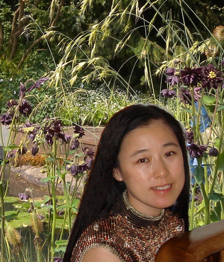
Dr Lilian Tang, University of Surrey, UK

Dr Lilian Tang, University of Surrey, UKDr Lilian Tang received her PhD from the University of Cambridge in medical informatics and currently is working at the University of Surrey as a senior lecturer in AI. She is interested in applying machine learning and vision to image analysis, with a particular focus on the medical domain. Over the last decade, she has been working with a number of NHS hospitals, as well as hospitals in Europe and Mid and Far-east Asia, on a wide range of projects including: the development of automated tracking of fine movements of surgical instruments in eye and other microsurgeries for objective dexterity analysis; digital retinal image analysis to understand various eye and systematic conditions; and, time-lapse bacterial cell image analysis for understanding mycobacterial persistence. On all the AI software systems she has developed, scalability is always a core quality she aims to achieve. |
|
|---|---|---|
| 13:45 - 14:30 |
What can statistics do for the understanding of ophthalmic diseases? From measurement errors and bias to inference and risk estimation using complex data that contain images
In the era of collecting complex ophthalmic datasets we are more and more facing the questions of how to best utilise such rich information from patients. In this talk we will discuss three challenges that are sometimes not considered. For example, not considering the measurement errors can lead to lower power of statistical inference. The potential of sector-wise approach to inference from imaging data is often not considered. The accuracy of the traditional statistical approaches to the disease detection is just yet to be fully utilised. 
Dr Gabriella Czanner, University of Liverpool, UK

Dr Gabriella Czanner, University of Liverpool, UKDr Gabriela Czanner is a Chartered Statistician (Royal Statistical Society 2012) and a Lecturer in the Department of Biostatistics and Department of Eye and Vision Science at University of Liverpool. She works closely with ophthalmologists from the Clinical Research Eye Centre at Royal Liverpool and the Broadgreen University Hospitals NHS Trust. She develops methods for feature selection and classification using complex ophthalmic datasets that contain imaging data. She regularly contributes to workshops for Royal College of Ophthalmologists and she led the biostatistics course for ARVO in 2017. She has been an invited speaker, among other, at the Royal Statistical Society GAS, the Neuroscience Statistics Research Laboratory of MIT and Harvard, and the NIHR Statistics Group Imaging UK. She is an associate editor in biostatistics for BMJ Open Ophthalmology and member of editorial board of Graefe’s Archive for Clinical and Experimental Ophthalmology. She is a member of steering committee for the national Ophthalmic Statistics Group and NIHR Statistics Group Ophthalmology Research Section. |
|
| 14:30 - 15:00 |
Mining large-scale sets of OCT images
Ophthalmology is among the most technology-driven of the all the medical specialties, with treatments utilising high-spec medical lasers and advanced microsurgical techniques, and diagnostics involving ultra-high resolution imaging. Ophthalmology is also at the forefront of many trailblasing research areas in healthcare, such as stem cell and gene therapies. Moorfields Eye Hospital in London is the oldest eye hospital in the world. Every year, >700,000 patients attend Moorfields - more than double the number of the largest eye hospitals in North America. Together with the adjacent UCL Institute of Ophthalmology, Moorfields is among the largest centres for vision science research in the world. In July 2016, Moorfields announced a formal collaboration and data sharing agreement with DeepMind Health. This collaboration involves the sharing of >1,000,000 anonymised retinal optical coherence tomography (OCT) scans with DeepMind to allow for the automated diagnosis of diseases such as age-related macular degeneration (AMD) and diabetic retinopathy (DR). This presentation, will describe the motivation - and urgent need - to apply deep learning to ophthalmology, the processes required to establish a research collaboration between the NHS and a company like DeepMind, the goals of our research, and finally, why I believe that ophthalmology could be first branch of medicine to be fundamentally reinvented through the application of deep learning. 
Dr Pearse Keane, Moorfields Eye Hospital, UK

Dr Pearse Keane, Moorfields Eye Hospital, UKPearse A Keane, MD, FRCOphth, is a consultant ophthalmologist at Moorfields Eye Hospital, London and an NIHR Clinician Scientist, based at the Institute of Ophthalmology, University College London (UCL). Dr Keane specialises in applied ophthalmic research, with a particular interest in retinal imaging and new technologies.
|
|
| 15:00 - 15:20 | Coffee break | |
| 15:20 - 16:30 |
Facilitated open discussion: Final focus
Facilitated open discussion: Final focus - agreeing activities and actions to address key issues. Led by Michael Bowen, College of Optometrists 
Michael Bowen, College of Optometrists, UK

Michael Bowen, College of Optometrists, UKMichael is the Director of Research at the College of Optometrists, where he is responsible for the strategic development of the College's Research activities. Michael has led various research projects within the sector, including: PrOVIDe - which gathered data on the prevalence of visual impairment among people living with dementia in England; the National Minimum Data Sets Project to develop harmonised data sets for hospital eye service clinics; and the Optometric Public Health Project, exploring data sets for primary care setting in optometry. Prior to joining the College in 2008, Michael worked at the professional and regulatory body for Psychotherapy in the UK, running their research, education and standards, and fitness to practice functions. Michael's academic background is in psychology, biology and medical ethics. |
|
| 16:30 - 16:45 | Concluding remarks from organisers |

