Links to external sources may no longer work as intended. The content may not represent the latest thinking in this area or the Society’s current position on the topic.
New technologies in cancer mechanobiology
Satellite meeting organised by Dr Chris Bakal and Dr Julia Sero
The development of new technologies has long been a driving force in the study of mechanobiology. This workshop will bring together an interdisciplinary group of early to senior level researchers to discuss cutting-edge in vivo, in vitro, and in silico techniques and how these methods can be applied to cancer research, diagnostics, and therapeutics.
Recorded audio of the presentations will be available on this page after the meeting has taken place. The speaker biographies and abstracts are available below.
Enquires: Contact the Scientific Programmes team.
Organisers
Schedule
| 09:05 - 09:15 |
Mechanobiology-based technology for rapid cancer diagnosis and prognosis
Metastasis requires cells to dynamically adapt to changing microenvironments and apply forces. Daphne Weihs shows that subpopulations of metastatic cells rapidly (<2 hours) and forcefully indent elastic, physiological stiffness, synthetic, impenetrable gels to depths of 1–20 µm, whereas benign/normal cells do not indent. The indenting subpopulation is highly migratory and invasive (Boyden chamber) and includes chemotherapy-resistant and cancer stem cells. Indentation capacity is governed by mechanobiology and is applicable to various solid cancers. Daphne will demonstrate with breast, pancreas, and skin cancers, rapid, same-day diagnosis and prognosis that matches the metastatic risk and clinical outcome in patients. 
Professor Daphne Weihs, Technion-Israel Institute of Technology, Israel
Professor Daphne Weihs, Technion-Israel Institute of Technology, IsraelAssociate Professor Daphne Weihs is a Professor of Biomedical Engineering at the Technion-Israel Institute of Technology since 2006. She obtained her BSc, MSc, and PhD (2004) at the Faculty of Chemical Engineering, Technion. She did her post-graduate research at the Department of Pathology and Lab Medicine, Medical School, University of California at Los Angeles (UCLA, 2004–2006). Professor Weihs is the head of the Scientific Committee of the Israel Society of Medical and Biological Engineering (ISMBE) since 2015. Her focus is the mechanobiology of cells; the stiffness and dynamics of cells and their mechanical interactions with the microenvironment, in the contexts of cancer progression and in wound prevention and healing. She has recently developed a mechanobiology-based approach to diagnose cancer and predict metastases. |
|
|---|---|---|
| 09:15 - 09:30 |
Using mesoscopic models to understand the physical behaviours of cells and tissues
Living cells generate and transmit mechanical forces over diverse time-scales and length-scales to determine the dynamics of cell and tissue shape during both homeostatic and pathological processes. On the molecular scale, Mike Murrell’s group uses active gels as a framework to understand how mechanical stresses are transmitted within the cell cytoskeleton. On the scale of cells and tissues, they abstract these stresses to surface tensions in a liquid film and draw analogies between the dynamics of wetting to the shape dynamics of simple tissues. Together, they develop comprehensive descriptions for how cytoskeletal stresses translate to the physical behaviours of cells and tissues. 
Dr Mike Murrell, Yale University, USA
Dr Mike Murrell, Yale University, USAMurrell’s interests are in understanding the mechanical principles that drive major cellular life processes through the design and engineering of novel biomimetic systems. To this end, he develops simplified and tractable experimental models of the mechanical machinery within the cell with the goal of reproducing complex cellular behaviour, such as cell division and cell migration. Murrell then combines these ‘bottom-up’ experimental models with concepts from soft matter physics to gain a fundamental understanding of the influence of mechanics on cell and tissue behaviour. In parallel, he hopes to identify new design principles from biology which can be used to create novel technologies. |
|
| 09:30 - 09:45 |
Development of biomimetic stroma for 3D tumouroids
There has been a drive to develop 3D models to test mechanisms of cancer progression. Such models aim to recapitulate specific aspects of the native microenvironment of cancer tissue. Umber Cheema’s group has developed 3D models of solid cancers, termed tumouroids. Using tissue engineering techniques, they control the spatial positioning of a cancer mass and its surrounding stroma. The group are able to engineer specific components into each component. They have engineered hypoxic gradients within the 3D model as well as engineering an intact primitive vascular network in the stromal component. They have observed interaction of cancer cells with engineered vascular networks- and measured angiogenic remodelling of the networks. By incorporating cancer associated fibroblasts from patient samples Umber is further investigating how these cells enhance cancer invasion and the mechanisms by which this is done. 
Dr Umber Cheema, University College London, UK
Dr Umber Cheema, University College London, UKDr Cheema is a senior lecturer in tissue engineering, and head of education for the Division of Surgery at UCL. Her research interests include engineering vascular networks in 3D engineered tissues to further understand the role native matrix density and composition have on this biological process. Her research also focuses on controlling elements of collagen architecture to build biomimetic tissues in vitro. In particular, by studying mechanisms which control collagen fibril alignment, diameter and density. Her work on engineering tissues has resulted in the development of 3D in vitro models of solid tumours or tumouroids. Here the spatial architecture of a tumour and its surrounding stroma has been reproduced in vitro, with evidence of tumour invasion into surrounding 'normal' tissue. Engineered tumouroids include epithelial cancers and sarcoma's. Her collaborative work includes engineering basic tissue models which are subjected to decompression regimes to mimic the 'bends' in diving. Experimental data has been modeled to further our understanding of how bubbles nucleate, grow and coalesce in tissues during decompression. |
|
| 09:45 - 10:00 |
Mechanobiology in a microplate – opportunities for high throughput screening
Mechanobiology focuses on understanding how physical forces correlate with protein, cell and tissue dynamics and organization through mechano-transduction. Single molecule force measurements have revealed how force is used in biological systems, from individual proteins to complexes. These changes occur through alteration of biochemical properties, yet such properties cannot be readily measured alongside force manipulation experiments. To this end Chris Toseland has developed a novel mechanobiology assay format to encompass mechanical measurements with biochemical and cellular assays using a modified microplate system. He demonstrates his group’s approach using force-induced in vitro enzymatic activity and conformation changes, along with receptor activation of signalling pathways in live cells. 
Dr Chris Toseland, University of Kent, UK
Dr Chris Toseland, University of Kent, UKChris Toseland (CT) is an MRC Career Development Award holder at the University of Kent. The research group was established in 2015 with a focus upon the regulation of motor proteins in transcription. CT gained his PhD from the MRC National Institute for Medical Research in biochemistry/biophysics before applying single molecule methods during post-doctoral positions at the LMU Munich and Max Planck Institute for Biochemistry. The Toseland group now applies a range of biophysical and single molecule imaging methods. The group also develops novel methodologies and was recently awarded a Cancer Research UK Pioneer Award for the development of new mechanobiology tools. |
|
| 10:00 - 10:30 |
Discussion
Professor Daphne Weihs, Technion-Israel Institute of Technology, Israel
Professor Daphne Weihs, Technion-Israel Institute of Technology, IsraelAssociate Professor Daphne Weihs is a Professor of Biomedical Engineering at the Technion-Israel Institute of Technology since 2006. She obtained her BSc, MSc, and PhD (2004) at the Faculty of Chemical Engineering, Technion. She did her post-graduate research at the Department of Pathology and Lab Medicine, Medical School, University of California at Los Angeles (UCLA, 2004–2006). Professor Weihs is the head of the Scientific Committee of the Israel Society of Medical and Biological Engineering (ISMBE) since 2015. Her focus is the mechanobiology of cells; the stiffness and dynamics of cells and their mechanical interactions with the microenvironment, in the contexts of cancer progression and in wound prevention and healing. She has recently developed a mechanobiology-based approach to diagnose cancer and predict metastases.
Dr Mike Murrell, Yale University, USA
Dr Mike Murrell, Yale University, USAMurrell’s interests are in understanding the mechanical principles that drive major cellular life processes through the design and engineering of novel biomimetic systems. To this end, he develops simplified and tractable experimental models of the mechanical machinery within the cell with the goal of reproducing complex cellular behaviour, such as cell division and cell migration. Murrell then combines these ‘bottom-up’ experimental models with concepts from soft matter physics to gain a fundamental understanding of the influence of mechanics on cell and tissue behaviour. In parallel, he hopes to identify new design principles from biology which can be used to create novel technologies.
Dr Umber Cheema, University College London, UK
Dr Umber Cheema, University College London, UKDr Cheema is a senior lecturer in tissue engineering, and head of education for the Division of Surgery at UCL. Her research interests include engineering vascular networks in 3D engineered tissues to further understand the role native matrix density and composition have on this biological process. Her research also focuses on controlling elements of collagen architecture to build biomimetic tissues in vitro. In particular, by studying mechanisms which control collagen fibril alignment, diameter and density. Her work on engineering tissues has resulted in the development of 3D in vitro models of solid tumours or tumouroids. Here the spatial architecture of a tumour and its surrounding stroma has been reproduced in vitro, with evidence of tumour invasion into surrounding 'normal' tissue. Engineered tumouroids include epithelial cancers and sarcoma's. Her collaborative work includes engineering basic tissue models which are subjected to decompression regimes to mimic the 'bends' in diving. Experimental data has been modeled to further our understanding of how bubbles nucleate, grow and coalesce in tissues during decompression.
Dr Chris Toseland, University of Kent, UK
Dr Chris Toseland, University of Kent, UKChris Toseland (CT) is an MRC Career Development Award holder at the University of Kent. The research group was established in 2015 with a focus upon the regulation of motor proteins in transcription. CT gained his PhD from the MRC National Institute for Medical Research in biochemistry/biophysics before applying single molecule methods during post-doctoral positions at the LMU Munich and Max Planck Institute for Biochemistry. The Toseland group now applies a range of biophysical and single molecule imaging methods. The group also develops novel methodologies and was recently awarded a Cancer Research UK Pioneer Award for the development of new mechanobiology tools. |
|
| 10:30 - 11:00 | Coffee break |
| 00:00 - 00:00 |
Sparks, waves, well-plates and 3D tissue culture: high speed light sheet fluorescence microscopy in space and time
Light sheet fluorescence microscopy (LSFM) provides high speed low out-of-plane photobleaching and phototoxicity, but standard LSFM requires two microscope objective lenses orientated at 90° to one another to image the sample and this prevents imaging of conventional microscope slides and 96-well plates. Oblique plane microscopy (OPM) is an alternative approach that uses a single high numerical aperture microscope objective to provide both fluorescence excitation and detection, whilst maintaining the advantages of LSFM. Results of video-rate 3D imaging of calcium dynamics in cardiac myocytes and of cell migration in a 3D 96-well plate assay will be presented. 
Dr Chris Dunsby, Imperial College London, UK
Dr Chris Dunsby, Imperial College London, UKChris Dunsby is a Reader with a joint post between Photonics, Department of Physics and the Centre for Histopathology, Department of Medicine at Imperial. His research interests are centred on the application of photonics and ultrafast laser technology to biomedical imaging with an emphasis on quantitative fluorescence imaging, and include light sheet microscopy, multiphoton microscopy, multiparameter fluorescence imaging and fluorescence lifetime imaging. |
|
|---|---|---|
| 11:00 - 11:15 |
Toward systems biophotonics: imaging biology across high spatio-temporal dimensions and scales
The distribution and interactions of single molecules in three-dimensionally organised cellular networks are fundamental to the function of living systems. Today, we still lack a complete understanding of how local molecular mechanisms are integrated and dynamically mapped over larger scales on to functional activities. The challenges pose high demands for imaging technologies to provide molecular specificity, nanometre-scale resolution, ultrafast speed, and accessibility across larger volumes of tissues. In this presentation, Shu Jia will talk about his laboratory’s recently developed super-resolution and light-field microscopy, and functional imaging tools for high-throughput extraction of molecular information in cells and tissues with ultrahigh-spatiotemporal resolution and accessibility. 
Dr Shu Jia, Stony Brook University, the State University of New York (SUNY), USA
Dr Shu Jia, Stony Brook University, the State University of New York (SUNY), USADr Jia received his BS in electrical engineering from Tsinghua University and PhD in electrical engineering with a minor in physics from Princeton University. He was a postdoctoral fellow in the Department of Chemistry and Chemical Biology at Harvard University. Dr Jia has been an assistant professor in the Department of Biomedical Engineering at SUNY Stony Brook since 2015. Dr Jia is an recipient of DARPA’s Young Faculty Award and NIH's MIRA Award for Early-Stage Investigators. |
|
| 11:30 - 11:45 |
Multiparametric in vivo microscopy of tumour-microenvironment interactions
Third harmonic generation microscopy (THGM) is a versatile label free imaging modality for intravital microscopy, revealing discontinuities of the refractive index in the specimen and suited to visualise microvesicles, nerves, basement membranes, collagen structures and muscle fibres. Here, it is shown how optimised TGHM can be combined with second harmonics generation and multiphoton fluorescence microscopy. It is demonstrated that tissue structures of the tumour microenvironment assist guided migration, but also limit tumour cell migration by induction of nuclear damage. Finally, THGM imaging depth is extended into previously hidden areas of the tumour, by application of high pulse energy excitation sources. 
Dr Gert-Jan Bakker, Radboud Institute for Molecular Life Sciences, The Netherlands
Dr Gert-Jan Bakker, Radboud Institute for Molecular Life Sciences, The NetherlandsAlready from his early days Gert-Jan Bakker enjoyed to reveal the unseen, by taking complex machines apart and creating new devices by re-assembling the collected bits and pieces. In 2011, he obtained his PhD in Biophysics at the University of Twente, the Netherlands. He currently works in the group of Peter Friedl at the Radboud Institute for Molecular Life Sciences (RIMLS, the Netherlands), where he is part of the preclinical and microscopic imaging centre. His current focus is development and application of novel intravital imaging strategies such as third harmonics generation microscopy, to visualize tumour cell dynamics into the context of their surrounding tissue structures in vivo. Other developments are the application of high-energy pulsed excitation sources, for deeper imaging of multiphoton excited fluorescently labelled tumour cells and their micro-environment. Furthermore, he is interested in the application of molecular probes and probes that measure physical properties within the tumour micro-environment. |
|
| 11:45 - 12:15 |
Discussion
Dr Shu Jia, Stony Brook University, the State University of New York (SUNY), USA
Dr Shu Jia, Stony Brook University, the State University of New York (SUNY), USADr Jia received his BS in electrical engineering from Tsinghua University and PhD in electrical engineering with a minor in physics from Princeton University. He was a postdoctoral fellow in the Department of Chemistry and Chemical Biology at Harvard University. Dr Jia has been an assistant professor in the Department of Biomedical Engineering at SUNY Stony Brook since 2015. Dr Jia is an recipient of DARPA’s Young Faculty Award and NIH's MIRA Award for Early-Stage Investigators.
Dr Chris Dunsby, Imperial College London, UK
Dr Chris Dunsby, Imperial College London, UKChris Dunsby is a Reader with a joint post between Photonics, Department of Physics and the Centre for Histopathology, Department of Medicine at Imperial. His research interests are centred on the application of photonics and ultrafast laser technology to biomedical imaging with an emphasis on quantitative fluorescence imaging, and include light sheet microscopy, multiphoton microscopy, multiparameter fluorescence imaging and fluorescence lifetime imaging.
Dr Gert-Jan Bakker, Radboud Institute for Molecular Life Sciences, The Netherlands
Dr Gert-Jan Bakker, Radboud Institute for Molecular Life Sciences, The NetherlandsAlready from his early days Gert-Jan Bakker enjoyed to reveal the unseen, by taking complex machines apart and creating new devices by re-assembling the collected bits and pieces. In 2011, he obtained his PhD in Biophysics at the University of Twente, the Netherlands. He currently works in the group of Peter Friedl at the Radboud Institute for Molecular Life Sciences (RIMLS, the Netherlands), where he is part of the preclinical and microscopic imaging centre. His current focus is development and application of novel intravital imaging strategies such as third harmonics generation microscopy, to visualize tumour cell dynamics into the context of their surrounding tissue structures in vivo. Other developments are the application of high-energy pulsed excitation sources, for deeper imaging of multiphoton excited fluorescently labelled tumour cells and their micro-environment. Furthermore, he is interested in the application of molecular probes and probes that measure physical properties within the tumour micro-environment. |
| 13:15 - 13:30 |
Mass spectrometry proteomics for mechanobiology
Proteins are the molecular machines that perform fundamental functional, structural and signalling roles in our cells and extracellular matrix. Protein regulation is therefore essential to the maintenance of tissue homeostasis, and the ability to measure the proteome can aid our understanding of ageing and disease processes. This presentation will examine how advances in label-free mass spectrometry proteomics can be applied to identify and quantify proteins in complex cell or tissue samples, and explore the relationship between proteins, mechano-signalling and mechanical properties. 
Dr Joe Swift, Wellcome Trust Centre for Cell-Matrix Research, University of Manchester, UK
Dr Joe Swift, Wellcome Trust Centre for Cell-Matrix Research, University of Manchester, UKJoe holds a BBSRC David Phillips Fellowship at the Wellcome Trust Centre for Cell-Matrix Research (University of Manchester, UK). He is recognized for his post-doctoral work examining the effects of nuclear mechanical properties on the regulation of stem cells (Swift et al. Science 2013) and cell migration (Harada et al. J. Cell Biol. 2014), highlighting the key role of lamin proteins. He has developed applications of mass spectrometry proteomics to study whole-cell, nuclear and extracellular components (Swift et al. Nucleus 2013; Majkut et al. Curr. Biol. 2013). The recently established Swift lab currently examines how tissue mechanics and communication between cells and matrix change in ageing, applying combinations of biophysics and ‘-omics’ methodologies. |
|
|---|---|---|
| 13:30 - 13:45 |
Motility, mechanics and microscopes: local activation of TRPV4 channel at focal adhesions mediates cell mechanosensing and migration
The ability of cells to migrate toward areas of higher extracellular matrix (ECM) rigidity, durotaxis, plays a key role in various physiological processes ranging from immune responses to embryo development. This ability is also central to pathological conditions such as inflammatory diseases and cancer. To understand how cells sample ECM rigidity to migrate along ECM stiffness gradients, Sergey Plotnikov characterised calcium signalling at integrin-based focal adhesions. Sergey shows that focal adhesions are centres of transient calcium sparks via mechano-gated ion channel TRPV4. He demonstrates that these sparks are driven by cellular actomyosin contractility and dynamic traction forces exerted by focal adhesions on the ECM. Sergey found that calcium sparks are increased in cells plated on soft ECM allowing local ECM stiffness probing. He also shows that calcium sparks regulate focal adhesion turnover as well as directed migration towards stiffer ECM. He concludes that local activation of TRPV4 channels regulated by ECM stiffness promotes selective disassembly of the focal adhesions attached to softer ECM and thus provides a gradient of focal adhesion turnover across a migrating cell, which mediates durotaxis.
Dr Sergey Plotnikov, University of Toronto, Canada
Dr Sergey Plotnikov, University of Toronto, Canada |
|
| 13:45 - 14:00 |
Probing epithelial mechanics in 3D
Biological processes such as morphogenesis, tissue regeneration, and cancer invasion are driven by collective migration, division, and folding of epithelial tissues. Each of these functions is tightly regulated by mechanochemical networks and ultimately driven by physical forces. Technologies to map of cell-cell and cell-extracellular matrix (ECM) forces during cell migration and division in a variety of epithelial models, from the expanding MDCK cluster to the regenerating zebrafish epicardium, will be presented. Direct measurements of epithelial traction, tension, and luminal pressure in three-dimensional epithelia of controlled size and shape will also be presented. 
Professor Xavier Trepat, Institute for Bioengineering of Catalonia, Spain
Professor Xavier Trepat, Institute for Bioengineering of Catalonia, SpainXavier Trepat was trained in Physics and Engineering at the University of Barcelona. In 2004 he obtained his PhD from the Medical School at the University of Barcelona. He then joined the Program in Molecular and Integrative Physiological Sciences at Harvard University as a postdoctoral researcher. In January 2011 he became an ICREA Research Professor at the Institute for Bioengineering of Catalonia (IBEC). Trepat’s research aims to understand how cells and tissues grow, move, invade and regenerate in a variety of processes in health and disease. To achieve this, he has developed and patented different technologies to measure cellular properties at the micro- and nanoscales. He has then applied these technologies to identify fundamental mechanisms in cell biology and biophysics. |
|
| 14:00 - 14:30 |
Discussion
Dr Joe Swift, Wellcome Trust Centre for Cell-Matrix Research, University of Manchester, UK
Dr Joe Swift, Wellcome Trust Centre for Cell-Matrix Research, University of Manchester, UKJoe holds a BBSRC David Phillips Fellowship at the Wellcome Trust Centre for Cell-Matrix Research (University of Manchester, UK). He is recognized for his post-doctoral work examining the effects of nuclear mechanical properties on the regulation of stem cells (Swift et al. Science 2013) and cell migration (Harada et al. J. Cell Biol. 2014), highlighting the key role of lamin proteins. He has developed applications of mass spectrometry proteomics to study whole-cell, nuclear and extracellular components (Swift et al. Nucleus 2013; Majkut et al. Curr. Biol. 2013). The recently established Swift lab currently examines how tissue mechanics and communication between cells and matrix change in ageing, applying combinations of biophysics and ‘-omics’ methodologies.
Dr Sergey Plotnikov, University of Toronto, Canada
Dr Sergey Plotnikov, University of Toronto, Canada
Professor Xavier Trepat, Institute for Bioengineering of Catalonia, Spain
Professor Xavier Trepat, Institute for Bioengineering of Catalonia, SpainXavier Trepat was trained in Physics and Engineering at the University of Barcelona. In 2004 he obtained his PhD from the Medical School at the University of Barcelona. He then joined the Program in Molecular and Integrative Physiological Sciences at Harvard University as a postdoctoral researcher. In January 2011 he became an ICREA Research Professor at the Institute for Bioengineering of Catalonia (IBEC). Trepat’s research aims to understand how cells and tissues grow, move, invade and regenerate in a variety of processes in health and disease. To achieve this, he has developed and patented different technologies to measure cellular properties at the micro- and nanoscales. He has then applied these technologies to identify fundamental mechanisms in cell biology and biophysics. |
|
| 14:30 - 15:00 | Tea |
| 15:00 - 15:15 |
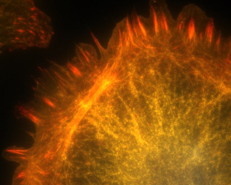
Tracing actin polarity in cellular networks
The ability of a eukaryotic cell to resist deformation, to transport intracellular cargo and to change shape depends on the cytoskeleton, an interconnected network of filamentous polymers and regulatory proteins. In order to understand these processes, the directionality of actin filaments and their interactions with binding partners are of major importance. In this talk, Matthias Eibauer discusses how his team developed the means to detect actin directionality within unlabelled filamentous networks. This approach is based on cryo-electron tomography in conjunction with image processing. This method can be adopted to reveal the polarity and binders of actin filaments in cells and at physiological cellular processes.
Dr Matthias Eibauer, University of Zurich, Switzerland
Dr Matthias Eibauer, University of Zurich, SwitzerlandMatthias Eibauer received his diploma in Physics at Ludwig-Maximilians-University, Germany in 2007. In 2011 he obtained his PhD from the Max Planck Institute of Biochemistry, Germany, under the supervision of Professor Baumeister. Between 2011 and 2016, he was a postdoctoral fellow under the guidance of Professor Medalia at the University of Zurich, Switzerland. Since 2016, Matthias has been a senior scientist at the University of Zurich. |
|
|---|---|---|
| 15:15 - 15:30 |
Multiscale models to uncover the systems-mechanobiology of cancer
The field of mechanobiology uncovered a large number of ways how mechanical properties or forces may affect intracellular signalling pathways, eg through conformal deformations of proteins or the opening of ion gates. Such pathways, in turn, may also regulate the cytoskeleton or other mechanically active components of cells, leading to mechanochemical feedbacks. In this talk, mathematical modelling approaches are discussed that describe different aspects of mechanobiology. For instance, ODE models are employed to understand the pathway dynamics, PDE models are used to understand the effect of spatial localisation of signalling molecules, and stochastic models are used to describe the force-dependent dynamics of adhesion binding. Fabian Spill finally outlines approaches to combine different models, eg adhesion dynamics with cell and tissue forces, to gain a systems-level understanding of the mechanobiology of cancer.
Dr Fabian Spill, University of Birmingham, UK
Dr Fabian Spill, University of Birmingham, UKFabian Spill trained as a mathematician and theoretical physicist and started to apply mathematical models to solve biological problems when he worked as a postdoc with Helen Byrne and Philip Maini. Since his second postdoc in the experimental bioengineering labs of Roger Kamm and Muhammad Zaman, Fabian started to collaborate closely with experimentalists on modelling mechanochemical pathways or the impact of cell shape on cell behaviour. He is now a Lecturer in Applied Mathematics at the University of Birmingham. Some of his recent work includes a stochastic-mechanical model of extravasation, a model of YAP/TAZ signal integration and a model of TGFbeta/TNFalpha synergies on MMP production in migrating cells. |
|
| 15:30 - 15:45 |
Reverse engineering cell competition

Professor Guillaume Charras, University College London, UK
Professor Guillaume Charras, University College London, UKProfessor Guillaume Charras obtained an MSc in mechanical engineering from Ecole Centrale de Paris and Georgia Institute of Technology in 1997. He then went on to study for a PhD in biochemistry from UCL between 1999 and 2002. Professor Charras did a post-doc with Tim Mitchison at Harvard Medical School between 2003 and 2007. Since 2007, he has been a group leader at the London Centre for Nanotechnology, UCL. |
|
| 15:45 - 16:00 |
Discussion
Dr Matthias Eibauer, University of Zurich, Switzerland
Dr Matthias Eibauer, University of Zurich, SwitzerlandMatthias Eibauer received his diploma in Physics at Ludwig-Maximilians-University, Germany in 2007. In 2011 he obtained his PhD from the Max Planck Institute of Biochemistry, Germany, under the supervision of Professor Baumeister. Between 2011 and 2016, he was a postdoctoral fellow under the guidance of Professor Medalia at the University of Zurich, Switzerland. Since 2016, Matthias has been a senior scientist at the University of Zurich.
Professor Guillaume Charras, University College London, UK
Professor Guillaume Charras, University College London, UKProfessor Guillaume Charras obtained an MSc in mechanical engineering from Ecole Centrale de Paris and Georgia Institute of Technology in 1997. He then went on to study for a PhD in biochemistry from UCL between 1999 and 2002. Professor Charras did a post-doc with Tim Mitchison at Harvard Medical School between 2003 and 2007. Since 2007, he has been a group leader at the London Centre for Nanotechnology, UCL. |
|
| 16:00 - 18:00 | Breakout sessions I |
| 09:30 - 09:45 |
Cell motility and extracellular basement membrane development in a model of breast development and breast cancer
Loss of the basement membrane is a hallmark of invasive cancer. Claire Robertson’s group studies how basement membrane develops by culturing nonmalignant breast epithelial cells (BEC), which develop into polarized, growth arrested structures to malignant breast cancer cells (BCC), which form disorganised masses under the same conditions. These two cell types show different movement patterns in the ECM gels: BEC rotate around their centre of mass in a process termed coherent angular motion, whereas BCC migrate randomly. Coherent angular motion appears to affect concentration of exogenous ECM by physically pulling ECM proteins into an organised shell at the cell surface as an early aspect of formation of a basement membrane-like ECM structure in mammary epithelial cells in culture, whereas this process is defective in malignant cells undergoing random motility. 
Dr Claire Robertson, Lawrence Berkeley National Lab, USA
Dr Claire Robertson, Lawrence Berkeley National Lab, USAClaire Robertson is a postdoctoral fellow at Lawrence Berkeley National Lab. She studies regulation of extracellular matrix in breast development and cancer. |
|
|---|---|---|
| 09:45 - 10:00 |
CAFs: useful cellular models to investigate cancer mechanobiology
Cancer-associated fibroblasts (CAFs) are predominant non-malignant cells in tumours that actively remodel the extracellular matrix thereby affecting the physical properties of solid tumours. Fernando Calvo will show technologies to assess CAF function in cancer and how he has used them to unveil important aspects of cancer mechanobiology. In addition, he will discuss how he has used CAFs to decipher the molecular mechanisms governing mechanotransduction, and their impact in CAF behaviour and cancer progression. 
Dr Fernando Calvo, Institute of Biomedicine & Biotechnology of Cantabria, Spain
Dr Fernando Calvo, Institute of Biomedicine & Biotechnology of Cantabria, SpainDr Fernando Calvo uses a wide range of techniques from conventional cell and molecular biology, to state-of-the-art intravital imaging of in vivo models and analysis of clinical material to study the role of the tumour microenvironment in cancer progression and dissemination. He obtained his PhD with Piero Crespo in Santander (Spain) studying Ras GTPases. He then carried out post-doctoral work in London (Erik Sahai) investigating the role of cancer-associated fibroblasts (CAFs) in tumour progression and uncovered the importance of mechanotransduction in generating and maintaining the activated status of CAFs. In 2013 he set up his own group at the Institute of Cancer Research (ICR) before moving back to Spain in early 2017, where he leads a team at the Institute of Biomedicine & Biotechnology of Cantabria. |
|
| 10:00 - 10:15 |
YAP/TAZ in development, regeneration, and cancer
The YAP and TAZ transcriptional co-activators drive TEAD-dependent transcription to promote cell proliferation during normal development, tissue regeneration, and cancer. Barry Thompson will describe how YAP/TAZ are regulated by growth factor signalling, the Hippo pathway, mechanical forces and tissue damage in epithelia. He will further describe genetic loss-of-function and gain-of-function analysis of YAP/TAZ in both Drosophila and mice, and the implications of these findings for various types of human cancer. 
Dr Barry Thompson, Francis Crick Institute, UK
Dr Barry Thompson, Francis Crick Institute, UKDr Barry Thompson was a graduate student at the Medical Research Council Laboratory for Molecular Biology (MRC-LMB) in Cambridge, where he studied the Wnt signalling pathway in the fruit fly Drosophila, in the lab of Dr Mariann Bienz. Barry then moved to the European Molecular Biology Laboratory (EMBL) to the lab of Professor Stephen Cohen, where he used Drosophila genetics to study Hippo signalling and tumour formation. Subsequently, he visited the Institute for Molecular Pathology in Vienna, where he performed an in vivo RNAi screen in Drosophila in the lab of Dr Barry Dickson. The Thompson laboratory at the Crick (previously the Cancer Research UK London Research Institute) is now interested in studying genes that control tissue growth and form in Drosophila and translating these findings into mouse models. Dr Thompson received the EMBO Young Investigator Award in 2011 and was recently appointed as a Wellcome Trust Investigator. |
|
| 10:15 - 10:30 |
Discussion
Dr Claire Robertson, Lawrence Berkeley National Lab, USA
Dr Claire Robertson, Lawrence Berkeley National Lab, USAClaire Robertson is a postdoctoral fellow at Lawrence Berkeley National Lab. She studies regulation of extracellular matrix in breast development and cancer.
Dr Fernando Calvo, Institute of Biomedicine & Biotechnology of Cantabria, Spain
Dr Fernando Calvo, Institute of Biomedicine & Biotechnology of Cantabria, SpainDr Fernando Calvo uses a wide range of techniques from conventional cell and molecular biology, to state-of-the-art intravital imaging of in vivo models and analysis of clinical material to study the role of the tumour microenvironment in cancer progression and dissemination. He obtained his PhD with Piero Crespo in Santander (Spain) studying Ras GTPases. He then carried out post-doctoral work in London (Erik Sahai) investigating the role of cancer-associated fibroblasts (CAFs) in tumour progression and uncovered the importance of mechanotransduction in generating and maintaining the activated status of CAFs. In 2013 he set up his own group at the Institute of Cancer Research (ICR) before moving back to Spain in early 2017, where he leads a team at the Institute of Biomedicine & Biotechnology of Cantabria. |
|
| 10:30 - 11:00 | Coffee |
| 11:00 - 11:15 |
Fast super-resolution imaging without artefacts
Conventional localisation microscopy relies on sparse activation of flurophores to allow accurate data fitting, meaning acquisition is slow and live cell experiments difficult or impossible. Several algorithms have been developed to cope with high emitter density. However, these produce significant image artefacts as the density is increased, which are easily mistaken for high resolution. By examining known biological structures it is shown that artefacts can be largely eliminated by pre-processing the image sequence with a succession of Haar wavelet kernels (HAWK), improving the resolving power and ensuring that the image reflects the structure of the sample.
Dr Susan Cox, King's College London, UK
Dr Susan Cox, King's College London, UKSusan is a Royal Society University Research Fellow in the Randall Centre for Biophysics at King's College London. Her research is centred around developing novel super-resolution microscopy techniques, and using them to investigate the cytoskeleton. The methods she developed have enabled localisation microscopy to be carried out routinely in live cells. In recognition of this Susan was awarded both the RMS Medal for Light Microscopy and the President's Medal of the Society of Experimental Biology for the Cell Section in 2015. Recently she has become interested in the fundamental limitations of super-resolution microscopy techniques, and how to make sure data is reliable and accurate. |
|
|---|---|---|
| 11:15 - 11:30 |
Next-generation spatial RNA sequencing technologies
Tumour growth is regulated by cell autonomous as well as short- and long-range cell-cell interactions. The Lee group has been developing methods to label, track, and isolate single cells and their gene expression products in tumour tissues, including sequencing of somatic mutations, lineage reporters, and transcriptional markers. First, the talk will focus on a high-sensitivity in situ sequencing method for fluorescently labelling single cells and their applications in cancer medicine. Second, the talk will present paired-end DNA sequencing chemistry for 3D transcriptome reconstruction of developing tissues, concluding with discussing ways to study genotype-phenotype correlations in vivo. 
Dr Je H Lee, Cold Spring Harbor Laboratory, USA
Dr Je H Lee, Cold Spring Harbor Laboratory, USADr Lee received his MD PhD in Cell and Molecular Physiology and trained in Medicine. As a post-doc in Dr George Church’s laboratory at Harvard, he developed technologies to reconstruct transcriptome signatures with single-cell resolution in 3D to study cellular behaviour in their native environment. He joined CSHL in 2014, and his lab is primarily focused on how cell-cell interactions produce a gradient of transcriptionally competent states during embryo development and morphogen signalling, linking mechanotransduction, cell cycle progression and cell fate specification. To spatially reconstruct the genetic, epigenetic, and regulatory basis of embryonic development, the Lee lab develops various in situ sequencing, single-cell, and image reconstruction technologies. |
|
| 11:30 - 12:00 |
Discussion
Dr Susan Cox, King's College London, UK
Dr Susan Cox, King's College London, UKSusan is a Royal Society University Research Fellow in the Randall Centre for Biophysics at King's College London. Her research is centred around developing novel super-resolution microscopy techniques, and using them to investigate the cytoskeleton. The methods she developed have enabled localisation microscopy to be carried out routinely in live cells. In recognition of this Susan was awarded both the RMS Medal for Light Microscopy and the President's Medal of the Society of Experimental Biology for the Cell Section in 2015. Recently she has become interested in the fundamental limitations of super-resolution microscopy techniques, and how to make sure data is reliable and accurate.
Dr Je H Lee, Cold Spring Harbor Laboratory, USA
Dr Je H Lee, Cold Spring Harbor Laboratory, USADr Lee received his MD PhD in Cell and Molecular Physiology and trained in Medicine. As a post-doc in Dr George Church’s laboratory at Harvard, he developed technologies to reconstruct transcriptome signatures with single-cell resolution in 3D to study cellular behaviour in their native environment. He joined CSHL in 2014, and his lab is primarily focused on how cell-cell interactions produce a gradient of transcriptionally competent states during embryo development and morphogen signalling, linking mechanotransduction, cell cycle progression and cell fate specification. To spatially reconstruct the genetic, epigenetic, and regulatory basis of embryonic development, the Lee lab develops various in situ sequencing, single-cell, and image reconstruction technologies. |