Links to external sources may no longer work as intended. The content may not represent the latest thinking in this area or the Society’s current position on the topic.
Dynamic in-situ microscopy relating structure and function
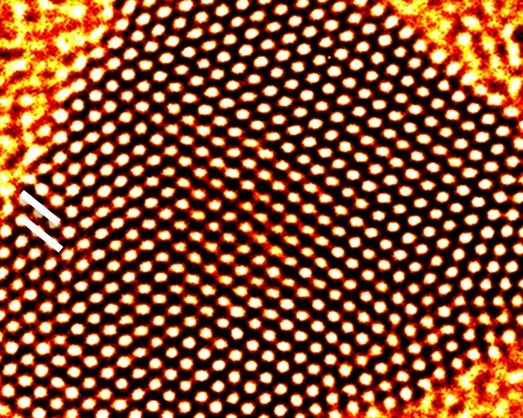
Fast-track discussion meeting organised by Dame Pratibha Gai FREng FRS, Professor Edward Boyes, Professor Rik Brydson and Professor Richard Catlow FRS.
This meeting evidenced and advanced development in dynamic in-situ environmental electron, scanning probe, optical and fast time resolved microscopy and computer modelling studies of vital interest in the chemical, physical and life sciences, underpinning technologies of high commercial and societal value. It focused on the pivotal role of imaging and spectroscopy for dynamic processes across the sciences to access previously invisible aspects of real world processes.
The schedule of talks and speaker biographies are available below. Speaker abstracts are also available below. Recorded audio of the presentations is available below. An accompanying journal issue for this meeting was published in Philosophical Transactions of the Royal Society A.
Attending the event
This meeting has taken place.
Enquiries: contact the Scientific Programmes team
Organisers
Schedule
Chair

Professor Dame Pratibha Gai FREng FRS, University of York, UK

Professor Dame Pratibha Gai FREng FRS, University of York, UK
Pratibha Gai is a Fellow of the Royal Society and Fellow of the Royal Academy of Engineering. She is Professor and Chair of Electron Microscopy in the Departments of Chemistry and Physics, and founding Co-Director of the York Nanocentre, at University of York. She joined York from the USA where she held senior positions as DuPont Research Fellow and concurrently as adjunct Professor at the University of Delaware. Previously, she established and led the surface reactions and in-situ microscopy group at University of Oxford, after graduating with a PhD in Physics from Cambridge. With E D Boyes she co-invented the atomic resolution-environmental transmission electron microscope (ETEM) to image dynamic gas-solid reactions at the atomic level, which is used by researchers worldwide, electron microscope manufacturers and chemical companies; and developed single atom resolution-E(Scanning)TEM with E D Boyes. Her key contributions include in-situ EM of catalysts, cleaner processes for energy, food, healthcare and pigment coatings, and stabilised ceramics. Her awards include Institute of Physics Gabor Prize and L’Oréal-UNESCO International Women in Science Award as the 2013 Laureate for Europe. In 2018 she was appointed a Dame (DBE) for services to chemical sciences and technology.
| 08:00 - 08:30 |
Native state analysis of complex, beam-sensitive systems by electron microscopy
Complex, electron-beam sensitive systems include organic crystals, hybrid organic-inorganic materials, some inorganic materials such as hydrates as well as multiphase solid/liquid and solid/gas materials. Arguably they constitute the majority of systems of current interest across a wide range of scientific and engineering disciplines including chemistry and chemical engineering, engineering materials, food science, biology and increasingly physics and electronic engineering. For the purposes of this presentation Professor Brydson defines in-situ as the native-state analysis of such complex, beam sensitive materials and systems, in that they remain unmodified by interaction with the electron beam which is well-known to alter both the structure and chemistry of the specimen. Here, he also concentrates almost exclusively on transmission electron microscopy (TEM) of thin specimens. Electron dose is related to the energy absorbed by the specimen during electron irradiation and can be a function of both the electron fluence and fluence rate in the TEM. Professor Brydson discusses a number of useful concepts for TEM imaging and/or chemical analysis, including a dose budget for experiments and also a dose-limited resolution. We also highlight the current state-of-the-art in dose control during imaging and spectroscopy including the use of scanning TEM (STEM) as opposed to conventional TEM (CTEM), compressed sensing methods and the cryo-preservation of samples. Finally, he also discusses dynamic studies of such complex materials and systems including the use of in-situ liquid and gas cells TEM holders, which have their own additional limitations due to dose, as well as the use of time-resolved cryo-methods to provide snapshots of dynamic systems. 
Professor Rik Brydson, University of Leeds, UK

Professor Rik Brydson, University of Leeds, UKRik Brydson holds a chair in Nanomaterials Characterisation at the University of Leeds. He was a member of the Executive Board of the European Microscopy Society until 2016 and is currently Honorary Secretary Physical Sciences of the Royal Microscopy Society. He helped found the SuperSTEM advanced electron microscopy facility at Daresbury (the EPSRC National Facility) and chairs their Consortium Advisory Board. He has over 30 years research experience in advanced analytical electron microscopy of engineering materials particularly focused around both inorganic and organic nanoparticles and nanostructured materials and their dispersions.
|
|
|---|---|---|
| 08:30 - 08:35 | Discussion | |
| 08:35 - 09:05 |
Spatio-temporal dynamics of active catalysts observed by multi-scale in-situ electron microscopy
Catalysts are operating far from thermodynamic equilibrium in an open system that exchanges heat and matter with the surrounding. In their active state, they are supposed to repeatably facilitate sequences of reaction steps of a catalytic cycle. The combination of non-equilibrium conditions and cyclic behaviour can result in complex dynamics and formation of dissipative structures. Such structures are difficult to study because they only exist under reactive conditions. Furthermore, they are difficult to predict using computational methods because of the non-linearity of the underlying kinetics. Structure-function relationships have thus to be studied under relevant reaction conditions. While in situ TEM enables observation of atomistic details under conditions of slow kinetics, in situ SEM delivers complementary information about catalyst dynamics at reduced lateral resolution and provides insight about collective phenomena that are controlled by heat and mass transport. As will be shown in the presentation, the combination of in situ SEM with in situ TEM enables a multi-scale approach for the study of catalyst dynamics from the Å to the mm range and across pressures ranging from 10-5 to 10+5 Pa. Combined with simultaneous detection of reaction products by RGA it is possible to correlate structural dynamics with catalytic activity. 
Dr Marc Willinger, ETH Zürich, Switzerland

Dr Marc Willinger, ETH Zürich, SwitzerlandDr Willinger studied physics at the Technical University in Vienna and obtained his PhD from the Technical University in Berlin for the investigation of the electronic structure of vanadium phosphorous oxides using a combination of electron energy loss spectroscopy and DFT based simulations. After one and a half years post-doc at the Fritz-Haber-Institute, he moved to the University of Aveiro in Portugal, where he worked as an independent researcher for four years. In 2011 he went back to the Fritz-Haber-Institute as leader of the electron microscopy group at the Department of Inorganic Chemistry, where he used in-situ microscopy to study catalysts in their active state. Since 1 February 2018, he has led the TEM group at the 'Eidgenössische Technische Hochschule' (ETH) in Zürich. |
|
| 09:05 - 09:15 | Discussion | |
| 09:15 - 09:30 | Contributed talk | |
| 09:30 - 10:00 | Coffee | |
| 10:00 - 10:30 |
Using light to see at the atomic scale using pico-cavities
Recent advances in using noble metals to confine light to the sub-nm3 scale through advanced plasmonics is opening up a new realm of 'picoscopy'. Professor Baumberg will show how a wide range of dynamic motions of atoms and molecules can be studied on microsecond timescales under ambient conditions. This explores crucial realms of catalytic surface reconstructions and electrochemical interfaces that have been inaccessible. Professor Baumberg will show how simple nano-architectures enable such trapping of visible light, and how elastic and inelastic light scattering reveal detailed spectral information about extremely small volumes of space. A few examples will highlight the light-induced motion of individual gold atoms, of flickering defects in metal interfaces, and of individual bonds in single molecules moving in real time. 
Professor Jeremy J Baumberg FRS, University of Cambridge, UK

Professor Jeremy J Baumberg FRS, University of Cambridge, UKProfessor Jeremy J Baumberg FRS, directs a UK Nano-Photonics Centre at the University of Cambridge and has extensive experience in developing optical materials structured on the nano-scale that can be assembled in large volume. He is also Director of the Cambridge Nano Doctoral Training Centre, a key UK site for training PhD students in interdisciplinary Nano research. Strong experience with Hitachi, IBM, his own spin-offs Mesophotonics and Base4, as well as strong industrial engagement give him a unique position to combine academic insight with industry application in a two-way flow. With over 20,000 citations, he is a leading innovator in Nano. This has led to awards of the Institute of Physics (IoP) Faraday Gold Medal (2017), Royal Society Rumford Medal (2014), IoP Young Medal (2013), Royal Society Mullard Prize (2005), the IoP Charles Vernon Boys Medal (2000) and the IoP Mott Lectureship (2005). He frequently talks on NanoScience to the media, and is a strategic advisor on NanoTechnology to the UK Research Councils. He is a Fellow of the Royal Society, the Optical Society of America, and the Institute of Physics. His recent popular science book The Secret Life of Science: How Science Really Works and Why it Matters is just published by PUP. |
|
| 10:30 - 10:40 | Discussion | |
| 10:40 - 11:10 |
The Light Years: combined optical and environmental electron microscopy to visualise plasmonic processes with atomic-scale resolution
Pearl Jam’s hit, 'The Light Years', declares "We were but stones, light made us stars". Bringing light to the transmission electron microscope (TEM) promises to transform our understanding of materials, enabling both direct observation of light-mediated processes and detection of optical emission with atomic scale resolution. To help achieve this goal, Professor Dionne's lab is developing capabilities for concurrent optical and electron microscopy within an environmental TEM. This presentation will describe their efforts to visualise a variety of plasmonic processes in situ with nanometre-to-atomic scale resolution. The group focuses in particular on i) observing plasmon-driven chemical transformations in nanoparticles; ii) detecting quantum light emission from two-dimensional materials; and iii) developing TEM-Raman with nanometre-scale resolution. First, they study the photocatalytic dehydrogenation of Au-Pd systems, in which the Au acts as a plasmonic light absorber and Pd serves as the catalyst. They find that plasmons modify the rate of distinct reaction steps differently, increasing the overall rate more than ten-fold. Plasmons also open a new reaction pathway that is not observed without illumination, laying a foundation for site-selective and product-specific photocatalysts. Secondly, the group uses scanning transmission electron microscopy and cathodoluminescence spectroscopy to investigate colour centres in two-dimensional hexagonal boron nitride, a wide bandgap material capable of room-temperature, high-brightness visible quantum emission. They find that sharp, single-photon emission peaks are usually associated with regions of multiple fork dislocations, facilitating design of next-generation two-dimensional materials and devices. Finally, they describe a new technique for high-resolution spectroscopy in the TEM: electron-and-light-induced stimulated Raman scattering (ELISR). In particular, theyleverage the electron beam as a highly-localised Angstrom-scale source to locally excite the plasmonic resonances of individual nanoparticles, which stimulate local Raman scattering. They show how ELISR can be used to map molecular signatures with nanometre-scale resolution. 
Professor Jennifer Dionne, Stanford University, USA

Professor Jennifer Dionne, Stanford University, USAJennifer Dionne is an associate professor of Materials Science and Engineering at Stanford, and an affiliate faculty of the Stanford Neurosciences Institute, TomKat Center for Sustainable Energy, and Bio-X. Jen received her PhD in Applied Physics at the California Institute of Technology, advised by Harry Atwater, and BS degrees in Physics and Systems & Electrical Engineering from Washington University in St. Louis. Prior to joining Stanford, she served as a postdoctoral researcher in Chemistry at Berkeley, advised by Paul Alivisatos. Jen’s research develops new nanophotonic materials and microscopies to observe chemical and biological processes as they unfold with nanometer scale resolution. She then uses these observations to help improve energy-relevant processes (such as photocatalysis and energy storage) and medical diagnostics. Her work has been recognised with a Moore Inventor Fellowship (2017), the Materials Research Society Young Investigator Award (2017), Adolph Lomb Medal (2016), and Sloan Foundation Fellowship (2015), and was featured on Oprah’s list of '50 Things that will make you say ‘Wow’!'. She currently serves as director of the DOE-funded Photonics at Thermodynamic Limits Energy Frontier Research Center and faculty co-director of Stanford’s Photonics Research Center. |
|
| 11:10 - 11:20 | Discussion | |
| 11:20 - 11:35 | Contributed talk |
Chair

Professor Jennifer Dionne, Stanford University, USA

Professor Jennifer Dionne, Stanford University, USA
Jennifer Dionne is an associate professor of Materials Science and Engineering at Stanford, and an affiliate faculty of the Stanford Neurosciences Institute, TomKat Center for Sustainable Energy, and Bio-X. Jen received her PhD in Applied Physics at the California Institute of Technology, advised by Harry Atwater, and BS degrees in Physics and Systems & Electrical Engineering from Washington University in St. Louis. Prior to joining Stanford, she served as a postdoctoral researcher in Chemistry at Berkeley, advised by Paul Alivisatos. Jen’s research develops new nanophotonic materials and microscopies to observe chemical and biological processes as they unfold with nanometer scale resolution. She then uses these observations to help improve energy-relevant processes (such as photocatalysis and energy storage) and medical diagnostics. Her work has been recognised with a Moore Inventor Fellowship (2017), the Materials Research Society Young Investigator Award (2017), Adolph Lomb Medal (2016), and Sloan Foundation Fellowship (2015), and was featured on Oprah’s list of '50 Things that will make you say ‘Wow’!'. She currently serves as director of the DOE-funded Photonics at Thermodynamic Limits Energy Frontier Research Center and faculty co-director of Stanford’s Photonics Research Center.
Professor Peter van Aken, Max Planck Institute for Solid State Research, Germany

Professor Peter van Aken, Max Planck Institute for Solid State Research, Germany
Professor Peter A van Aken leads the Stuttgart Center for Electron Microscopy (StEM), adding exceptional strength to the analytical capabilities of the Max Planck Institute for Solid State Research. StEM possesses outstanding expertise in scanning and transmission electron microscopy (TEM), focused ion-beam applications, and methodology development. Professor van Aken’s research focuses on the atomic-scale characterisation of interfaces, functional complex oxide hetero-structures, strained semiconductors, of optical properties of nanostructured thin films and plasmonic-active nanostructures, nanoparticles and nanomaterials, as well as of molecules on 2D materials. He uses and further develops the advanced TEM techniques electron energy-loss spectroscopy, energy-dispersive X-ray spectroscopy, inline electron holography, in-situ TEM methods, strain mapping, quantitative high-resolution TEM analysis, different quantitative electron diffraction techniques, image processing and simulation, and electron ptychography and tomography. Professor van Aken’s research mission is the advancement of the in-depth microscopic understanding of materials with respect to their functionalities and structure–property relationships.
| 12:30 - 13:00 |
Present status and future prospects of environmental HVEM for nanomaterials
Environmental transmission electron microscopy (E-TEM) attracts a strong interest of materials scientists, particularly, those of batteries and catalysts, because the actual chemical reaction processes in gases and liquids should be clarified in real space and in atomic level. E-TEM observations of thick samples also become important for cutting-edge materials in practical use. 3D observation is also necessary for clarifying morphologies of practical catalysts. Professor Tanaka and colleagues have developed 1MV TEM/STEM equipped with an open-type environmental cell which enables observations in 100 Torr of hydrogen, oxygen, nitrogen and carbon mono-oxide(CO) gases, named 'Reaction Science HVEM'(RSHVEM). In this talk, Professor Tanaka would like to present new data obtained by the instrument such as (1) in-situ observation of porous gold catalysts, whose inner surfaces with zigzag atom arrangement enhance oxidation of CO gas, (2) observation of diesel soot oxidized in air, and (3) in-situ observation of fractures related to 'hydrogen brittleness' for a Cu/Si interface and grain boundaries in Ni3Al alloys. Observation of lithium battery materials is also available using a non-exposure transfer holder as well as QMAS ability. Finally, advantages and future prospects of E-HVEM are discussed. 
Professor Nobuo Tanaka, Nagoya University, Japan

Professor Nobuo Tanaka, Nagoya University, JapanDr Tanaka obtained a PhD in Applied Physics from Nagoya University in 1978 on the research of growth of metal clusters on crystals. He has majored in high-resolution and in-situ electron microscopy and diffraction of small particles and clusters. He is currently a designated professor of Institute of Materials and Systems for Sustainability (IMaSS) of Nagoya University, Japan. He was the director of the institute from 2012 to 2015 and also the president of the Japanese Society of Microscopy (JSM) from 2015–2017. Dr Tanaka studies nano-structure science, thin film physics, catalysis science, and surface/interface physics by using aberration-corrected high-resolution electron microscopy including phase imaging, environmental electron microscopy and electron tomography. He is also interested in magnetic properties of transition metal clusters and semiconductor micro-devices studied by using pulsed, spin-polarized and vortex electron beams as well as electron energy-loss spectroscopy (EELS). |
|
|---|---|---|
| 13:00 - 13:10 | Discussion | |
| 13:10 - 13:35 |
Atoms in action for energy, healthcare and environment using dynamic in-situ atomic resolution environmental (S)TEM: advancing the frontiers of chemical research
Many heterogeneous chemical reactions utilise gas-solid catalyst reactions at elevated temperatures and they play a pivotal role in the production of energy, healthcare, pollution control and food. The dynamic catalytic chemical reactions take place at the atom level. Understanding atomic structure evolution and the ensuing catalyst function are vital to developing novel materials and improved chemical processes. However due to limitations of the instrumentation these atomic processes were not well understood. The development of the first atomic resolution-Environmental Transmission Electron Microscope (atomic resolution-ETEM) [Boyes and Gai, Ultramicroscopy 67 (1997) 197], enables the visualisation and analysis of gas-solid catalyst reactions at the atomic level leading to key discoveries and the development is used globally. The ETEM has been advanced to analytical environmental scanning TEM (ESTEM) with single atom resolution and full analytical capabilities. Novel E(S)TEM imaging and analysis in real-time in controlled real-world reaction environments reveal single atom dynamics in fuel cell catalysts, fuels including sustainable biofuels, healthcare applications and ammonia synthesis for agricultural products in food production. The development of a liquid holder enables imaging and analysis of chemical reactions in liquid environments. The dynamic studies, in combination with chemical methods, provide unique insights into atoms in action and atomic scale reaction pathways in chemical reactions, opening new opportunities for the control of the dynamic atomic structure for beneficial catalytic and functional properties. Smarter synthesis leading to improved materials and processes with significantly enhanced performance are possible. Benefits include new knowledge and cleaner processes for renewable energy and healthcare as well as improved or replacement mainstream technologies in the chemical and energy industries. 
Professor Dame Pratibha Gai FREng FRS, University of York, UK

Professor Dame Pratibha Gai FREng FRS, University of York, UKPratibha Gai is a Fellow of the Royal Society and Fellow of the Royal Academy of Engineering. She is Professor and Chair of Electron Microscopy in the Departments of Chemistry and Physics, and founding Co-Director of the York Nanocentre, at University of York. She joined York from the USA where she held senior positions as DuPont Research Fellow and concurrently as adjunct Professor at the University of Delaware. Previously, she established and led the surface reactions and in-situ microscopy group at University of Oxford, after graduating with a PhD in Physics from Cambridge. With E D Boyes she co-invented the atomic resolution-environmental transmission electron microscope (ETEM) to image dynamic gas-solid reactions at the atomic level, which is used by researchers worldwide, electron microscope manufacturers and chemical companies; and developed single atom resolution-E(Scanning)TEM with E D Boyes. Her key contributions include in-situ EM of catalysts, cleaner processes for energy, food, healthcare and pigment coatings, and stabilised ceramics. Her awards include Institute of Physics Gabor Prize and L’Oréal-UNESCO International Women in Science Award as the 2013 Laureate for Europe. In 2018 she was appointed a Dame (DBE) for services to chemical sciences and technology.
|
|
| 13:35 - 13:40 | Discussion | |
| 13:40 - 14:10 |
Seeing is believing: atomic-scale imaging of catalysts under reaction conditions
The atomic-scale structure of a catalyst under reaction conditions determines its activity, selectivity, and stability. Recently it has become clear that essential differences can exist between the behaviour of catalysts under industrial conditions (high pressure and temperature) and the (ultra)high vacuum conditions of traditional laboratory experiments. Differences in structure, composition, reaction mechanism, activity, and selectivity have been observed. These observations made it clear that meaningful results can only be obtained at high pressures and temperatures. Therefore, the last years have seen a tremendous effort in designing new instruments and adapting existing ones to be able to investigate catalysts in situ under industrially relevant conditions. In this talk, Professor Groot will give an overview of the in situ imaging techniques the group uses to study the structure of model catalysts under atmospheric pressures and elevated temperatures. The group has developed set-ups that combine an ultrahigh vacuum environment for model catalyst preparation and characterisation with a high-pressure flow reactor cell, integrated with either a scanning tunneling microscope or an atomic force microscope. With these set-ups the group is able to perform atomic-scale investigations of well-defined model catalysts under industrial conditions. Additionally, they combine the structural information from scanning probe microscopy with mass spectrometry measurements. In this way, the group can correlate structural changes of the catalyst due to the gas composition with its catalytic performance. Furthermore, they use other in situ imaging techniques such as transmission electron microscopy, surface X-ray diffraction, and optical microscopy, all combined with mass spectrometry. 
Professor Irene Groot, Leiden University, The Netherlands

Professor Irene Groot, Leiden University, The NetherlandsIrene Groot obtained her PhD degree at Leiden University investigating the dissociation of hydrogen on single crystals, both experimentally using supersonic molecular beams and theoretically performing density functional theory and quantum dynamics calculations. After a postdoc at the Fritz Haber Institute in Berlin, she went to the Leiden Institute of Physics with a personal Veni fellowship from the Netherlands Organization for Scientic Research. Currently, Irene Groot is associate professor (tenured) at the Leiden Institute of Chemistry where she heads the operando catalysis research group. She investigates the structure-activity relationship of catalysts under industrial conditions using operando scanning probe microscopy, transmission electron microscopy, Raman spectroscopy, and X-ray-based techniques. She focuses on industrial processes related to sustainable energy and materials production. Current topics in her group are hydrodesulfurization, Fischer-Tropsch synthesis, methanol steam reforming, automotive catalysis, chlorine production, methanol steam reforming, and graphene growth on liquid copper. |
|
| 14:10 - 14:20 | Discussion | |
| 14:20 - 14:50 | Coffee | |
| 14:50 - 15:20 |
Micro to macro-scopic in vivo imaging including magnetic resonance imaging: using advanced medical imaging technologies as a ‘macroscope’ to determine pathophysiologies of complex human brain diseases
The rapid advances in understanding and treatment of common human brain diseases have (arguably) been made possible by advances in development of in vivo diagnostic imaging technologies in the last few decades. The human brain is complex, with no truly comparable model species, limiting the ultimate clinical value of many rodent models. Advances in non-invasive medical imaging technologies particularly magnetic resonance imaging, mean that detailed data on brain structure and function and pathological changes in disease can now be determined and tracked in large human samples including population studies and disease cohorts. However, these methods also have limitations of resolution, and tissue often only becomes available post mortem, reflecting end stage disease. Hence, to tackle a complex disorder that develops insidiously in life requires a multimodal approach that includes in vivo MRI and related ‘macroscopic’ methods in patients and volunteers, and parallel and complementary microscopic methods applied to post-mortem brain tissue and in vivo and in vitro static and dynamic imaging methods in rodent models such as MRI, 2-PI, histological and electron microscopic methods. This lecture will illustrate these principles using the example of cerebral small vessel disease, a major cause of both stroke and dementia and of ageing-related multimorbidities. 
Professor Joanna Wardlaw CBE FMedSci, University of Edinburgh and NHS Lothian, UK

Professor Joanna Wardlaw CBE FMedSci, University of Edinburgh and NHS Lothian, UKProfessor Joanna Wardlaw, CBE FMedSci, is Professor of Applied Neuroimaging at the University of Edinburgh and Consultant Neuroradiologist for NHS Lothian. She has worked for many years to understand the brain and its blood supply, and on treatments to improve blood flow to the brain, including thrombolytic drugs that are now used routinely to treat stroke. She now focuses on the much more complicated problem of ‘small vessel disease’, which as well as being a common cause of stroke, is also a common cause of dementia and physical frailty as people get older. Working with many colleagues, she has been instrumental in advancing understanding of the causes of small vessel disease and is now testing possible treatments in clinical trials. She has published over 500 papers, set up national research imaging facilities, co-ordinated international research networks, and advanced stroke care worldwide. Recent recognition for her work includes the European Stroke Organisation President’s Award (2017), the American Heart Association Feinberg Award (2018), and the Karolinska Stroke Award (2018). A Fellow of the Royal Society of Edinburgh and of the UK’s Academy of Medical Sciences, she was the first woman to be awarded the American Heart Association’s Feinberg Award for Clinical Advances in Stroke in 2018, was one of 35 Women in Medicine recognized by the UK Royal Medical Colleges in 2017-18, and was made a Commander of the Order of the British Empire (CBE) for services to Medicine and Neuroscience in 2016, a high honour in the UK. |
|
| 15:20 - 15:30 | Discussion | |
| 15:30 - 15:55 |
Modelling the structures of crystals, surfaces and nano-particles
Structure prediction is a long standing challenge in solid state and nano-science, with a variety of approaches including the use of topological principles and the search of energy landscapes employing a range of procedures and algorithms. Professor Catlow will review briefly the methodologies currently in use and illustrate both their efficacy and the current challenges by describing recent applications, concentrating mainly on inorganic materials and their surfaces and on both metallic and oxide nano-particles. He will discuss how computational structure modelling and prediction can guide and assist the interpretation of experimental studies. 
Sir Richard Catlow FRS, Cardiff University and University College London, UK

Sir Richard Catlow FRS, Cardiff University and University College London, UKRichard Catlow is developing and applying computer models to solid state and materials chemistry: areas of chemistry that investigate the synthesis, structure and properties of materials in the solid phase. By combining his powerful computational methods with experiments, Richard has made considerable contributions to areas as diverse as catalysis and mineralogy. His approach has also advanced our understanding of how defects (missing or extra atoms) in the structure of solids can result in non-stoichiometric compounds. Such compounds have special electrical or chemical properties since their contributing elements are present in slightly different proportions to those predicted by chemical formula. Richard’s work has offered insight into mechanisms of industrial catalysts, especially involving microporous materials and metal oxides. In structural chemistry and mineralogy. Simulation methods are now routinely used to predict the structures of complex solids and silicates, respectively, thanks to Richard’s demonstrations of their power. Richard was Foreign Secretary of the Royal Society from 2016 until 2021. He has for many years been involved in the exploitation of High Performance Computing in Modelling Materials. |
|
| 15:55 - 16:00 | Discussion |
Chair
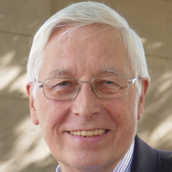
Professor Edward D Boyes, University of York, UK

Professor Edward D Boyes, University of York, UK
Ed Boyes has a PhD in Materials Science from Cambridge University, followed by research fellowships in the Oxford Materials Dept and at Wolfson College, Oxford. The almost 20 year move to the Experimental Station of DuPont CR&D in Wilmington, DE, USA, produced the original catalyst applications oriented atomic lattice resolution environmental TEM (ETEM, with P L Gai) and the ultrahigh resolution (~1nm at 1kV), highly surface sensitive, low voltage SEM (+LVEDX). He was appointed to the technical advisory group (TAG) of the US President’s Council of Advisors on Science and Technology (PCAST) for NNI review. In 2007 he returned to the UK as Professor of Physics and Electronics and founding co-director (with P L Gai) of the York JEOL Nanocentre at the University of York; with development and applications of aberration corrected sub-Angstrom ETEM and introduction of the single atom sensitive analytical Environmental STEM (ESTEM).

Professor Rik Brydson, University of Leeds, UK

Professor Rik Brydson, University of Leeds, UK
Rik Brydson holds a chair in Nanomaterials Characterisation at the University of Leeds. He was a member of the Executive Board of the European Microscopy Society until 2016 and is currently Honorary Secretary Physical Sciences of the Royal Microscopy Society. He helped found the SuperSTEM advanced electron microscopy facility at Daresbury (the EPSRC National Facility) and chairs their Consortium Advisory Board. He has over 30 years research experience in advanced analytical electron microscopy of engineering materials particularly focused around both inorganic and organic nanoparticles and nanostructured materials and their dispersions.
Chair
Professor Archie Howie CBE FRS, University of Cambridge, UK

Professor Archie Howie CBE FRS, University of Cambridge, UK
Archie Howie has worked on physics problems of electron microscopy for over 60 years. He started out with Peter Hirsch and Mike Whelan on diffraction contrast imaging of dislocations and developed with Whelan the dynamical theory of electron diffraction to explain the images. This was extended to deal with weak beam images in collaboration with Pratibha Gai. Archie then spent a few years attempting with Ondrej Krivanek and John Rodenburg to determine the structure of 'amorphous' materials from high resolution electron microscope images. With his colleague Mick Brown he obtained the second VG STEM which supported pioneering work with Mike Treacy and Steve Pennycook on high angle annular dark field imaging of catalysts. The first aloof beam energy loss spectra obtained with this instrument by Laurie Marks opened the door to the dielectric theory of localised valence spectroscopy and the study of plasmon excitations on complex nanostructures.
Dynamic processes stimulated by the electron beam have been a frequent feature in this work though usually something to be understood so that they could be avoided or at least minimised. He previously investigated the possible role in thin crystal image contrast of thermally excited flexural modes. In cryo electron microscopy where the specimen may be subject to buckling stresses from the freezing process or due to charge accumulation, flexural movement may be significant.

Sir Richard Catlow FRS, Cardiff University and University College London, UK

Sir Richard Catlow FRS, Cardiff University and University College London, UK
Richard Catlow is developing and applying computer models to solid state and materials chemistry: areas of chemistry that investigate the synthesis, structure and properties of materials in the solid phase. By combining his powerful computational methods with experiments, Richard has made considerable contributions to areas as diverse as catalysis and mineralogy. His approach has also advanced our understanding of how defects (missing or extra atoms) in the structure of solids can result in non-stoichiometric compounds. Such compounds have special electrical or chemical properties since their contributing elements are present in slightly different proportions to those predicted by chemical formula. Richard’s work has offered insight into mechanisms of industrial catalysts, especially involving microporous materials and metal oxides. In structural chemistry and mineralogy. Simulation methods are now routinely used to predict the structures of complex solids and silicates, respectively, thanks to Richard’s demonstrations of their power. Richard was Foreign Secretary of the Royal Society from 2016 until 2021. He has for many years been involved in the exploitation of High Performance Computing in Modelling Materials.
| 08:00 - 08:30 |
Molecular to cellular mechanics probed by high-speed atomic force microscopy
Mechanical forces in biological processes operate at the level of individual proteins, macromolecular complexes and the whole cell. Moreover, the dynamic response of biological processes span over several orders of magnitude in time, from sub-microseconds to several minutes. Thus, deciphering the role of mechanical forces in biological systems requires probing their mechanical properties over a wide range of length and time scales. High-speed atomic force microscopy (HS-AFM) is a unique technology allowing sub-second, nanometric imaging. Professor Rico's group has adapted HS-AFM to perform high-speed force spectroscopy (HS-FS) both on single molecules and living cells with microsecond time resolution. They probed protein unfolding and receptor/ligand unbinding up to the velocity of molecular dynamics simulations. HS-FS gives access to previously hidden events and, combined with MD simulations, provides atomistic understanding of biomolecular processes based on experimental results. These recent results revealed heterogeneous and rate dependent unfolding and unbinding pathways on different biomolecules. At the cellular level, the group has developed high-frequency microrheology to probe the mechanics of cells at short timescales, down to ~10 µs. The group probed the viscoelastic response of different cell types and upon cytoskeleton perturbation. At microsecond timescales, cells exhibit rich viscoelastic responses that depend on the state of the cytoskeleton and on the cell type. Mechanical measurements over a wide dynamic range provides a mechanistic understanding of cell mechanics based on the dynamics of individual filaments. Thus, HS-FS opens an avenue to better understand the dynamics of proteins and cells at previously inaccessible short timescales. 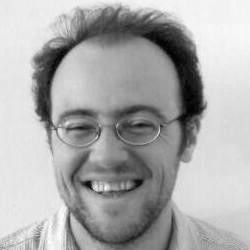
Professor Felix Rico, U1067 Aix-Marseille University, CNRS and Inserm, France

Professor Felix Rico, U1067 Aix-Marseille University, CNRS and Inserm, FranceFelix Rico has been Associate Professor in the Department of Physics of Aix-Marseille University since 2013. His research in force microscopy is developed at the LAI U1067, a joint Aix-Marseille University, CNRS and Inserm laboratory. He studied Physics at the University Autonoma of Barcelona and received his PhD in Biophysics from the University of Barcelona. Before moving to Marseille, he was postdoc at the University of Miami, USA and then at Institut Curie in Paris. He has been working in force measurements with atomic force microscopy (AFM) since 2001. His research track is focused on the mechanics and adhesion properties of biological systems. He has developed various AFM based approaches to investigate the mechanical response of single molecules, membranes and cells. Recently, he adapted high speed AFM (HS-AFM) to work as a force spectroscopy tool, probing the mechanics and dynamics of single biomolecules and living cells at the microsecond timescales. |
|
|---|---|---|
| 08:30 - 08:40 | Discussion | |
| 08:40 - 08:55 | Contributed talk | |
| 08:55 - 09:25 |
Emerging imaging technologies to study cell architecture, dynamics and function
Powerful new ways to image the internal structures and complex dynamics of cells are revolutionising cell biology and bio-medical research. Here, Professor Lippincott-Schwartz will focus on how emerging fluorescent technologies are increasing spatio-temporal resolution dramatically, permitting simultaneous multispectral imaging of multiple cellular components. Using these tools, it is now possible to begin constructing an 'organelle interactome' describing the interrelationships of different cellular organelles as they carry out critical functions. The same tools are also revealing how genetic mutations disrupting these organelles underlie various human diseases. 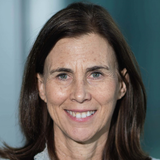
Dr Jennifer Lippincott-Schwartz, Janelia Research Campus, USA

Dr Jennifer Lippincott-Schwartz, Janelia Research Campus, USADr Jennifer Lippincott-Schwartz is a Senior Group Leader at the Howard Hughes Medical Institute’s Janelia Research Campus. She has pioneered the use of green fluorescent protein technology for quantitative analysis and modelling of intracellular protein traffic and organelle dynamics in live cells. Her innovative techniques to label, image, quantify and model specific live cell protein populations and track their fate have provided vital tools used throughout the research community. Her own findings using these techniques have reshaped thinking about the biogenesis, function, targeting, and maintenance of various subcellular organelles and macromolecular complexes and their crosstalk with regulators of the cell cycle, metabolism, aging, and cell fate determination. She is an elected member of the National Academy of Sciences, the National Academy of Medicine, the American Society of Arts and Sciences and the European Molecular Biology Organization. She is also a Fellow of The Biophysical Society, The Royal Microscopical Society and The American Society of Cell Biology. Her awards include the E.B. Wilson Medal of the American Society of Cell Biology, the Newcomb Cleveland Prize of the American Association for the Advancement of Science, the Van Deenen Medal, the Keith Porter Award of the American Society of Cell Biology, the Feodor Lynen Medal, and the Feulgen Prize of the Society of Histochemistry. She co-authored of the textbook Cell Biology and was President of the American Society of Cell Biology. Dr Lippincott-Schwartz attended Swarthmore College, received her MS from Stanford University, and obtained her PhD in Biochemistry from Johns Hopkins University in 1986. |
|
| 09:25 - 09:35 | Discussion | |
| 09:35 - 10:05 | Coffee | |
| 10:05 - 10:35 |
Radiation damage in electron images of biological structures
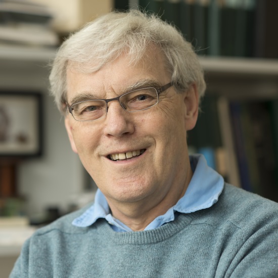
Professor Richard Henderson CH FMedSci FRS, MRC Laboratory of Molecular Biology, UK

Professor Richard Henderson CH FMedSci FRS, MRC Laboratory of Molecular Biology, UKRichard Henderson is a structural biologist with a background in physics, doctoral work on enzyme structure and mechanism, and most recently an interest in membrane proteins. Working with Nigel Unwin in the 1970s, he used electron microscopy to determine the structure of the membrane protein bacteriorhodopsin in two-dimensional crystals, initially at low resolution and 15 years later at atomic resolution. For the last 20 years, he has been working to improve the methodology of single particle electron cryomicroscopy (cryoEM), which recently reached the stage where atomic structures of a wide variety of macromolecular complexes can be obtained routinely without crystals. There are a number of time-dependent structural rearrangements that affect the cryoEM images, and these are under active investigation. |
|
| 10:35 - 10:45 | Discussion | |
| 10:45 - 11:00 | Contributed talk | |
| 11:00 - 11:15 | Contributed talk |
Chair
Sir Colin Humphreys CBE FREng FRS, Queen Mary University of London, UK

Sir Colin Humphreys CBE FREng FRS, Queen Mary University of London, UK
Colin is Professor of Materials Science at Queen Mary University of London, Distinguished Research Fellow at the University of Cambridge, and a Fellow of Selwyn College, Cambridge. He founded the Cambridge Centre for Gallium Nitride and set up two spin-off companies to exploit the research of his group on low-cost LEDs for lighting. The companies were acquired by Plessey, which is manufacturing LEDs based on this technology at their factory in Plymouth, UK. He founded the Cambridge/Rolls-Royce Centre for Advanced Materials for Aerospace. Materials developed in the Centre are now flying in Rolls-Royce engines. He recently set up a new company, Paragraf, to exploit the research of his group on next-generation large-area graphene, which promises to revolutionise a wide range of products including sensors, solar cells and electronic devices. Paragraf moved into premises in 2018 and is already employing 21 people.
| 12:15 - 12:45 |
Far-field radiation of three-dimensional plasmonic gold tapers and of dynamic toroidal moments
Elementary electromagnetic sources with well-defined near- and far-field properties form the fundamentals to systematically describe and understand observed electromagnetic phenomena of nanometre-sized objects, as well as their interactions and potential applications. Electron energy-loss spectroscopy (EELS) and cathodoluminescence spectroscopy (CL) can access these optical properties at nanometre spatial resolution, where the contrasting juxtaposition of EELS and CL information on the identical object helps to discern radiative from non-radiative optical modes. This presentation will exemplify the applications of both EELS and CL for enhancing the perception in nano-optics by showing two case studies, namely the far-field radiation of three-dimensional plasmonic gold taper and of dynamic toroidal moments. The studies on the gold tapers revealed that the excited surface plasmons either are guided away from the apex along the taper or couple to the far-field radiation depending on the tapers’ opening angle. The radiative nature of dynamic toroidal dipole modes supported in oligomer nanocavities in thin silver films has been addressed to distinguish them from static toroidal dipoles and plasmonic dark modes. Moreover, numerical finite-difference time-domain simulations were performed for comprehensive understanding and strengthening the conclusions. This project has received funding from the European Union’s Horizon 2020 research and innovation programme under grant agreement No. 823717 – ESTEEM3. 
Professor Peter van Aken, Max Planck Institute for Solid State Research, Germany

Professor Peter van Aken, Max Planck Institute for Solid State Research, GermanyProfessor Peter A van Aken leads the Stuttgart Center for Electron Microscopy (StEM), adding exceptional strength to the analytical capabilities of the Max Planck Institute for Solid State Research. StEM possesses outstanding expertise in scanning and transmission electron microscopy (TEM), focused ion-beam applications, and methodology development. Professor van Aken’s research focuses on the atomic-scale characterisation of interfaces, functional complex oxide hetero-structures, strained semiconductors, of optical properties of nanostructured thin films and plasmonic-active nanostructures, nanoparticles and nanomaterials, as well as of molecules on 2D materials. He uses and further develops the advanced TEM techniques electron energy-loss spectroscopy, energy-dispersive X-ray spectroscopy, inline electron holography, in-situ TEM methods, strain mapping, quantitative high-resolution TEM analysis, different quantitative electron diffraction techniques, image processing and simulation, and electron ptychography and tomography. Professor van Aken’s research mission is the advancement of the in-depth microscopic understanding of materials with respect to their functionalities and structure–property relationships. |
|
|---|---|---|
| 12:45 - 12:55 | Discussion | |
| 12:55 - 13:20 |
Opportunities and challenges in Environmental Scanning Transmission Electron Microscopy (ESTEM) for gas reaction catalysis
Heterogeneous gas-solid catalysis depends on interactions down to single atoms, separately or in ensemble, with selected parts of gas molecules. The ESTEM has been developed to provide atom-by-atom levels of sensitivity and context in catalyst analysis. The preparation, transfer, operation and analysis of meaningful chemically active catalytic surfaces all need to be under controlled conditions of continuous gas atmosphere and temperature, rather than using a standard high vacuum instrument after transfer of sensitive surfaces through air from a conventional chemical reactor. The ESTEM supports dynamic in-situ reaction studies in a custom designed instrument combining reaction chemistry with uncompromised analysis, including (heavier) single atom resolution STEM. It supports fundamental studies of the key processes of activation, reaction, deactivation and reactivation, by combining continuous chemical reaction capabilities with minimally invasive full function single atom resolution STEM. The presentation includes reports of activation, structurally selective reaction, deactivation and reactivation mechanisms, together with discussion of the contribution the method can make to the underlying science and management of catalytic reactions for societal good, including in emission controls and the energy sector. 
Professor Edward D Boyes, University of York, UK

Professor Edward D Boyes, University of York, UKEd Boyes has a PhD in Materials Science from Cambridge University, followed by research fellowships in the Oxford Materials Dept and at Wolfson College, Oxford. The almost 20 year move to the Experimental Station of DuPont CR&D in Wilmington, DE, USA, produced the original catalyst applications oriented atomic lattice resolution environmental TEM (ETEM, with P L Gai) and the ultrahigh resolution (~1nm at 1kV), highly surface sensitive, low voltage SEM (+LVEDX). He was appointed to the technical advisory group (TAG) of the US President’s Council of Advisors on Science and Technology (PCAST) for NNI review. In 2007 he returned to the UK as Professor of Physics and Electronics and founding co-director (with P L Gai) of the York JEOL Nanocentre at the University of York; with development and applications of aberration corrected sub-Angstrom ETEM and introduction of the single atom sensitive analytical Environmental STEM (ESTEM). |
|
| 13:20 - 13:25 | Discussion | |
| 13:25 - 13:40 | Contributed talk | |
| 13:40 - 14:10 | Coffee | |
| 14:10 - 14:40 |
Femtosecond TEM: status, challenges and outlook
Using an ultrashort-pulsed laser to both trigger specimen excitation in situ and to generate discrete packets of photoelectrons in the TEM gun region enables temporal resolutions to be extended to femtosecond timescales. Such an approach – dubbed ultrafast electron microscopy (UEM) – has been explored in earnest for the last 15 years, and the number of operational systems has steadily increased over this time. Though still somewhat lacking and system dependent, benchmarking generally has shown that coherence can be retained within a particular parameter space, resulting in spatial and energy resolutions that are on par with those achievable with the conventional instrument. Accordingly, numerous discoveries and paradigm tests have been reported, including (for example) the direct visualisation of acoustic-phonon dynamics in plasmonic metallic nanocrystals and archetypal semiconducting materials. Despite noteworthy advances, significant challenges remain with respect to combined spatiotemporal resolutions, accessible fundamental phenomena, and practical ease of use. Here, Professor Flannigan will provide an overview of the current status of UEM instrumentation and capabilities, illustrated in part with specific examples detailing femtosecond bright-field imaging of coherent structural dynamics. He will then outline and describe challenges and limitations of the current approaches, particularly from the perspective of a materials scientist. This will include a discussion of the importance of balancing instrument repetition rate with specimen recoverability and spatiotemporal resolution. Finally, he will conclude with a discussion of the promising new approaches being pursued, and I will share my perspective on the outlook of the field. 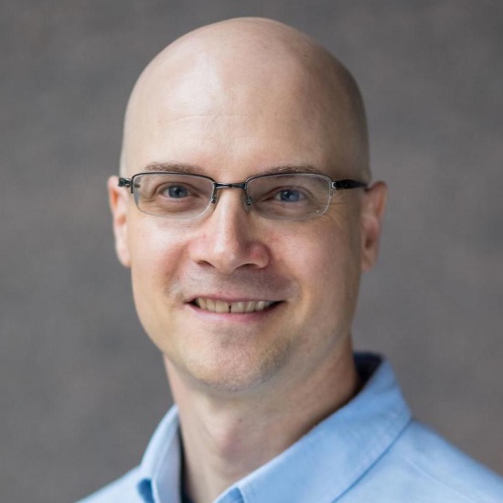
Professor David J Flannigan, University of Minnesota, USA

Professor David J Flannigan, University of Minnesota, USADavid Flannigan is an Associate Professor of Materials Science at the University of Minnesota, and he is currently a McKnight Presidential Fellow and the Director of Undergraduate Studies for Materials Science. He received his BS and PhD in Chemistry from the University of Minnesota and the University of Illinois at Urbana-Champaign, respectively. At Illinois, he studied the physical conditions and chemical processes of sonoluminescence under the guidance of Professor Ken Suslick. Following this, he was a Postdoctoral Scholar at the California Institute of Technology, where he worked on the development and application of ultrafast electron microscopy with Professor Ahmed Zewail. He joined the faculty at Minnesota in 2012, where his research focuses on the study of materials dynamics with ultrafast electron imaging, diffraction, and spectroscopy. His group’s work has been recognized with a Beckman Young Investigator Award, an NSF CAREER Award, and a DOE Early Career Award. |
|
| 14:40 - 14:50 | Discussion | |
| 14:50 - 15:05 | Contributed talk | |
| 15:05 - 15:20 | Contributed talk | |
| 15:20 - 16:00 | Panel discussion |
