Links to external sources may no longer work as intended. The content may not represent the latest thinking in this area or the Society’s current position on the topic.
Mass spectrometry imaging – challenges and opportunities for next-generation capabilities
Theo Murphy scientific meeting organised by Professor Ian Gilmore, Professor John Vickerman, Professor R. Graham Cooks, Professor Anne Dell CBE FRS and Professor Richard Caprioli
Event details
Mass spectrometry imaging is a rapidly growing field to study molecules from the macro to nanoscale in 2D and 3D and in real-time. There are major challenges ahead to meet demanding requirements in biosciences and emerging electronics. This meeting brought together leaders in the principal techniques, innovators in instrumentation and data interpretation for in-depth scientific debate.
Download the meeting programme (PDF).
Enquiries: please contact Kavli.Events@royalsociety.org
Organisers

Schedule
Chair
Professor Nicholas Winograd, Penn State University, USA
Professor Nicholas Winograd, Penn State University, USA
Professor Winograd is Evan Pugh Professor of Chemistry at Penn State University. He received his PhD degree from Case Western Reserve University and began his academic career at Purdue University in 1970. He moved to Penn State in 1979. Winograd has been fascinated with the fundamental aspects of ion/solid collisions, developing both theoretical and experimental techniques that allow a more detailed understanding at the atomic and molecular level. During the last decade, he has been focused upon the use of various cluster ion beams, including buckminsterfullerene, for bioimaging applications. He lives with his wife, Professor Barbara Garrison, in Spring Mills, Pennsylvania where he enjoys running, gardening and sampling fine wine.
| 09:05 - 09:35 |
Ion microscopy with pixelated detectors: new avenues in MSI research
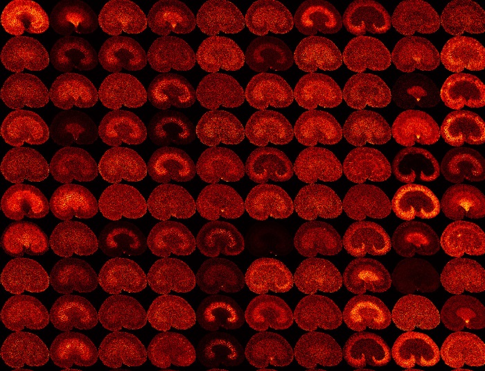
Mass spectrometry based imaging (MSI) has matured to a high throughput tool in biomedical research. MALDI-MSI is routinely used to study protein and peptide distributions on tissue sections for the analysis of molecular signalling processes in various diseases. Studies have demonstrated excellent diagnostic and prognostic value that can be directly translated to the clinic. It is key to employ a multimodal biomolecular imaging approach combined with quantitative analytical proteomics and metabolomics. Innovations in the field target breaking boundaries in resolution, sensitivity and speed. The development of novel approaches such as microscope mode imaging combined with innovative detector technology and cluster based SIMS is one of these development areas. Here we discuss how the novel pixelated detectors can extend speed, spatial resolution and the molecular weight range that can sensitively be imaged. The imaging MS community is driving translational molecular imaging research and these needed developments rapidly forward. The results of the implementation of a pixelated detector array with extremely high charge sensitivity on a MALDI-equipped linear time-of-flight mass spectrometer or a q-ToF MS system are described. This coupling is shown to allow a significant increase in detection efficiency for large macromolecules (i.e., intact antibodies) having m/z values up and above 400,000 and thereby providing a means to make the detection of large macromolecules more accessible in MALDI-mass spectrometry. In addition, the direct imaging capabilities of this detector are shown allow visualisation of mass-dependent spatial distributions in the MALDI ion cloud and the effect the ion optics exerts on the ion cloud. Such capabilities are demonstrated to provide new insights into dynamic chemical phenomena occurring in the ion source, ion optics and resulting from fragmentation. 
Professor Dr Ron Heeren, FOM Institute AMOLF, The Netherlands
Professor Dr Ron Heeren, FOM Institute AMOLF, The NetherlandsProf. Dr. Ron M.A. Heeren obtained a PhD degree in technical physics in 1992 at the University of Amsterdam on plasma-surface interactions. In the period 1995-2014 he has been developing new approaches towards high spatial resolution and high throughput molecular imaging mass spectrometry at FOM-AMOLF. In 2001 he was appointed professor at the chemistry faculty of Utrecht University lecturing on the physical aspects of biomolecular mass spectrometry. In 2014 he was appointed as distinguished professor and Limburg Chair at the University of Maastricht where he now is the director of M4I, the Maastricht MultiModal Molecular Imaging institute and heads the division of imaging MS. His academic research interests are the fundamental studies of the energetics of macromolecular systems, conformational studies of non-covalently bound protein complexes, high-throughput bioinformatics and the development and validation of new mass spectrometry based proteomic imaging techniques for the life sciences. |
|
|---|---|---|
| 09:35 - 09:50 |
Nanospray desorption electrospray ionization (nano-DESI) imaging of biological systems
Mass spectrometry imaging (MSI) is ideally suited for the label-free spatially-resolved analysis of molecules in complex biological samples with high sensitivity, speed and unprecedented chemical specificity. We have developed nanospray desorption electrospray ionisation (nano-DESI) for ambient pressure imaging of biological samples without special sample pre-treatment. Furthermore, we have developed approaches for quantitative nano-DESI imaging of lipids and metabolites. In these experiments, quantification is performed by doping the nano-DESI solvent with appropriate standards and normalising the ion image of the endogenous lipids and metabolites to the ion image of the standard. This approach enables online quantification of biomolecules extracted from the biological sample without introducing any complexity into the experimental protocol. Nano-DESI has been used for or studying metabolite exchange between living microbial communities and quantitative imaging of lipids and metabolites in tissues. 
Dr Julia Laskin, Pacific Northwest National Laboratory, USA
Dr Julia Laskin, Pacific Northwest National Laboratory, USAJulia Laskin is currently a scientist at the Pacific Northwest National Laboratory. She received her M.Sc. in Physics from the Leningrad Polytechnical Institute in 1990 and her Ph.D. in Physical Chemistry from the Hebrew University of Jerusalem in 1998. Her research is focused on a fundamental understanding of physical and chemical phenomena underlying interactions between complex ions and surfaces important for controlling activation, dissociation and deposition of complex ions following ion-surface collisions. Another area of Dr. Laskin’s research is focused on understanding the effect of the chemical composition on the physical properties of organic aerosols relevant to climate change. Finally, she is involved in the development of nanospray desorption electrospray ionization (nano-DESI) mass spectrometry for imaging of fully hydrated biological samples in their native environment and for analysis of complex mixtures directly from solid substrates. |
|
| 09:50 - 10:05 |
Advances in MALDI MSI
MALDI MS is a versatile technique suitable for direct analysis and imaging of small and large molecules at atmosphere and in vacuo. Achievable sensitivity and laser focusing has improved significantly since MALDI MSI was first introduced and pixel sizes of ∼ 1 – 10 µm have been reported for some analytes in both microprobe and microscope modes of analysis. Fast stage operation and continuous movement and control of high-repetition rate lasers have been reported providing the increased throughput essential for wider use. The routine application of MALDI MSI in drug development and clinical settings is not yet widespread but steady development and innovation over the last 20 years has moved the technique ever closer to these goals. In this presentation the current state of the art in methods and instrumentation for MALDI MS imaging will be discussed. 
Dr Josephine Bunch, National Physical Laboratory, UK
Dr Josephine Bunch, National Physical Laboratory, UKDr Josephine Bunch is a Principal Scientist at National Physical Laboratory (NPL) and Associate Professor in the School of Pharmacy, University of Nottingham. At NPL Josephine leads research in MALDI and ambient MS within the recently established National Centre for Excellence in Mass Spectrometry Imaging (NiCE MSI). Josephine previously led a research group in mass spectrometry imaging at the University of Birmingham. She has expertise in a range of mass spectrometry imaging techniques and has published articles on mass spectrometry imaging of lipids, drugs, proteins, peptides and metabolites using a range of dedicated MS techniques (MALDI, LA-ICP-MS, SIMS and LESA). |
|
| 10:05 - 10:20 |
Orbitrap mass spectrometry for imaging
Imaging imposes very stringent and ever increasing requirements on throughput and quality of mass spectrometric analysis. This talk is devoted to evolution of Orbitrap mass spectrometry from a non-relevance to imaging toward its current status as the one of the most important mass spectrometric technique for high-resolution, high mass accuracy imaging. This evolution was greatly facilitated by increasing variety and power of atmospheric pressure imaging ion sources as well as drastic increase of transmission of atmospheric-to-vacuum interfaces. As the result, paradigm shift from in-vacuum imaging to imaging at atmospheric pressure took place over recent years. It was accelerated by recent improvements in throughput for Q Exactive and Orbitrap Fusion families of instruments, with numerous new modes of operation and quantitation enabled by parallelisation of detection and ion processing and concerted operation of different ion-optical devices. In conclusion, future trends and perspectives of Orbitrap mass spectrometry for imaging applications are discussed. 
Professor Dr Alexander Makarov, Thermo Fisher Scientific, Germany
Professor Dr Alexander Makarov, Thermo Fisher Scientific, GermanyAlexander Makarov was born in the Siberian town of Irkutsk in 1966 and went to study at the Moscow Engineering Physics institute, where he also obtained his PhD. After 2 post-doc years at Warwick University, he joined a small high-tech company, HD Technologies, in Manchester (UK). There he started his work on the Orbitrap mass analyzer. Following the acquisition of the firm by Thermo Electron Corp. in 2000, Alexander provided scientific leadership of the Orbitrap R&D, which led to the commercial launch of LTQ Orbitrap mass spectrometer in 2005 and subsequent numerous extensions of this technology. He has received the Award for Distinguished Contribution in Mass Spectrometry of ASMS, the Science and Technology Award of HUPO, the Thomson medal of IMSF and others. His hobbies include travelling, mountain skiing and roller-blading. He lives and works in Bremen, Germany and holds the position of Director of Research, Life Science Mass Spectrometry. |
|
| 10:20 - 11:00 | Discussion |
Chair
Professor Dr Ron Heeren, FOM Institute AMOLF, The Netherlands
Professor Dr Ron Heeren, FOM Institute AMOLF, The Netherlands
Prof. Dr. Ron M.A. Heeren obtained a PhD degree in technical physics in 1992 at the University of Amsterdam on plasma-surface interactions. In the period 1995-2014 he has been developing new approaches towards high spatial resolution and high throughput molecular imaging mass spectrometry at FOM-AMOLF. In 2001 he was appointed professor at the chemistry faculty of Utrecht University lecturing on the physical aspects of biomolecular mass spectrometry. In 2014 he was appointed as distinguished professor and Limburg Chair at the University of Maastricht where he now is the director of M4I, the Maastricht MultiModal Molecular Imaging institute and heads the division of imaging MS. His academic research interests are the fundamental studies of the energetics of macromolecular systems, conformational studies of non-covalently bound protein complexes, high-throughput bioinformatics and the development and validation of new mass spectrometry based proteomic imaging techniques for the life sciences.
| 13:30 - 14:00 |
The open microscopy environment: open image informatics for the life and biomedical sciences
Despite significant advances in biological imaging and analysis, major informatics challenges remain unsolved: file formats are proprietary, storage and analysis facilities are lacking, as are standards for sharing image data and results. The Open Microscopy Environment (OME) is an open-source software framework developed to address these challenges. OME has three components—an open data model for biological imaging: OME data model; standardised file formats (OME-TIFF) and software libraries for file conversion (Bio-Formats); and a software platform for image data management and analysis (OMERO). The Java-based OMERO client-server platform comprises an image metadata store, an image repository, visualisation and analysis by remote access, enabling sharing and publishing of image data. OMERO’s model-based architecture has enabled its extension into a range of imaging domains, including light and electron microscopy, high content screening and recently into applications using non-image data from clinical and genomic studies Our current version, OMERO-5 improves support for large datasets and reads images directly from their original file format, allowing access by third party software. OMERO and Bio-Formats run the JCB DataViewer, the world’s first on-line scientific image publishing system and several other institutional image data repositories. 
Professor Jason Swedlow, University of Dundee, UK
Professor Jason Swedlow, University of Dundee, UKDr Swedlow received his Ph.D. in Biophysics from the University of California, San Francisco after completing a B.A. in Chemistry at Brandeis University in Massachusetts. He joined the Wellcome Trust Biocentre at the University of Dundee in the United Kingdom following postdoctoral training at UCSF and Harvard Medical School in the U.S. Dr Swedlow is currently Professor of Quantitative Cell Biology at the University of Dundee. His research interests are the mechanisms and regulation of chromosome segregation during mitotic cell division, and the development of software tools for accessing, processing, sharing and publishing large scientific image datasets. Dr Swedlow is a co-founder of the Open Microscopy Environment (OME), an international consortium that develops and releases open source software for biological imaging. He also cofounded Glencoe Software, Inc., which commercializes and customizes OME technology for use in biopharma and data publishing, and BioImagingUK, a consortium of UK imaging scientsts who develop, use, or administer imaging solutions for life sciences research. In 2011, Dr Swedlow was named Social and Overall Innovator of the Year by the BBSRC. In 2012, Dr Swedlow was named a Fellow of the Royal Society of Edinburgh. |
|
|---|---|---|
| 14:00 - 14:15 |
Does LC/MS metabolomics metabolite annotation make sense for imaging MS?
Metabolite profiling via LC/MS can reveal ‘interesting’ features, and subsequent tandem MS experiments provide powerful structural hints for the elucidation of these unknown mass spectral features. Reference libraries like MassBank and in-silico methods such as MetFrag help to identify compounds with tandem MS among candidate structures obtained from general purpose compound libraries. I won't give an answer to the question in the title; that will be part of the discussion. 
Dr Steffen Neumann, Leibniz Institute of Plant Biochemistry, Germany
Dr Steffen Neumann, Leibniz Institute of Plant Biochemistry, GermanySteffen Neumann studied computer science and bioinformatics in Bielefeld, and now his group focuses on the development of tools and databases for metabolomics and computational mass spectrometry. The group develops algorithms for data processing of metabolite profiling experiments, which are available in several Open Source Bioconductor packages, and addresses the most pressing bottleneck in Metabolomics: the identification of unknowns in mass spectrometry data. The institute is part of the MassBank consortium and operated the first MassBank server in Europe. The MetFrag and MetFusion tools allow the identification of compounds where no reference spectra are available. To compare such identification methods on common challenge data, Steffen Neumann initiated the CASMI contest in 2012, which is now going into its third year. Within the EU project COSMOS, he develops and promotes the use of data standards to enable data sharing and -archiving. |
|
| 14:15 - 14:30 |
Statistical methods for mass spectrometry-based imaging
Statistical methods are key for detecting systematic signal (e.g., caused by an intervention or a disease) in presence of variation and uncertainty, and for making objective and reproducible conclusions. This is particularly important for mass spectrometry-based imaging, where signals are obscured by 3 types of variation: the variation between different biological replicates, the spatial variation within images of a same biological replicate, and the technical variation due to sample handling and spectral acquisition. Moreover, the large-scale nature of mass spectrometry imaging experiments presents an additional challenge. As spatial and mass resolution increase, the experiments become more prone to generating spurious associations, and to amplifying bias and confounding. This talk will discuss the importance of statistical inference when designing and analysing mass spectrometry-based imaging experiments, as well as statistical methods and open-source software designed to facilitate the statistical inference tasks. 
Dr Olga Vitek, Northeastern University, USA
Dr Olga Vitek, Northeastern University, USADr. Vitek holds a B.S. degree from University of Geneva, Switzerland, and a M.S., a PhD in Statistics from Purdue University, and a post-doctoral training in the Aebersold lab at the Institute for Systems Biology in Seattle. Between 2006-20014 Dr. Vitek was a faculty in the Departments of Statistics and Computer Science at Purdue University. She is currently a Sy and Laurie Sternberg Interdisciplinary Associate Professor College of Science and College of Computer and Information Science at Northeastern University. Dr. Vitek‘s group develops statistical and computational methods and software for high-throughput quantitative experiments in molecular biology that rely on mass spectrometric workflows. The contributions from the Vitek lab help increase the accuracy of these experiments and their insight into the biological function. Dr. Vitek serves on the Editorial Boards of Molecular & Cellular Proteomics and of the Journal of Statistical Planning and Inference. She was a recipient of the NSF CAREER award (2011), and was distinguished as Purdue University Faculty Scholar. |
|
| 14:30 - 14:45 |
Imaging mass spectrometry: unique approaches for the structural identification of biomolecules
Imaging mass spectrometry (IMS) is a rapidly advancing technology, however the identification of species detected from tissue remains a significant challenge. Biomolecular identification strategies for IMS fall into two general categories: on-tissue fragmentation and indirect identification approaches. Since IMS analysis often ablates all material from the measurement area, on-tissue identification is typically performed using serial tissue sections or unmeasured regions of the sample. This can prove problematic because, for many ions, optical inspection alone is insufficient to determine their location, making manual prediction of where to focus fragmentation experiments impractical. Indirect identification is performed by using secondary information such as mass accuracy to link separate IMS and LC-ESI MS/MS experiments. This approach is often hampered by insufficient mass resolving power and accuracy for the imaging experiment to correlate results with high confidence. Here we describe novel methods for the identification of metabolites, lipids and proteins in molecular imaging experiments using high performance instrumentation and advanced computational approaches. 
Dr Jeffrey Spraggins, Vanderbilt University, USA
Dr Jeffrey Spraggins, Vanderbilt University, USAJeffrey M. Spraggins received his B.A. in Chemistry from the College of Wooster and his Ph.D. in Analytical Chemistry from the University of Delaware (2009), where he studied gas-phase fragmentation mechanisms of modified biomolecules and metal-sulfide cluster ion-molecule reactions. Following graduate school, Dr. Spraggins continued his training as a postdoctoral research fellow in Richard Caprioli’s research group and is currently a Research Assistant Professor in the Department of Biochemistry and a member of the Mass Spectrometry Research Center at Vanderbilt University. His research focuses on developing new mass spectrometric technologies to enhance biomolecular imaging experiments. Specifically, he is working on expanding the application of FTICR MS for the spatial analysis and structural identification of metabolites, lipids, peptides and proteins in biological tissues. |
|
| 14:45 - 15:30 | Discussion | |
| 16:00 - 17:00 | Further discussion |
Chair
Professor John Vickerman FRSC, The University of Manchester, UK
Professor John Vickerman FRSC, The University of Manchester, UK
Building on an early career focused on basic surface chemistry and catalysis I have pioneered the development of molecular analysis by secondary ion mass spectrometry (SIMS). My Group has stimulated ground-breaking advances in ion beam and MS instrumentation that have transformed the application of SIMS in molecular analysis from a surface analysis technique able to probe at most 1% of a surface in the 'static' SIMS mode, to a technique that can probe 100% of a material, carry out depth profiles with around 15 nm depth resolution and generate 3D images of the chemical composition of complex organic and bio-organic systems. These successes have been recognised by the Theophillus Redwood Award by the Royal Society of Chemistry in 2009 and the Médaille Chevenard by the French Society of Metals and Materials in 2012.
| 09:00 - 09:30 |
Quantification in mass spectrometry imaging
Now that mass spectrometry imaging has come of age, it is appropriate to ask more sophisticated questions about our images and to move away from the ‘show and tell’ modality. Do the mass spectra really provide an accurate view of the material, or do we see only what the technique wants us to see? A closer look at ionisation and a renewed effort at quantification seems appropriate at this point. Matrix effects, ion suppression and molecular desorption probabilities are all well-known factors that influence ion yields. For SIMS experiments, the chemical and physical nature of the projectile often leads to very different endpoints. Our use of laser post-ionisation of neutral molecules is helpful in disentangling the origin of matrix effects during imaging. Here we discuss how these factors interact and suggest approaches to mitigate these dreaded effects.
Professor Nicholas Winograd, Penn State University, USA
Professor Nicholas Winograd, Penn State University, USAProfessor Winograd is Evan Pugh Professor of Chemistry at Penn State University. He received his PhD degree from Case Western Reserve University and began his academic career at Purdue University in 1970. He moved to Penn State in 1979. Winograd has been fascinated with the fundamental aspects of ion/solid collisions, developing both theoretical and experimental techniques that allow a more detailed understanding at the atomic and molecular level. During the last decade, he has been focused upon the use of various cluster ion beams, including buckminsterfullerene, for bioimaging applications. He lives with his wife, Professor Barbara Garrison, in Spring Mills, Pennsylvania where he enjoys running, gardening and sampling fine wine. |
|
|---|---|---|
| 09:30 - 09:45 |
Digitizing the chemistry of microbes and people through molecular 3D cartography
How does one map the chemistry of people as well as their associations with microbes? This is the fundamental question that will be addressed in this lecture. The key approach to understanding large volumes of mass spectrometry data that is generated generate is through visualisation. We will show how we map the chemistry of microbes on people and how to tease apart the origin of this information. Such chemistries are highly specialised. It turns out that people leave behind chemical traces, which we refer to as lifestyle chemistries, which can be detected on personal objects such as phones. Phones are great reporters of lifestyle chemistries. Similar human derived lifestyle chemistries can be detected in buildings, highlighting how people affect the chemistries in the build environment. 
Professor Pieter Dorrestein, University of California - San Diego, USA
Professor Pieter Dorrestein, University of California - San Diego, USADr Dorrestein, a scientist with Dutch heritage (born in Utrecht, December 1974), is a Professor at the University of California - San Diego. He is the Director of the Collaborative Mass Spectrometry Innovation Center and a Co-Director, Institute for Metabolomics Medicine in the Skaggs School of Pharmacy & Pharmaceutical Sciences and is a member of the Center for Marine Biotechnology and Biomedicine at the Scripps Institution of Oceanography. Dr Dorrestein joined UCSD September 2006 as an Assistant Professor. He was promoted to Associate Professor in July 2010 and in July 2014 he was promoted to full Professor. Professor Dorrestein has been pioneering the development of mass spectrometry methods to study the chemical ecological crosstalk between populations of organisms for agricultural, diagnostic and therapeutic applications. |
|
| 09:45 - 10:00 |
Matrix effects in secondary ion mass spectrometry of biologicals
Matrix effects are a concern for most types of mass spectrometric approaches and are especially problematic for desorption ionisation techniques that employed for imaging. The lack of a separation step prior to ionisation of the analyte means that competition for charge during the desorption process can lead to extreme challenges for quantitation as one analyte may significantly suppress or enhance the ionisation of another. In biological analysis using both MALDI and SIMS the competition for charge has been shown to be related to the gas phase basicity of the analyte for the formation of [M±H]± ions although a wide range of different ion species can also be formed. In SIMS matrix effects can also arise from changes in the local sputter yield in the sample. Examples of matrix effects in SIMS analysis, particularly imaging, will be presented and the challenges associated with understanding and hence overcome these challenges will be discussed. 
Dr John Fletcher, University of Gothenburg, Sweden
Dr John Fletcher, University of Gothenburg, SwedenJohn Fletcher is currently a research fellow at the University of Gothenburg in Sweden. John completed an MChem degree at the UMIST in 2000 and undertook a PhD using secondary ion mass spectrometry (SIMS) and FTIR to study aerosols and thin film aerosol mimics. Since gaining his PhD in 2004 John has worked in the development and application of SIMS for biological analysis including single cell and tissue imaging. John has worked extensively on the development and implementation of polyatomic and gas cluster ion beams and their use with new mass spectrometry approaches for 2D and 3D molecular imaging using SIMS. Johns current research activities include continued development of imaging mass spectrometry approaches for the study of biological changes related to disease progression and treatment. |
|
| 10:00 - 10:15 |
Spontaneous conversion of proteins to highly protonated gas-phase ions using aprotic matrices
A newly discovered matrix-assisted ionisation (MAI) method is capable of spontaneously transferring compounds at least as large as the 66 KDa bovine serum albumin protein into multiply protonated gas-phase ions when the matrix:analyte sample is simply exposed to sub-atmospheric pressure conditions. Sub-atmospheric pressure can be assessed from atmospheric pressure at the inlet of a mass spectrometer or by introduction of the sample directly into vacuum using an intermediate pressure MALDI source. Further, gas-phase ions are observed multiply protonated even using aprotic matrices. 3-Nitrobenzonitrile was the first spontaneous MAI matrix discovered, but over 40 additional compounds with no specific structural motifs have subsequently been discovered. All spontaneous MAI matrices sublime in vacuum and thus are no more likely to contaminate the mass spectrometer ion optics than solvents used with ESI or APCI. The method has excellent sensitivity producing clean full-scan mass spectra using low femtomoles of, for example, peptides. 
Professor Sarah Trimpin, Wayne State University, USA
Professor Sarah Trimpin, Wayne State University, USADr Sarah Trimpin, Associate Professor, Wayne State University (WSU), is developing methods and instrumentation for the structural characterization and imaging of cells. She obtained her PhD from the MPI-Polymer Research Institute where she pioneered solvent-free MALDI-MS for insoluble materials. After completing a joint postdoctoral appointment between OSU and OHSU, she joined Professor Clemmer’s group at IU to study IMS-MS. Her group at WSU discovered new matrix-assisted ionization processes that convert molecules as large as the 66 kDa BSA protein to gas-phase ions with excellent sensitivity and astonishing simplicity for analysis by MS. Dr Trimpin has published 59 peer-reviewed articles, 8 review articles, 7 book chapters, co-edited a book on IMS-MS, and received a number of honors including DGMS Wolfgang-Paul-Studienpreis and Wolfgang-Paul-Promotionpreis, Genome Technology Rising PI, NSF CAREER, WSU Schaap Faculty Fellow, and awards from ASMS, DuPont, Eli Lilly, and Pittcon. Her laboratory is designated a Waters Center of Innovation. |
|
| 10:15 - 11:00 | Discussion | |
| 11:30 - 12:30 | Further discussion |
Chair

Professor Anne Dell CBE FMedSci FRS, Imperial College London, UK
Professor Anne Dell CBE FMedSci FRS, Imperial College London, UK
Anne Dell was a chemistry undergraduate at the University of Western Australia before undertaking a PhD at the University of Cambridge supported by an 1851 Exhibition Research Scholarship. She moved to Imperial College London for her postdoc where she rose through the ranks to a Personal Chair in 1991. She was Head of the Biochemistry Department from 1999-2001. She was elected to the Fellowship of the Royal Society in 2002 and was awarded a CBE in recognition of her services to science in 2009. Anne was amongst the first investigators to apply soft ionization mass spectrometry to biopolymers. Over the past thirtyfive years she has been at the leading edge of the development of mass spectrometric methods for glycomic and glycoproteomic analyses, especially in the area of biomedical research. Her laboratory provides structural underpinning for programmes of research seeking to define the biological roles that carbohydrates play in health and disease.
| 13:30 - 14:00 |
Rapid stratification of cancer patients using imaging and in-vivo mass spectrometric approaches
Current state-of-the-art in cancer diagnostics is centred around the histological examination of tumour tissue using classical histopathology complemented by immunochemical and molecular genetic methods. Due to the complexity of the diagnostic procedures, the reporting times range from few days to a few weeks, depending on the disease and the healthcare system. Excessively long reporting times combined with the high inter-observer variability of results lead to sub-optimal performance both for pre-interventional and intra-interventional diagnostics. Imaging mass spectrometry has been proposed more than a decade ago as an alternative to classical histopathology, however the technology has only recently reached sufficient maturity for clinical translation regarding both hardware and data interpretation algorithms. Desorption Electrospray Ionization is one of the most widely used imaging mass spectrometric modality showing certain advantages with regard to the ease of sample processing. We have used DESI MS imaging to analyse solid tumour specimens obtained from tumour resection procedures performed on ovarian cancer, colorectal cancer, breast cancer and oesophageal cancer patients. Imaging datasets were subjected to data pre-processing and were co-registered with optical image of the tissue sections stained following imaging MS. Tumour and peritumoral spectral data was associated with the histological type of the tumours, status of various biomarkers (including genetic and protein expression markers) and the stage of the disease. Additionally, data obtained by the analysis of gastrointestinal tumours was also associated with 16S rRNA sequencing-based microbiome composition data. Results revealed that lipidomic profiles of both solid tumours and their histological environment carry information about the diagnostic classification, stage and prognosis of the disease. Using this information, an MS imaging-based histological workflow have been developed for the diagnostics and stratification of cancer patients. 
Professor Zoltan Takats, Imperial College London, UK
Professor Zoltan Takats, Imperial College London, UKDr Zoltan Takats obtained his M.Sc. degree in Chemistry from the Eotvos Lorand University, Budapest, in 1998. He carried out his Ph.D. research at the Chemical Research Centre of the Hungarian Academy of Sciences and at Purdue University. He obtained his Ph.D.degree in analytical chemistry in 2003. He has been doing mass spectrometry-related research for more than 15 years, with a primary focus on the development of novel atmospheric pressure ionization methods. He is the primary inventor of electrosonic spray ionization, desorption electrospray ionization, jet desorption ionization and rapid evaporative ionization mass spectrometry methods. Besides pursuing a scientific career, he has been deeply involved in the introduction of a mass spectrometry-based neonatal screening programme in Hungary and served as the head of one of the national screening laboratories. Following a couple of years spent at Justus-Liebig-Universität in Gießen, Germany, he currently works as Professor of Analytical Chemistry at the Department of Surgery and Cancer, Faculty of Medicine, Imperial College London. Present research interests include the application of ambient ionization methods in surgical metabonomics and development of mass spectrometric imaging techniques for the rapid phenotyping of cancer patients. |
|
|---|---|---|
| 14:00 - 14:15 |
High performance MALDI MS imaging at cellular resolution and beyond
Recent improvements in spatial resolution and sensitivity will be reported for atmospheric-pressure scanning microprobe MALDI mass spectrometry imaging (AP-SMALDI MSI). The method has gained significant attention especially due to its capability to disclose morphologic distributions of substances in complex biological samples with high sensitivity, without vacuum-induced analyte losses or morphological artefacts. High resolution in mass and space has been used to derive distinct molecular information from sub-cellular structures. A commercial imaging ion source (AP-SMALDI10, TransMIT GmbH, Giessen, Germany) was modified in order to further improve spatial resolution. A new objective lens was developed and integrated into the ion source. Both, the commercial and the modified imaging ion sources were coupled to a Q ExactiveTM (Thermo Fisher Scientific) orbital trapping mass spectrometer, operated at a mass resolution of 140000 at m/z 200. Biological tissue samples were prepared from fresh frozen material and matrix-coated by various methods, for comparison of analytical sensitivity. Results demonstrate routine capability of the instrumentation to obtain high-quality, highly sensitive images without oversampling down to a step size (pixel resolution) of 2 µm. 
Professor Dr Bernhard Spengler, Justus Liebig University Giessen, Germany
Professor Dr Bernhard Spengler, Justus Liebig University Giessen, GermanyBernhard Spengler is Professor of Analytical Chemistry at Justus Liebig University of Giessen, Germany. He has contributed to the development of MALDI MS in the 1980's, developed the Postsource Decay technique and in 1994 introduced the MALDI Imaging methodology. He and his group work in the field of mass spectrometry imaging for many years, in several fields of atmospheric pressure in situ MS techniques and in aerosol analysis. |
|
| 14:15 - 14:30 |
Ion microscopy at the nanoscale
I will sketch directions for instrument improvements and extensions of biomedical applications. The increase of ionisation yield with low energy cesium deposition. The elimination of analytical dead space and vignetting by replacing dynamic transfer and step scanning of the primary beam with nanometric stage scanning. The identification of individual cell types within a population, of cell organelles within a cell, and of molecules within organelles; this can be accomplished with correlated microscopy, SEM or TEM, and immuno-typing; it can be performed either on alternate sections or on the sample studied with several methodologies. Finally, the characterization of depth resolution using a model of a transmembrane protein labeled at a variety of positions with different tags. 
Professor Claude Lechene, Harvard Medical School and Brigham and Women's Hospital, USA
Professor Claude Lechene, Harvard Medical School and Brigham and Women's Hospital, USAProfessor Lechene studied at the University of Paris Special Mathematics, Atomic, Structural and Kinetic Chemistry, Thermodynamics, and Medicine. He worked at the French AEC, at U. Sherbrooke, and finally Harvard Medical School (Professor of Medicine). He is developing Multi-isotope Imaging Mass Spectrometry (MIMS) combining ion microscopy, SIMS, isotope tracers and custom software. MIMS can measure proteins, sugars, lipids and nucleotides metabolism within cellular volumes smaller than a cubic micron. He also studied the mechanism of urine concentration, the physiological role of the Na-K pump, the mechanisms of cellular homeostasis, the role of gels in cellular volume maintenance, the quantal packaging of pancreatic enzymes, the role of integrins and the viability of early mammalian embryo. |
|
| 14:30 - 15:30 | Discussion | |
| 16:00 - 17:00 |
Summary of discussions and closing remarks
Professor Ian Gilmore, National Physical Laboratory, UK
Professor Ian Gilmore, National Physical Laboratory, UKIan S Gilmore is a National Physical Laboratory Fellow in Surface & Nanoanalysis and a Visiting Professor in the School of Pharmacy at the University of Nottingham. His research focus is on the analysis of molecules at surfaces and interfaces in a wide range of sciences, including the intracellular distribution of pharmaceuticals, drug delivery systems, medical devices, organic electronics and devices using 2D materials. Ian has recently launched a UK National Centre of Excellence in Mass Spectrometry Imaging (NiCE-MSI). This world-class centre has over 15 staff scientists who are experts in the three principal techniques: secondary ion mass spectrometry (SIMS); ambient mass spectrometries (AMS); and matrix assisted laser desorption ionisation (MALDI MS). The aims of NiCE-MSI are to advance the fundamentals of the techniques and innovate measurement capabilities; establish metrology for reliability and standardisation; and support the uptake of the techniques in industry and academia. For example, Ian is leading a major new project, 3D nanoSIMS, in collaboration with GlaxoSmithKline, to create the next generation SIMS instrument for label-free imaging of drugs in cells with a spatial resolution at the organelle scale. He has authored and co-authored over 100 papers on surface analysis and co-edited the popular teaching book 'Surface Analysis - The Principal Techniques'. Ian is a Fellow of the Institute of Physics (UK) and was awarded the Institute's Paterson Medal (2004) for his innovation of G-SIMS. He is a Fellow of the American Vacuum Society, a major science and technology society in the USA, and served on the Board of Directors in 2013-14 where he led the development of the Society's strategy. Ian has made a major contribution to international leadership in surface analysis, recognised by the Rivière Prize in 2013 from the UK Surface Analysis Forum. He is chair of the International SIMS conference series and is chairing a Royal Society Scientific Meeting on Mass Spectrometry Imaging in 2015. He has previously successfully co-chaired the SIMS XV international conference (Manchester, 2005) and chaired the IUVSTA workshop on sputtering and ion emission by cluster ion beams (Scotland, 2007). Ian provides leadership in international standardisation through chairing the committee for SIMS in ISO TC 201 and he is chair of VAMAS for surface chemical analysis, which conducts interlaboratory studies in electron spectroscopies, scanning probe microscopies and mass spectrometries to advance the techniques, support uptake into industry and standardisation. |