Links to external sources may no longer work as intended. The content may not represent the latest thinking in this area or the Society’s current position on the topic.
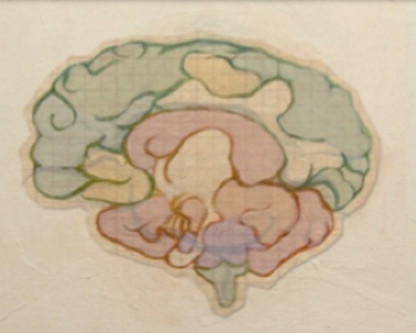
Applying computational modelling to clinical neuroscience
Theo Murphy meeting organised by Dr Randy McIntosh, Dr Karl Friston FRS and Dr Cathy Price and Dr Viktor Jirsa and Dr Petra Ritter.
Computational neuroscience can play a pivotal role in the translation of research to clinical practice. Big data initiatives aspire to solve critical clinical issues, but data alone is insufficient. Computational modelling links theory and data, providing key mechanistic insights into brain dysfunction. The meeting will highlight practical examples and foster discussion on further advances.
Attending this event
This is a residential conference, which allows for increased discussion and networking. It is free to attend, however participants need to cover their accommodation and catering costs.
Enquiries: Contact the events team
Organisers
Schedule
Chair
Dr Randy McIntosh, Baycrest Research Centre, Canada
Dr Randy McIntosh, Baycrest Research Centre, Canada
Dr. Randy McIntosh is vice-president of Research at Baycrest and director of Baycrest's Rotman Research Institute, and Professor of Psychology at the University of Toronto. Dr. McIntosh is a pioneer in the study of how different parts of the brain work together to bring about the wide range of human mental operations. He is leading a team of international scientists on a mammoth project —The Virtual Brain (thevirtualbrain.org) — which has the potential to revolutionize how clinicians assess and treat various brain disorders, including cognitive impairment caused by stroke and Alzheimer’s disease. The computerized model will deliver the first real, usable and open simulation of the human brain. For researchers, surgeons, neuroscientists and therapists, The Virtual Brain promises improved patient outcomes by letting clinicians simulate cognitive interventions – right from a Web browser.
| 09:05 - 09:30 |
Personalised clinical decisions based on computational brain model predictions: What do we need and how can we get it?
Dr. Ritter will speak about The Virtual Brain, a framework for computational modelling of full-brain activity that can be related to the data generated in typical neuroimaging protocols. The key innovation is that an individual’s data on brain structure is used to set the initial constraints for model parameters, such as network connection strength and conduction rates. This has profound implications for the understanding and treatment of brain processes and disorders such as those occurring in aging. Extensive simulations on supercomputers allow building a comprehensive library of dynamical regimes that systematically catalogues functional brain modes and underlying candidate mechanisms of neuronal interactions and their age or disease related changes. Such a comprehensive data base is prerequisite for making The Virtual Brain a useful clinical tool. 
Dr Petra Ritter, The Charité Universitätsmedizin Berlin, Germany
Dr Petra Ritter, The Charité Universitätsmedizin Berlin, GermanyDr. Ritter is a clinician trained in Neurology and Research Group leader at the Dept. of Neurology, Charité University Medicine Berlin. She is leader of a German research network focusing on biological underpinnings of learning. Dr. Ritter’s research program addresses multi-scale computations in the human brain by combining multimodal neuroimaging and computational modeling. With the recently awarded renowned ERC consolidator grant Dr. Ritter continues to contribute to the integration of knowledge about the brain in a comprehensive unifying computational framework – The Virtual Brain (TVB) of which she is co-founder. TVB is a unique simulation tool that integrates experimental data in a mathematical model of the brain and allows for personalized predictions of healthy and pathological brain function. Dr. Ritter successfully pushes forwards the transfer of these new technologies into usable products. Jointly with Dr. Randy McIntosh (Toronto) she leads a public engagement BCI project – My Virtual Dream. Dr. Ritter’s scientific achievements have been published in several high-profile publications. |
|
|---|---|---|
| 09:30 - 10:00 |
Translational neuromodeling
For many brain diseases, particularly in psychiatry, we lack objective diagnostic tests and cannot predict optimal treatment for individual patients. This presentation outlines a translational neuromodeling framework which aims at establishing “computational assays” for inferring subject-specific mechanisms of brain disease from non-invasive measures of behaviour and neuronal activity. Such assays, derived from a generative modelling approach, may provide a formal basis for differential diagnosis and treatment predictions in individual patients and, eventually, facilitate the construction of pathophysiologically grounded disease classifications. The framework presented emphasises the importance of prospective validation studies in patients and of concrete clinical problems for providing benchmarks for model validation. The presentation will show some early (and mostly very simple) proof of concept studies in patients, and outline the opportunities and challenges that lie ahead. 
Professor Klaas Enno Stephan, University of Zurich and ETH Zurich, Switzerland
Professor Klaas Enno Stephan, University of Zurich and ETH Zurich, SwitzerlandKlaas Enno Stephan is Full Professor at the University of Zurich & ETH Zurich and Founding Director of the Translational Neuromodeling Unit (TNU), Zurich. Integrating computational scientists and clinicians under one roof, the mission of the TNU is to develop computational assays for inferring individual disease mechanisms and for predicting individual treatment response. Following doctoral degrees in Medicine and Neuroinformatics, Klaas’ scientific track record includes the construction of the connectivity database CoCoMac, development of various neuroinformatics tools and computational modeling techniques, and numerous studies on psychiatric and neurological conditions. In addition to his work at Zurich, he is Honorary Principal at the Wellcome Trust Centre for Neuroimaging, London, and External Scientific Member at the Max Planck Institute for Metabolism Research, Cologne. His work has been recognized by several awards and honours, including the Wiley Young Investigator Award for Human Brain Mapping and election to the Max Planck Society. |
|
| 10:00 - 10:30 | Discussion | |
| 10:30 - 11:00 | Coffee | |
| 11:00 - 11:30 |
Channelopathies: linking ions to cognition through Dynamic Causal Models
Dr Rosalyn Moran, Virginia Tech, USA
Dr Rosalyn Moran, Virginia Tech, USADr. Moran received her PhD in Electrical Engineering at University College Dublin in 2007 and joined the methods group at the Wellcome Trust Centre for Neuroimaging at University College London. There she worked on the development of dynamic causal models for electrophysiology with Professor Karl Friston. In 2012 she moved to Virginia Tech to direct a research group who’s focus includes aging, neurodegenerative disease and neuroimaging methods development. She is a section editor at Neuroimage: Clinical. |
|
| 11:30 - 12:00 |
From differential diagnosis to cognitive restoration in Parkinson’s disease and dementia
Better treatments for neurodegenerative disorders require a clear mechanistic understanding of the cascade of effects from cell biology through (dys)functional neural networks to complex behavioural syndromes. Computational modelling is critical to this endeavour. I will use examples from several disorders, including Parkinson’s disease, progressive supranuclear palsy and frontotemporal dementia to illustrate our approach. I will argue for a dimensional approach to cognition and behaviour that cuts across traditional diagnostic boundaries, building on the RDoC framework for mental health disorders, and illustrating this with impulsivity and disorders of response inhibition. I will show how formalising action and inhibition as accumulation-to-threshold decisions can provide insights into disease that are not apparent with traditional cognitive outcomes like mean RT and accuracy. I will then examine a network for response inhibition, and its abnormalities in Parkinson’s disease and Frontotemporal dementia. Model-based and data-driven analysis of functional brain networks will be compared, including dynamic causal models for fMRI and MEG. I will present the results of randomised placebo-controlled double-blind crossover functional imaging studies that assess the restoration of network function by noradrenergic and serotonergic reuptake inhibition in clinical disorders associated with noradrenaline and serotonin deficiency respectively. Finally I will touch on models to predict treatment response in highly heterogeneous disorders. Together, these studies confirm that computational models with patient data can facilitate the development of novel therapeutic interventions and individualized therapy while validating preclinical models of brain function and disease. 
Professor James Rowe, University of Cambridge, UK
Professor James Rowe, University of Cambridge, UKAs Professor of Cognitive Neurology, James Rowe studies the cognitive physiology of neurodegenerative diseases, including frontotemporal dementia, Parkinson's disease and primary tauopathies. He trained in medical sciences and experimental psychology at Cambridge, before clinical studies in Oxford and his PhD at the Functional Imaging Laboratory, UCL. After further training in Copenhagen, he returned to Cambridge in 2005. With the support of the Wellcome Trust, MRC and NIHR his team combines advanced methods in magnetoencephalography, positron emission tomography, magnetic resonance imaging and computational modelling in order to measure the impact of disease and treatment on human brain function. The integration of multimodal brain imaging with computational modelling provides a powerful platform to understand the mechanisms of cognitive and behavioural change that result from neurological disease, and also provides sensitive methods to evaluate the efficacy and mechanisms of novel therapeutics. |
|
| 12:00 - 12:30 | Discussion |
Chair
Dr Petra Ritter, The Charité Universitätsmedizin Berlin, Germany
Dr Petra Ritter, The Charité Universitätsmedizin Berlin, Germany
Dr. Ritter is a clinician trained in Neurology and Research Group leader at the Dept. of Neurology, Charité University Medicine Berlin. She is leader of a German research network focusing on biological underpinnings of learning. Dr. Ritter’s research program addresses multi-scale computations in the human brain by combining multimodal neuroimaging and computational modeling. With the recently awarded renowned ERC consolidator grant Dr. Ritter continues to contribute to the integration of knowledge about the brain in a comprehensive unifying computational framework – The Virtual Brain (TVB) of which she is co-founder. TVB is a unique simulation tool that integrates experimental data in a mathematical model of the brain and allows for personalized predictions of healthy and pathological brain function. Dr. Ritter successfully pushes forwards the transfer of these new technologies into usable products. Jointly with Dr. Randy McIntosh (Toronto) she leads a public engagement BCI project – My Virtual Dream. Dr. Ritter’s scientific achievements have been published in several high-profile publications.
| 13:30 - 14:00 |
Computational nosology and precision psychiatry
This presentation provides an illustrative treatment of psychiatric morbidity that offers an alternative to the standard nosological model in psychiatry. It considers what would happen if we treated diagnostic categories not as putative causes of signs and symptoms – but as diagnostic consequences of psychopathology and pathophysiology. This reconstitution (of the standard model) opens the door to a more natural formulation of how patients present – and their likely response to therapeutic interventions. In brief, we describe a model that generates symptoms, signs and diagnostic outcomes from latent psychopathological states. In turn, psychopathology is caused by pathophysiological processes that are perturbed by (aetiological) causes; such as predisposing factors, life events and therapeutic interventions. The key advantages of this nosological formulation include: (i) the formal integration of diagnostic (e.g. DSM) categories and latent psychopathological constructs (e.g., the dimensions of RDoC); (ii) the provision of a hypothesis or model space that accommodates formal evidence-based hypothesis testing or model selection (using Bayesian model comparison); (iii) the ability to predict therapeutic responses (using a posterior predictive density), as in precision medicine and (v) a framework that allows one to test hypotheses about the interactions between pharmacological and psychotherapeutic interventions. These and other advantages are largely promissory at present: the purpose of this talk is to suggest what might be possible, through the use of idealised simulations. These simulations can be regarded as a (conceptual) prospectus that motivates a computational nosology for psychiatry. 
Professor Karl Friston FMedSci FRS, University College London, UK
Professor Karl Friston FMedSci FRS, University College London, UKKarl Friston is a theoretical neuroscientist and authority on brain imaging. He invented statistical parametric mapping (SPM), voxel-based morphometry (VBM) and dynamic causal modelling (DCM). These contributions were motivated by schizophrenia research and theoretical studies of value-learning – formulated as the dysconnection hypothesis of schizophrenia. Mathematical contributions include variational Laplacian procedures and generalized filtering for hierarchical Bayesian model inversion. Friston currently works on models of functional integration in the human brain and the principles that underlie neuronal interactions. His main contribution to theoretical neurobiology is a free-energy principle for action and perception (active inference). Friston received the first Young Investigators Award in Human Brain Mapping (1996) and was elected a Fellow of the Academy of Medical Sciences (1999). In 2000 he was President of the international Organization of Human Brain Mapping. In 2003 he was awarded the Minerva Golden Brain Award and was elected a Fellow of the Royal Society in 2006. In 2008 he received a Medal, Collège de France and an Honorary Doctorate from the University of York in 2011. He became of Fellow of the Society of Biology in 2012 and received the Weldon Memorial prize and Medal in 2013 for contributions to mathematical biology. |
|
|---|---|---|
| 14:00 - 14:30 |
Brain network disturbances in mood disorders
Mood serves to adjust our internal somatic state to match the expectations we hold about our social milieu. This requires a dynamic interplay between cognition and interoception. Mood disorders – major depression and bipolar disorder – are characterised by episodic disturbances in arousal, appraisal and socialization. In this talk, evidence will be provided that mood disorders are associated with structural, functional and effective disturbances in brain networks that integrate interoception, social appraisal and cognitive control. However, unlike schizophrenia, the structural “core” of the brain is preserved. These findings position mood disorders as disturbances in the precision with which we hold interoceptive beliefs, and the degree to which we adjust our social expectations following social surprise. Networks supporting broader cognitive and perceptual dynamics are relatively preserved. 
Professor Michael Breakspear, QIMR Berghofer Medical Research Institute, Australia
Professor Michael Breakspear, QIMR Berghofer Medical Research Institute, AustraliaMichael Breakspear leads the Systems Neuroscience Group at the QIMR Berghofer Medical Research Institute in Australia. His group uses nonlinear dynamics to integrate computational psychiatry and systems neuroscience with applications to basic neuroimaging science as well as mood disorders, psychoses, epilepsy and dementia. He is also a psychiatrist in the Queensland Prison Mental Health Service. |
|
| 14:30 - 15:00 | Discussion | |
| 15:00 - 15:30 | Tea | |
| 15:30 - 16:00 |
Predicting outcome and seizure freedom in drug-resistant epilepsy
Starting from first principles of the theory of slow-fast systems in nonlinear dynamics, we conceptualize seizure dynamics mathematically and establish a taxonomy of seizures based on seizure onset and offset bifurcations. It will be demonstrated that only five state variables linked by integral-differential equations are sufficient to describe the onset, time course and offset of ictal-like discharges as well as their recurrence. These state variables define the model system called the Epileptor, where two state variables are responsible for generating rapid discharges (fast time scale), two for spike and wave events (intermediate time scale) and one permittivity variable (slow time scale). The permittivity variable captures effects evolving on slow timescales, including extracellular ionic concentrations and energy metabolism, with time delays of up to seconds as observed clinically. Extending this generic approach towards human brain networks, personalized connectivity matrices of human epileptic patients using Diffusion Tensor weighted Imaging (DTI) will be reconstructed. Subsets of brain regions generating seizures in patients with refractory partial epilepsy are referred to as the epileptogenic zone (EZ). During a seizure, paroxysmal activity is not restricted to the EZ, but may recruit other brain regions and propagate activity through large brain networks, which comprise brain regions that are not necessarily epileptogenic. The identification of the EZ is crucial for candidates for neurosurgery and requires unambiguous criteria that evaluate the degree of epileptogenicity of brain regions. Stability analyses of propagating waves provide a set of indices quantifying the degree of epileptogenicity and predict conditions, under which seizures propagate to nonepileptogenic brain regions, explaining the responses to intracerebral electric stimulation in epileptogenic and nonepileptogenic areas. The predictive value of our seizure propagation model will be demonstrated by validating it against empirical patient data. In conjunction, our results provide guidance in the presurgical evaluation of epileptogenicity based on electrographic signatures in intracerebral electroencephalograms. 
Dr Viktor Jirsa, University Aix-Marseille, France
Dr Viktor Jirsa, University Aix-Marseille, FranceViktor Jirsa is a physicist and computational neuroscientist who works on the mathematical principles how brain networks function and process information. His major contributions are on the resting state dynamics of the brain and understanding how it relates to function and dysfunction. He is Director of Research at the Centre National de la Recherche Scientifique (CNRS) and Director of the System Neuroscience Institute in Marseille, France. He has been awarded various national and international honors including the Francois Erbsmann Prize in 2001 and the CNRS Award of Scientific Excellence in 2011. |
|
| 16:00 - 16:30 |
Brain network dynamics in epilepsy: diagnosis, prognosis and heritability

Professor Mark Richardson, King’s College London, UK
Professor Mark Richardson, King’s College London, UKProfessor Mark Richardson's lab studies people with epilepsy using neuroimaging and neurophysiological techniques. Current work aims to understand the brain networks underlying epilepsy, how these network abnormalities affect cognition, and whether interventions targeting specific components of the network can improve epilepsy. Mark's lab collaborates with mathematicians and engineers in developing computational models to explain phenomena associated with epilepsy and cognition. |
|
| 16:30 - 17:00 | Discussion |
Chair

Professor Cathy Price, University College London, UK
Professor Cathy Price, University College London, UK
Cathy Price is an expert in the use of structural and functional brain imaging for understanding cognitive processing in the neurologically healthy and damaged brain. Her basic training was in Physiology and Psychology and she has a PhD in cognitive neuropsychology. Since 1997, she has been funded by the Wellcome Trust to build a functional anatomical model of auditory and visual word processing that will predict language outcome after brain damage. In parallel, she is developing data-led approaches that provide probabilistic lesion-symptom mapping associations. Her goal is to develop a clinical tool that uses lesion site to predict the most likely outcome and recovery of language abilities after stroke.
| 09:00 - 09:30 |
Network control theory offers a fundamental mechanism of executive function
Dr Danielle Bassett, University of Pennsylvania, USA
Dr Danielle Bassett, University of Pennsylvania, USADanielle S. Bassett is the Skirkanich Assistant Professor of Innovation in the Department of Bioengineering at the University of Pennsylvania. She is most well-known for her work blending neural and systems engineering to identify fundamental mechanisms of cognition and disease in human brain networks. She received a B.S. in physics from the Pennsylvania State University and a PhD in physics from the University of Cambridge, UK. Following a postdoctoral position at UC Santa Barbara, she was a Junior Research Fellow at the Sage Center for the Study of the Mind. In 2012, she was named American Psychological Association's `Rising Star' and given an Alumni Achievement Award from the Schreyer Honors College at Pennsylvania State University for extraordinary achievement under the age of 35. In 2014, she was named an Alfred P Sloan Research Fellow and received the MacArthur Fellow Genius Grant. In 2015, she received the IEEE EMBS Early Academic Achievement Award, and was named an ONR Young Investigator. She is the founding director of the Penn Network Visualization Program, a combined undergraduate art internship and K-12 outreach program bridging network science and the visual arts. Her work has been supported by the National Science Foundation, the National Institutes of Health, the Army Research Office, the Army Research Laboratory, the Alfred P Sloan Foundation, the John D and Catherine T MacArthur Foundation, and the University of Pennsylvania. She lives with her husband and two sons in Wallingford, Pennsylvania. |
|
|---|---|---|
| 09:30 - 10:00 |
Testing proposed mesocircuit mechanisms underlying recovery of conciousness with analysis of local and global dynamics of the EEG
This presentation will discuss the “mesocircuit hypothesis” for the key role of the human anterior forebrain mesocircuit in recovery from disorders of consciousness following multi-focal brain injuries. The mesocircuit model makes several specific predictions for the co-variation of measures reflecting progressive restoration of cellular and circuit-level functional integrity following severe deafferentation produced by brain injuries. The talk will focus primarily on the testing of model predictions using quantitative measurements of local and global dynamics of the human electroencephalogram (EEG). Related predictions and measurements from neuroimaging studies of patients with disorders of consciousness and experimental animal studies will also be discussed. Validation of the EEG predictions derived from this large-scale brain network model may provide a set of measures useful to track recovery and predict the impact of different therapeutic inventions in the injured brain on an individual basis. 
Dr Nicholas Schiff, Cornell University, USA
Dr Nicholas Schiff, Cornell University, USADr Nicholas Schiff is a physician-scientist with an internationally recognized expertise in neurological disorders of consciousness. He is The Jerold B. Katz Professor of Neurology and Neuroscience in the Feil Family Brain and Mind Research Institute at Weill Cornell Medical College. Dr. Schiff’s research efforts bridge basic neuroscience and clinical investigative studies of the pathophysiology of impaired consciousness using state-of-the art neuroimaging and neurophysiological measurements. This research program has provided several landmark contributions to the understanding of cerebral activity in severely brain injured-patients. Most notably, Dr Schiff and colleagues have taken insights into the neurophysiological mechanisms of arousal regulation and of deep brain electrical stimulation techniques to demonstrate evidence that long-lasting, severe cognitive disability may be influenced by electrical stimulation of the central thalamus. Based on extensive and long-range longitudinal human subject studies of recovery of consciousness, Dr Schiff has formulated a testable hypothesis for the contribution of the anterior forebrain mesocircuit to recovery of consciousness that organizes the findings from work in central thalamic deep brain stimulation into a wider context. This work is an important foundation for developing further understanding of both the mechanisms of recovery of consciousness and basic mechanisms underlying consciousness in the human brain. Dr Schiff is an elected member of the American Neurological Association and the recipient of several awards, including the 2007 Research Award for Innovation in Neuroscience from the Society for Neuroscience. Dr Schiff's research is currently supported by the US President’s BRAIN Initiative, NIH-NINDS, The Jerold B. Katz Foundation and the James S. McDonnell Foundation. Past support has included NIH-NICHD, NEI-NIDDR, NIH-NIMH, Swartz Foundation, Lounsbery Foundation, Charles A. Dana Foundation, and IntElect Medical, Inc. |
|
| 10:00 - 10:15 | Discussion | |
| 10:15 - 10:30 | Coffee | |
| 10:30 - 11:00 |
The effect of Deep Brain Stimulation in whole brain activity: a computational perspective
Deep brain stimulation (DBS) for Parkinson’s disease (PD) is a highly effective treatment in controlling otherwise debilitating symptoms yet the underlying brain mechanisms are currently not well understood. Whole-brain computational modelling was used to disclose the effects of DBS ON and OFF during collection of resting state fMRI in ten PD patients. Specifically, the local and global impact of DBS in creating asynchronous, stable or critical oscillatory conditions using a supercritical Hopf bifurcation model were explored. It was found that DBS shifts the global brain dynamics of patients nearer to that of healthy people by significantly changing the bifurcation parameters in very specific local brain regions. Further, higher communicability and coherence brain measures during DBS ON compared to DBS OFF were found. These results offer important insights into the underlying effects of DBS as well as in finding novel stimulation targets, which may in time offer a route to more efficacious treatments. 
Professor Gustavo Deco, Universitat Pompeu Fabra, Spain
Professor Gustavo Deco, Universitat Pompeu Fabra, SpainGustavo Deco is ICREA Research Professor and Full Professor at the Universitat Pompeu Fabra, where he heads the Computational Neuroscience Group and directs the Center for Brain and Cognition. He studied Physics at the National University of Rosario. In 1987, he received his PhD in Physics for his thesis on Relativistic Atomic Collisions. In 1997, he obtained his habilitation (maximal academical degree in Germany) in Computer Science at the Technical University of Munich for his thesis on Neural Learning. In 2001, he received his PhD in Psychology at the Ludwig-Maximilian-University of Munich for his thesis on Visual Attention. His research interests include computational neuroscience, neuropsychology, psycholinguistics, biological networks, statistical formulation of neural networks, and chaos theory. He has actively contributed to the modelling and integration of experimental measurements through theoretical frameworks, and collaborates with many experimentalists to confront theory and experiments. Recognised as a world leader in computational neuroscience, he has led pioneering work in dynamical modelling of human brain activity. He is an ERC Advanced grantee and member of the Human Brain Project. |
|
| 11:00 - 11:30 |
Reconstruction and simulation of neocortical microcircuitry
In a recent publication, the Blue Brain Project has presented its data-driven modeling strategy on the example of the neocortical microcircuit of a young rat. An intricate pipeline of tools has been developed, allowing the integration of multi-modal data and the resolution of constraints using algorithms. Using this pipeline, it is possible to go from sparse data to a model that can be systematically validated against data across multiple levels. A wide range of validation data lays the foundation for the model to generalize to interesting questions that have not been put into the model reconstruction process, thus allowing research on the inverse problem. 
Professor Felix Schurmann, Ecole Polytechnique Fédér ale de Lausanne, Switzerland
Professor Felix Schurmann, Ecole Polytechnique Fédér ale de Lausanne, SwitzerlandFelix Schürmann is adjunct professor at the Ecole Polytechnique Fédérale de Lausanne, co-director of the Blue Brain Project and involved in several research challenges of the European Human Brain Project. He studied physics at the University of Heidelberg, Germany, supported by the German National Academic Foundation. Later, as a Fulbright Scholar, he obtained his Master's degree (M.S.) in Physics from the State University of New York, Buffalo, USA, under the supervision of Richard Gonsalves. During these studies, he became curious about the role of different computing substrates and dedicated his master thesis to the simulation of quantum computing. He studied for his Ph.D. at the University of Heidelberg, Germany, under the supervision of Karlheinz Meier. For his thesis he co-designed an efficient implementation of a neural network in hardware. |
|
| 11:30 - 12:00 | Discussion |
Chair
Professor Karl Friston FMedSci FRS, University College London, UK
Professor Karl Friston FMedSci FRS, University College London, UK
Karl Friston is a theoretical neuroscientist and authority on brain imaging. He invented statistical parametric mapping (SPM), voxel-based morphometry (VBM) and dynamic causal modelling (DCM). These contributions were motivated by schizophrenia research and theoretical studies of value-learning – formulated as the dysconnection hypothesis of schizophrenia. Mathematical contributions include variational Laplacian procedures and generalized filtering for hierarchical Bayesian model inversion. Friston currently works on models of functional integration in the human brain and the principles that underlie neuronal interactions. His main contribution to theoretical neurobiology is a free-energy principle for action and perception (active inference). Friston received the first Young Investigators Award in Human Brain Mapping (1996) and was elected a Fellow of the Academy of Medical Sciences (1999). In 2000 he was President of the international Organization of Human Brain Mapping. In 2003 he was awarded the Minerva Golden Brain Award and was elected a Fellow of the Royal Society in 2006. In 2008 he received a Medal, Collège de France and an Honorary Doctorate from the University of York in 2011. He became of Fellow of the Society of Biology in 2012 and received the Weldon Memorial prize and Medal in 2013 for contributions to mathematical biology.
| 13:00 - 13:30 |
Mapping causal functional contributions in the brain from the game-theoretical analysis of stroke lesions
Strokes affect multiple brain regions and produce multiple functional deficits, acting as a natural experiment in brain perturbation. However, the interpretation of such perturbation data and the inference of regional functional contributions to brain function is made difficult by the multivariate interactions between different brain regions. To address this problem, we used a multivariate, game-theoretical approach to objectively quantify the causal functional contributions of brain regions, and applied it to animal model data, ground-truth simulations, as well as clinical data of stroke lesions and corresponding functional deficits. The approach reliably inferred regional contributions to brain function and revealed a wide quantitative range of contributions of multiple regions to multiple functions. It also indicated functional synergies and redundancies among brain regions and helped to identify potential targets for clinical rehabilitation 
Professor Claus Hilgetag, University of Hamburg, Germany
Professor Claus Hilgetag, University of Hamburg, GermanyHilgetag studied Biophysics in Berlin and Neuroscience in Edinburgh, Oxford, Newcastle and Boston. He is interested in fundamental principles of the organization of brain architecture, connectivity, and dynamics. In particular, his group investigates structural factors that are predictive of anatomical connections in the brain, and explores relations between topological features of neural connectivity, such as modules and hubs, and patterns of brain activity. He also develops methods for inferring brain functions from the impact of brain lesions. |
|
|---|---|---|
| 13:30 - 14:00 |
Using The Virtual Brain as an 'informatics microscope' to uncover biophysical parameters of stroke recovery
An exciting advance in the field of neuroimaging is the acquisition and processing of very large data sets (so called “big data”), permitting large-scale inferences that foster a greater understanding of brain function in health and disease. Yet what we are clearly lacking are quantitative integrative tools to translate this understanding to the individual level to lay the basis for personalized medicine. This challenge will be addressed through a new neuroInformatics modelling platform that has the capacity to track brain network function at different levels of inquiry, from microscopic to macroscopic and from the localized to the distributed. The multi-scale approach, The Virtual Brain (TVB), can effectively model individualized brain activity, provides unique biological interpretable data in stroke. The talk will show how our results establish the basis for a deliberate integration of computational biology and neuroscience into clinical approaches for elucidating cellular mechanisms of disease including on the individual level via personalized therapeutic interventions. 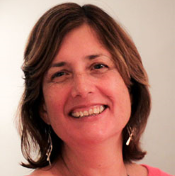
Dr Ana Solodkin, UC Irvine School of Medicine, USA
Dr Ana Solodkin, UC Irvine School of Medicine, USADr. Solodkin’s research interest has been centered from the beginning, around brain plasticity. During her Ph.D. at the "Center for Research and Advanced Studies of the National Polytechnic Institute" (Mexico) and NIH (Mentored by Dr. P Rudomin and Dr. MA Ruda) she focused on long-term plasticity in the developing spinal cord. Later on at the University of Iowa she joined the Cognitive Neurology group under the direction of Dr. A. Damasio receiving direct training with Dr. GW van Hoesen in human neuroanatomy. Her active and long-term research has targeted the relationship between basic neurobiology and cognitive neurology where using network analyses, she has focused on disease biomarkers on AD, stroke and spinocerebellar ataxias. Dr. Solodkin’s aim with this work has been to discover anatomical and physiological substrates of disease that have a reasonable likelihood of leading to therapeutic interventions. In this context, Dr. Solodkin joined Dr. McIntosh in 2006 at the early stages of the development of The Virtual Brain. Currently Dr. Solodkin utilizes The Virtual Brain platform to determine changes in brain dynamics as they relate to stroke. These efforts, in collaboration with Dr. McIntosh and Dr. Jirsa are providing a first glimpse on basic neural mechanisms associated with brain dynamics specifically related to long-term recovery. |
|
| 14:00 - 14:30 | Discussion | |
| 14:30 - 14:45 | Tea | |
| 14:45 - 15:15 |
Stroke dys-connectome: behaviour, structure and function
Since the early days of neuroscience the relative merit of structural vs. functional network accounts in explaining neurological deficits has been intensely debated. Using a large stroke cohort and a machine learning approach, we show that visual memory, and verbal memory deficits are better predicted by functional connectivity than by lesion location, while visual and motor deficits are better predicted by lesion location than functional connectivity. In addition, we show that disruption to a subset of cortical areas predicts general cognitive deficit (spanning multiple behavior domains). In a separate set of computational studies we show that these deficits affects the integration/segregation of brain regions. These results shed light on the complementary value of structural vs. functional accounts of stroke, and provide a physiological mechanism for general multi-domain deficits seen after stroke. 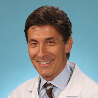
Dr Maurizio Corbetta, Washington University, USA
Dr Maurizio Corbetta, Washington University, USADr. Maurizio Corbetta is the Norman J. Stupp Professor of Neurology, and Professor of Radiology, Anatomy and Neurobiology, and Bioengineering at Washington University School of Medicine. He is the Chief of the Division of Neuro-Rehabilitation, and Director of Neurological Rehabilitation at the Rehabilitation Institute of St. Louis. As of 2016 Dr. Corbetta will be the Chair of Neurology in the Department of Neuroscience at the University of Padua, Italy. Dr. Corbetta has pioneered experiments on the neural mechanisms of human attention with Positron Emission Tomography (PET). He has discovered two brain networks dedicated to attention control, the dorsal and ventral attention networks, and developed, in collaboration with Dr. Gordon Shulman, a brain model of attention that has been cited in the literature more than 5,000 times. His clinical work has focused on the physiological correlates of focal injury. He has developed a pathogenetic model of the syndrome of hemispatial neglect. He is currently developing novel methods for studying the functional organization of the brain using functional connectivity MRI, magneto-encephalography (MEG), and electro-corticography (EcoG). He is also working on the effects of focal injuries on the network organization of brain systems with an eye to neuromodulation. |
|
| 15:15 - 15:45 |
Predicting outcome and recovery after stroke
Predicting outcome and recovery after stroke is notoriously difficult because the consequences of seemingly similar brain damage are inconsistent across patients. Clearly factors other than lesion site influence the level of recovery. To investigate the main causes of variability, machine learning on a wide variety of data acquired from large cohorts of stroke survivors has been used. This indicates the importance of lesion site, lesion size and years post stroke as the most informative variables for recovery. Although machine learning does not indicate how the predictions can be improved, it does place constraints on the variables that need to be controlled and considered in models of recovery. Hence, this talk will show how predicting outcome after brain damage can be optimised by a combination of model-based and data-led approaches.
Professor Cathy Price, University College London, UK
Professor Cathy Price, University College London, UKCathy Price is an expert in the use of structural and functional brain imaging for understanding cognitive processing in the neurologically healthy and damaged brain. Her basic training was in Physiology and Psychology and she has a PhD in cognitive neuropsychology. Since 1997, she has been funded by the Wellcome Trust to build a functional anatomical model of auditory and visual word processing that will predict language outcome after brain damage. In parallel, she is developing data-led approaches that provide probabilistic lesion-symptom mapping associations. Her goal is to develop a clinical tool that uses lesion site to predict the most likely outcome and recovery of language abilities after stroke. |
|
| 15:45 - 16:00 | Discussion | |
| 16:00 - 17:00 | Panel discussion/Overview (future directions) |