Links to external sources may no longer work as intended. The content may not represent the latest thinking in this area or the Society’s current position on the topic.
Molecular imaging and chemistry: defining the future
Satellite meeting organised by Professor David Parker, Professor Nicholas Long and Professor Stephen Faulkner
Breakthroughs in molecular imaging require cooperation from scientists across many disciplines. At the heart of this work, chemistry researchers are working to design more effective probes, understand their mode of action and help clinicians to pursue more accurate diagnosis of disease states.
The schedule of talks and speaker biographies are available below. Speaker abstracts will be available closer to the meeting date. Recorded audio of the presentations will be available on this page after the meeting has taken place.
Attending this event
This is a residential conference, which allows for increased discussion and networking.
- Free to attend
- Advance registration is essential (please request an invite)
- Catering and accommodation available to purchase during registration
Prior to this meeting, there will be a related discussion meeting Challenges for chemistry in molecular imaging held at the Royal Society London.
Enquiries: Contact the Scientific Programmes team
Organisers
Schedule
Chair
Professor Carolyn Anderson, University of Pittsburgh Bridgeside
Professor Carolyn Anderson, University of Pittsburgh Bridgeside
Carolyn J. Anderson, Ph.D. is a Professor of Medicine and is the Director of the Nuclear Molecular Imaging and Therapy Laboratory at the University of Pittsburgh (Pitt) and Co-Director of the In Vivo Imaging Facility at University of Pittsburgh Cancer Institute. Her research interests include development of novel radiometal tracers for diagnostic imaging and targeted radiotherapy of cancer and other diseases. Dr. Anderson pioneered the development of radiometal-labeled receptor-targeted PET imaging agents, leading the first human study of a 64Cu-labeled somatostatin analog in patients with neuroendocrine tumors. A current focus of her research lab is in the development of imaging agents for upregulated receptors on immune cells that are involved in inflammation related to lung diseases including tuberculosis, primary tumor growth and cancer metastasis. She has co-authored over 160 peer-reviewed and invited publications, mostly in the area of developing radiopharmaceuticals for oncological imaging and therapy.
| 09:45 - 10:15 |

Professor Franklin Aigbirhio, University of Cambridge
Professor Franklin Aigbirhio, University of CambridgeProfessor Franklin Aigbirhio trained in positron emission tomography (PET) radiochemistry and imaging at the MRC Cyclotron Unit, Hammersmith Hospital London. He joined the Wolfson Brain Imaging Centre, University of Cambridge in 1997 as a founding staff member establishing the PET radiochemistry infrastructure and programmes. Presently he is the Director of PET Sciences and Professor of Molecular Imaging Chemistry at the University of Cambridge Department of Clinical Neurosciences. He has a research focus on the development and application of PET radiotracers for preclinical and clinical research, primarily in neuroscience and cancer. Underpinning this is a research interest in developing novel radiosynthetic methods with carbon-11 and fluorine -18 radioisotopes. He also co-chairs the Dementia-Platform UK Imaging Network, which has established a network of PET-MR scanners in the UK and is the PET lead for the NIHR Translational Collaboration in Dementia. |
|
|---|---|---|
| 10:15 - 10:30 | Discussion | |
| 10:30 - 11:00 |
PET in India
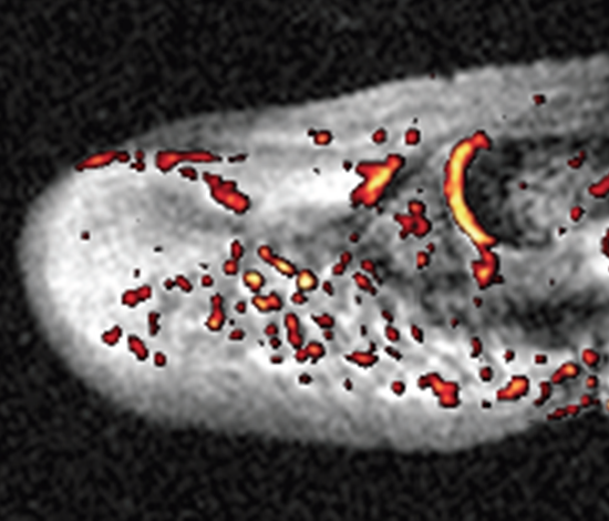
There are around 200 Nuclear Medicine Centers in India. PET based product mainly 18F-FDG is localized around big metropolitan cities where there is a centralized cyclotron facility for producing 18F and which can be transported quickly. At present around 18 cyclotrons are operational in India, which fulfill the need of 18F-FDG to greater part of the country. The first Medical cyclotron (16 MeV) was commissioned in the year 2002 at Radiation Medicine Centre, Mumbai. Since then a number of private set ups have installed cyclotron all over the country. The market for 18F-FDG is far superior to any other diagnostic product in India. Other than 18F-FDG, several other 18F based radiopharmaceuticals such as 18F-sodium fluoride for bone cancer detection (used since 2007), 18F-MISO for tumor hypoxia imaging and 18F-FLT for cancer detection (used since 2010) at the cellular level are now being used at limited nuclear medicine centers in India. FDG is the only PET or SPECT radiopharmaceutical being supplied commercially to many centers. Today in our country we have 98 Running PET/CT Centers and 70 centers are going be added in the next couple of years. Every center if doing average 5 cases a day and running 20 days then total FDG doses are 9800/month and 117600/year. Another promising PET radionuclide i.e. 68Ga which is available through a generator is gaining wide acceptance among the clinics. 68Ga labelled peptide such as DOTA-NOC, DOTA-TATE for neuroendocrine tumor imaging is used in number of Nuclear medicine centers in India. There is a small percentage of use of 11C-CO2, 13N-NH3, 15O-H2O and 82Rb which are in use for myocardial perfusion imaging. 
Professor Ajit Shinto, Kovai Medical Center and Hospital
Professor Ajit Shinto, Kovai Medical Center and HospitalDr Ajit Shinto,currently serves as the Chief of Nuclear Medicine services at KMCH, Coimbatore ; a large super specialty hospital of great repute in South India. He serves as the executive member and special advisor to the radio nuclide and targeted therapy committee .His special interests lie in PRRT, radio-synovectomy and intra-arterial liver cancer therapy. Being on the governing body of WARMTH(World Association of Radionuclide and Molecular Therapy), he also serves as the Task Force Chairman for Re 188 based therapy propagation .Currently ,he leads the Re 188 based Lipiodol therapy for inoperable HCC in Asia and is actively involved in setting up similar facilities in developing countries. He has more than 70 first author international publications, is reviewer for numerous peer reviewed international journals and serves as the Co editor in chief for International Journal of Nuclear Medicine and Research. He is one of the leaders of new brand of dynamic nuclear medicine professionals and has won numerous awards for his outstanding contribution to research and education. He serves as the proctor and academic advisor for various Asian pharmaceutical companies working with radio-pharmaceuticals and radionuclide industry and is a much sought after speaker at various national and international forums. |
|
| 11:00 - 11:15 | Discussion | |
| 11:45 - 12:15 |
PET for neuroimaging
Positron emission tomography has deep roots in neuroscience stemming from its first application in brain tumour and brain metabolism imaging. PET neuroimaging has evolved considerably since its birth and continues to play a prominent role in the study of neurochemistry in the living human brain-addressing research questions related to, among many others, protein density, drug occupancy, and endogenous neurochemical release. New radiotracers combined with new imaging protocol and equipment (e.g. simultaneous MR-PET) position PET neuroimaging for major breakthroughs in our understanding of human brain function and disease. Using recent studies of neuroinflammation and neuroepigenetics, I will attempt to highlight emerging opportunities with PET and the future needs of the field. Additionally, I will highlight the use of simultaneous MR-PET and dissecting temporal information from neuroimaging in an attempt to understand and validate models of neurochemical transmission. 
Professor Jacob Hooker, Martinos Center for Biomedical Imaging
Professor Jacob Hooker, Martinos Center for Biomedical ImagingJacob Hooker is an Associate Professor at Harvard Medical School, Associate Neuroscientist at Massachusetts General Hospital and Director of Radiochemistry at the Martinos Center for Biomedical Imaging. Dr. Hooker received his undergraduate degrees at NC State University and then completed his PhD in chemistry at UC Berkeley under the direction of Professor Matt Francis. In 2007, he was named Goldhaber Distinguished Fellow at Brookhaven National Laboratory and worked with Dr. Joanna Fowler to develop new imaging methods for neuroscience. In 2009, Dr. Hooker moved to Boston to begin is independent career at Harvard and MGH. His research group’s mission is to accelerate the study of the living human brain and nervous system through the development and application of molecular imaging agents. |
|
| 12:15 - 12:30 | Discussion |
Chair
Professor Stephen Faulkner, University of Oxford
Professor Stephen Faulkner, University of Oxford
Stephen Faulkner is a Professor of Chemistry at the University of Oxford, where he also carried out his undergraduate and postgraduate degrees. After an Addison Wheeler Fellowship at Durham University and a Lectureship at the University of Surrey he moved to the University of Manchester in 2001, before returning to Oxford in 2008. Having grown up near Keswick in the Lake District, he has now spent more than half of his life south of the Dunmail Raise. His interests encompass the chemistry and spectroscopy of the lanthanides and actinides, as well as their application in diagnostic imaging. His work combines fundamental studies on the behaviour of metal complexes with the development of new molecular probes.
| 13:30 - 14:00 |
PARACEST agents
Like other MRI contrast agents, CEST contrast from an endogenous species or an exogenous agent depends upon agent concentration, water or proton exchange rates, the relaxation rate of water protons, and other factors. CEST is governed by the exchange condition, kex <Dw, so if Dw is large, then faster exchanging systems can be used to initiate CEST contrast. It is relatively easy to modify Dw with paramagnetic complexes but manipulating kex is generally more difficult. Many types of paraCEST complexes have been reported over the past few years including those that can be CEST activated by irradiation at the frequency of a bound water molecule and others that are activated at the frequency of an exchanging ligand –NH or –OH proton. Even though water exchange rates in paramagnetic lanthanide complexes can be modified quite dramatically, water exchange in most paraCEST complexes remains too fast to achieve optimal CEST contrast. Conversely, many diaCEST agents that use an –OH or –NH resonance for CEST activation have proton exchange rates that are too slow for optimal CEST contrast. Proton exchange rates can of course be modified by temperature but simple chemical principles can also be used. For example, when a ligand –OH group coordinates directly to a metal ion, proton exchange from that bound –OH group is considerably faster. Using this principle, we have been exploring the possibility of using lanthanide shift reagents (LSR) to alter the frequency of slowing exchanging protons in endogenous metabolites such as glucose and lactate. The exchanging lactate –OH proton can potentially be used for CEST detection but the chemical shift of the lactate –OH proton is very close to water itself. Hence, it is quite difficult to activate the exchanging lactate –OH proton without substantially altering the water signal. Lactate is overproduced by most tumors even in the presence of abundant oxygen (the Warburg effect) so a method for direct imaging of lactate production by tumors could become a useful tool for monitoring tumor aggressiveness. To test this idea, we have used EuDO3A and other derivatives as aqueous shift reagents (SR) to shift the –OH resonance of lactate far away from water protons and used CEST to quantify the amount of lactate produced by tumor cells growing in tissue culture and, more recently, in vivo. Interestingly, the intensity of the lactate CEST signal is more intense in a slightly acidic environment and is more intense at 37°C than at 25°C. This new SR approach provides a framework for enabling metabolite-selective imaging of endogenous molecules by MRI. 
Professor Dean Sherry, UT Southwestern Medical Center
Professor Dean Sherry, UT Southwestern Medical CenterA. Dean Sherry, PhD, is Professor of Chemistry at the University of Texas at Dallas and Professor of Radiology at UT Southwestern Medical Center in Dallas. He joined UTD in 1972 as the 4th member of the chemistry department and was Program Head of Chemistry for 12 years before accepting a 50% appointment at UT Southwestern Medical Center in 1990. He currently holds a Cecil & Ida Green Distinguished Chair in Systems Biology at UTD and serves as Director of the Advanced Imaging Research Center at UT Southwestern. He was involved with the early development of gadolinium complexes as MRI contrast agents in the early 1980s and now works on development of next generation “smart” agents and MR technologies for reporting biological function in tissues by MRI. He has over 400 scientific publications and was recently honored with two Gold Medal Awards in recognition of his research contributions, one from the World Molecular Imaging Society in 2013 and another the International Society for Magnetic Resonance in Medicine in 2015. His research is supported by grants from the National Institutes of Health, the American Diabetes Association, the Robert A. Welch Foundation, and the Cancer Prevention & Research Institute of Texas (CPRIT). He is scientific founder of two Dallas-based biomedical technology & development companies, Macrocyclics in 1993 and VitalQuan in 2015. |
|
|---|---|---|
| 14:00 - 14:15 | Discussion | |
| 14:15 - 14:45 |
MRI in neuroscience
Among imaging techniques, MRI has great potential to enable mapping of neural events with excellent specificity, spatiotemporal resolution and unlimited tissue penetration depth. Molecular imaging approaches using neurotransmitter-sensitive MRI agents have appeared recently to study neuronal activity, along with the first successful in vivo MRI studies. The design of bioresponsive MRI agents for molecular targets such as neurotransmitters is highly challenging. So far, two approaches have been explored to tackle this problem, one involving host molecules developed by nature (i.e. proteins), and the second one involving supramolecular chemistry and artificial host molecules. In this second approach, our objective was to create a synthetic molecular platform, smaller than the robust proteins, with appropriate recognition moieties for neurotransmitters that generate an MR signal change upon neurotransmitter binding. We targeted zwitterionic amino acid neurotransmitters without necessarily seeking for selectivity to individual neurotransmitters. Our chemical design offers ditopic binding via interactions between a positively charged metal complex and the carboxylate and between the crown ether moiety and the amine of amino acids. The probes show selectivity towards zwitterionic over non-zwitterionic neurotransmitters. Neurotransmitter binding leads to a remarkable relaxivity decrease, due to a decrease in the hydration number. One of the probes was successfully used to monitor neural activity in acute mouse brain slices by MRI. 
Professor Eva Toth, Center for Moelcular Biophysics
Professor Eva Toth, Center for Moelcular BiophysicsEva Toth (Centre of Molecular Biophysics, Orléans, France) is interested in the design, synthesis and characterization of metal chelates related to imaging applications. After a PhD from the University of Debrecen, Hungary in lanthanide coordination chemistry, she occupied research positions at EPF Lausanne and was appointed as CNRS research director in 2005. Since 2012, she is director of the Centre of Molecular Biophysics, a CNRS research laboratory at the interface of chemistry, biology and physics. She is author of over 140 papers, 12 book chapters and 3 patents and edited “The Chemistry of Contrast Agents in Medical Magnetic Resonance Imaging”, Wiley, 2001 and 2013. She chaired the European COST Network “Metal-Based Systems for Molecular Imaging Applications”. Her recent research interest focuses on molecular imaging probes for the detection of enzymatic activities, neurotransmitters, metal ions and amyloid peptides. |
|
| 14:45 - 15:00 | Discussion | |
| 15:30 - 16:00 |
Functional MRI
Since functional magnetic resonance imaging (fMRI) was first demonstrated in 1991, the potential of the technique in cognitive neuroscience, neurology and psychiatry was apparent. Technical improvements in magnet and gradient hardware, combined with the work of many researchers to develop robust methods for experimental design, data acquisition and analysis have led to the method now being the pre-eminent technique in cognitive neuroscience research, with applications also in clinical neurology and in the understanding of psychiatric disease. The use of the technique has moved beyond the stage of relating simple functional tasks to anatomy, to investigating brain networks, neural connectivity and the anatomical substrates of complex neural processing. This work is nearly all based on exploiting the endogenous MRI contrast associated with deoxyhaemoglobin and the effect that local changes in neural activity have on blood oxygenation and consequently the local concentration of deoxyhaemoglobin (BOLD effect – blood oxygenation level dependent contrast). In addition to investigating cognitive and sensory challenges, fMRI can also be used to explore the brain’s reaction to neuro-active drugs by recording the BOLD response to pharmacological challenge (phMRI). The principles and application of phMRI will be discussed in this presentation, with examples of both human and animal implementations and the use of double challenges to elucidate neurotransmitter pathways and neurovascular coupling. There will be a focus on the serotonergic and glutamatergic neurotransmitter systems in the context of depression and schizophrenia. 
Professor Steve Williams, University of Manchester
Professor Steve Williams, University of ManchesterSteve Williams has been Professor of Imaging Science at Manchester University since 1999, following a career as a Lecturer and Reader at the Royal College of Surgeons and Institute of Child Health from 1983. He works across animal models and studies in humans. He has developed close collaborations with psychiatrists, GI scientists and basic scientists. With colleagues in appetite regulation and psychiatry he developed techniques for pharmacological challenge fMRI in both animal models and man. He has published >150 full papers, all concerned with MR and co-holds >£9M in grants. He is on the Editorial Board of the Journal of Neurochemistry, was a member of the College of Experts of the MRC, is in the EPSRC Review College and has served as Chair of the MR in Psychiatry Special Interest Group of the International Society of Magnetic Resonance in Medicine. He has worked with and consulted for a number of Pharma: AstraZeneca, Pfizer, GSK, J&J, Lilly, Servier, Novartis. |
|
| 16:00 - 16:15 | Discussion | |
| 16:15 - 16:45 |
Challenges in molecular imaging from an industry perspective
Most imaging applied within clinical drug development relies upon downstream markers of disease response (e.g. CT measurement of tumour size). Whilst such techniques remain important there is no doubt that molecular imaging has the potential to fill critical knowledge gaps in the early development of a drug. Important questions include- has the drug reached and engaged with the intended target and has there been some pharmacological response? Too often large, costly clinical trials proceed without these questions being adequately addressed. Whilst there are good examples of molecular imaging in pharmaceutical product development what are the impediments to a broader and more impactful use? In some cases molecular imaging may not be tractable based on current chemistry, it may be perceived as too costly or there may be insufficient time to develop a method. To ensure drug research benefits from molecular imaging tools, drug R&D organisations must develop strong multi-disciplinary partnerships with academic and commercial imaging groups. Through close collaboration we must share challenges and promote early scientific engagement with a focus on translation to the clinical experiment. If successful we will kill off poor drug candidates early, accelerate promising medicines towards clinical use and reduce the cost of drug development. All of these will improve industry productivity and deliver much needed medicines to patients. This talk will highlight the opportunities for molecular imaging and begin to identify the impediments towards broader use. 
Dr Philip Murphy, GSK
Dr Philip Murphy, GSKPhil heads clinical imaging for GSK supporting the application of methods from early clinical drug development through to regulatory filing. Whilst supporting standard methods the team believe the main benefits of imaging will be the application of innovative and highly informative methodologies to small, experimental medicine studies. Areas of application are broad, working across GSK therapeutic areas including Respiratory, Immuno-inflammation and Neuroscience. Our group partners extensively with academic and industrial collaborators to identify new imaging technologies, develop methods towards clinical application and then deploy within clinical trials. Methodology directions include better image processing and quantification, imaging tissue function, new molecular imaging probes and clinical optical methods. Phil has been applying clinical imaging in the pharmaceutical industry for over 15 years (at Pfizer and GSK) spanning from methodology development to large multi-centre trials. Before joining pharma he worked at Marconi Medical systems (which became Philips Medical Systems) developing one of the first commercial 3T MRI scanners. Phil’s academic background is in magnetic resonance with a Ph.D. in Biophysics (MR Spectroscopy of brain tumours) from the Institute of Cancer Research, University of London. |
|
| 16:45 - 17:00 | Discussion |
Chair
Professor Nicholas Long, Imperial College London, UK
Professor Nicholas Long, Imperial College London, UK
Nicholas Long is the Sir Edward Frankland BP Professor of Inorganic Chemistry at Imperial College London. He possesses wide-ranging experience and expertise in synthetic inorganic and organometallic chemistry and his novel compounds have found applications within catalysis, materials science and biomedical imaging. He has published 165 scientific papers, won the 2006 RSC Prize for Organometallic Chemistry, was a Leverhulme Trust Research Fellow in 2010 and became Fellow of the Royal Society of Chemistry in 2011. In 2014, Long became Deputy-Director of the new King’s/Imperial EPSRC Centre for Doctoral Training in Medical Imaging (www.imagingcdt.com) and in 2015, co-edited the textbook ‘The Chemistry of Molecular Imaging’ (Wiley). Recent biomedical imaging-related research highlights include (i) design of diabetes sensor – detecting zinc via MRI and optical imaging signalling, (ii) micro-reactors for metal-mediated radiolabelling in PET and (iii) ‘smart’ self-assembling iron oxide nanoparticles for cancer imaging.
| 09:00 - 09:30 |
Mulitmodual imaging: new probes for positron emission tomography combined with magnetic resonance or optical imaging
The design of multimodal imaging constructs requires consideration of the optimal method to combine components. Targeting to specific tissues or physiological processes introduces a further aspect to the system. In general two approaches have been taken: (1) Molecular constructs where components are combined in a stepwise manner allows more exquisite control of the imaging probe formed but can be laborious. This approach is most effective when components are multifunctional (i.e. a combined dye and targeting component). The development of molecular probes that combine positron emission tomography/optical imaging potential will be discussed. The use of molecular properties such as lipophilicity and charge to influence tissue/ cellular uptake and localisation will be exemplified in the determination of mitochondrial function and cell surface receptor targeting in vitro and in vivo. (2) Nanoparticles and polymers offer opportunity for conjugation of multiple types of imaging or targeting groups on the surface and the core can also have imaging or contrast agent properties. Examples will be presented of the construction of nanoparticles for multimodal imaging in tissue engineering applications. The design of probes from synthesis and characterisation through to in vivo imaging will be used to validate these approaches and demonstrate the potential for translational studies. 
Professor Steve Archibald, University of Hull
Professor Steve Archibald, University of HullSteve Archibald is the Director of the Positron Emission Tomography (PET) Research Centre and a Professor in Molecular Imaging Chemistry at the University of Hull. He has interests in PET probe development, chelator synthesis and chemokine receptor binding molecules. Recent research has explored multimodal imaging using nanoparticles to combine radiopharmaceuticals with MRI and/or optical contrast agents. He trained in the design and synthesis of macrocyclic chelators (PhD Edinburgh) and started his research into medical imaging and positron emission tomography on joining the University of Hull in 2000. Since 2011 he has led a research project to develop new lab-on-a-chip devices for integrated synthesis and quality control of radiopharmaceuticals (seven patent applications filed over 2015-2016). He has developed multimodal imaging probes for tissue engineering constructs as a part of an EU project. Steve is a company director of Daisy Medical Research Ltd, a not for profit that provides clinical PET scanning. |
|
|---|---|---|
| 09:30 - 09:45 | Discussion | |
| 09:45 - 10:15 |
Multi-modal nanparticles
Nanodrugs are self-assembled assembled structures for which drug loading stability is strongly influenced by the in vivo environment. Interactions between nanodrugs and blood components have been reported to cause drug leakage. Therefore, thoroughly understanding in vivo drug-carrier association stability and dissociation kinetics should improve delivery efficiency and, as a result, therapeutic efficacy. We have recently shown that optical imaging techniques, including Förster resonance energy transfer (FRET) imaging, can monitor the drug-carrier association and help identify key parameters that determine drug-carrier compatibility. These findings can serve as drug delivery efficiency guidelines that can be applied to improve nanodrug design. Despite nanomedicine’s promise and the field’s research activity, its potential is not being fully met and implementation in clinical care is falling behind. In part this is due to the technology’s immaturity, but – more importantly – ways to stratify patients that may benefit from nanodrug-based therapy are nonexistent. The ability to non-invasively evaluate targeting would greatly improve patient care by allowing swift adjustments in dosage and/or treatment regimen. Strategies in which nanodrugs are labeled for imaging-facilitated delivery are extensively studied. Unfortunately, such theranostic approaches have little clinical relevance. As has been shown for antibody therapy, an easy-to-prepare companion diagnostic for quantitative imaging of nanodrugs can overcome these issues. Practically, the companion diagnostic can be applied to screen for patient amenability, but can also be used as an agent that is co-injected with the actual nanodrug to aid in treatment continuation decision. In this presentation, imaging-facilitated optimization of nanodrugs and the “companion diagnostic’ concept, the latest advances in these fields, as well as translational considerations will be discussed. 
Professor Willem Mulder, Mount Sinai School of Medicine
Professor Willem Mulder, Mount Sinai School of MedicineDr Willem Mulder is a Professor at the Department of Radiology, Icahn School of Medicine at Mount Sinai (New York, USA) and a Professor of Cardiovascular Nanomedicine at the Academic Medical Center, Department of Medical Biochemsitry (Amsterdam, The Netherlands). His laboratory focuses on the development of nanoparticle libraries with differential specificity for immunological processes relevant to disease progression. Nanomaterials from these libraries can be loaded with drugs, enabling targeted therapy of a variety of pathophysiological processes, relevant to cardiovascular disease and cancer. For molecular imaging purposes, nanomaterials from immune cell screens are selected and labeled for highly sensitive and quantitative PET/MR imaging. Mulder has received several research grants from the National Institute of Health (NIH) to develop nanotechnology for vascular diseases and oncology. He has authored over 100 scientific articles, which are published in authoritative academic journals, including Nature Reviews Drug Discovery, Nano Letters, Nature Communications, Nature Nanotechnology and ACS Nano. |
|
| 10:15 - 10:30 | Discussion | |
| 11:00 - 11:30 |
Upconverting nanoarticles
Professor Chunhua Yan, Peking University
Professor Chunhua Yan, Peking UniversityChun-Hua Yan was born in Shanghai, China. He received his BS, MS and PhD degrees from Peking University (PKU), China. He became full Professor in 1991 and Cheung Kong Professor of Chemistry in 1999 at PKU. He is an elected Member of Chinese Academy of Sciences (2011) and a Fellow of the Academy of Sciences for the Developing World (TWAS, 2012). He is now the Director of the State Key Laboratory of Rare Earth Materials Chemistry and Applications; he also serves as an Associate Editor for Inorganic Chemistry (ACS) and the Managing Editor-in-Chief for J. Rare Earths (Elsevier). His main research fields are rare earth functional materials and rare earth separation chemistry and engineering. |
|
| 11:30 - 11:45 | Discussion | |
| 11:45 - 12:15 |
Lanthanide probes
One of the major limitations of the existing anticancer agents is their differentiation of cancer cells and normal cells. Recently, some studies in the literature have suggested that some overexpressed cancer cell cycle regulating proteins (e.g. Cyclin(s), Plk1 and EBNA1) can be the particular cancer targets. Several small molecules as their inhibitors have been reported; however, those reported inhibitors are not emissive and cannot directly evaluate their effectiveness, such as real time imaging, biodistribution and pharmacokinetic studies. Over the recent two decades, luminescent lanthanide materials have shown extensive applications in the detection of various bioactive molecules and for in vitro/in vivo imaging, addressing the problem of autofluorescence. For this reason, one of our driving forces for synthesizing new lanthanide materials for imaging these cell cycle regulated protein relates to their unique photophysical properties, such as long emission lifetimes (effective elimination of biological auto-fluorescence in time-resolved spectroscopy) and characteristic hypersensitive emissions, (providing real-time information about the effect on coordination environment by surrounding entities, especially with europium) which are attractive substitutes to the more commonly used organic fluorophores. In this seminar, I would like to share our development recently on emissive lanthanide materials as dual bioprobes for in vitro/in vivo imaging and inhibition of overexpressed tumor regulator proteins, such as Cyclin(s), Plk1 and EBNA1. It is hoped that the success in research could also lead to the success in practice, thereby providing more powerful tools to get more complete pictures of the roles of Cyclin(s)/Plk1/EBNA1. 
Professor Gary Wong, Hong Kong Baptist University
Professor Gary Wong, Hong Kong Baptist UniversityDr. Ka-Leung Wong (Gary) has his research field which mainly focuses on lanthanide chemistry for spectroscopy studies and molecular imaging. He completed a PhD degree in the University of Hong Kong in 2006, following two-year post-doctoral in the City University of Hong Kong and one-year Royal Society Post-doctoral fellowship with Professor David Parker in Durham. In September 2009, he returned to Hong Kong and joined the department of chemistry in Hong Kong Baptist University as a faculty member. He has published more than 80 papers and has been bestowed the ERES Junior award in 2015, an international triennial prize by the European Rare Earth Society and has selected international advisory board for Multidisciplinary Chemistry Journal in ChemPlusChem (Wiley Publisher). |
|
| 12:15 - 12:30 | Discussion |
Chair
Professor David Parker FRS
Professor David Parker FRS
David Parker grew up in the North-East of England and graduated from Oxford in 1978. After a D.Phil. with John M Brown in October 1980 he held a NATO fellowship with Jean-Marie Lehn (Strasbourg). He returned to Durham in January 1982 to a Lectureship in Chemistry and was promoted to a Chair in 1992. He has received several national and international awards, including the inaugural IBC Award for Supramolecular Science and Technology in 2000, a Tilden Silver Medal in 2003, the Ludwig Mond Medal in 2011 and the triennial Lecoq de Boisbaudran award for rare earth science in 2012. In 2002, he was elected a Fellow of the Royal Society , and in 2014 was recognized as a RISE Fellow by EPSRC. He has twice served as Chairman of Durham Chemistry.
Professor Stephen Faulkner, University of Oxford
Professor Stephen Faulkner, University of Oxford
Stephen Faulkner is a Professor of Chemistry at the University of Oxford, where he also carried out his undergraduate and postgraduate degrees. After an Addison Wheeler Fellowship at Durham University and a Lectureship at the University of Surrey he moved to the University of Manchester in 2001, before returning to Oxford in 2008. Having grown up near Keswick in the Lake District, he has now spent more than half of his life south of the Dunmail Raise. His interests encompass the chemistry and spectroscopy of the lanthanides and actinides, as well as their application in diagnostic imaging. His work combines fundamental studies on the behaviour of metal complexes with the development of new molecular probes.
Professor Nicholas Long, Imperial College London, UK
Professor Nicholas Long, Imperial College London, UK
Nicholas Long is the Sir Edward Frankland BP Professor of Inorganic Chemistry at Imperial College London. He possesses wide-ranging experience and expertise in synthetic inorganic and organometallic chemistry and his novel compounds have found applications within catalysis, materials science and biomedical imaging. He has published 165 scientific papers, won the 2006 RSC Prize for Organometallic Chemistry, was a Leverhulme Trust Research Fellow in 2010 and became Fellow of the Royal Society of Chemistry in 2011. In 2014, Long became Deputy-Director of the new King’s/Imperial EPSRC Centre for Doctoral Training in Medical Imaging (www.imagingcdt.com) and in 2015, co-edited the textbook ‘The Chemistry of Molecular Imaging’ (Wiley). Recent biomedical imaging-related research highlights include (i) design of diabetes sensor – detecting zinc via MRI and optical imaging signalling, (ii) micro-reactors for metal-mediated radiolabelling in PET and (iii) ‘smart’ self-assembling iron oxide nanoparticles for cancer imaging.
| 13:30 - 14:00 |
Near IR probes
Optical imaging is unequivocally the most versatile and widely used visualization modality in the life sciences. Yet it is significantly limited by photon scattering, which complicates imaging beyond a few hundred microns. For the past few years however there has been an emergence of powerful new optical imaging methods that can offer high resolution imaging beyond the penetration limits of microscopic methods. These methods can prove essential in cancer research. Of particular importance is the development of multi-spectral opto-acoustic tomography (MSOT) that brings unprecedented optical imaging performance in visualizing anatomical, physiological and molecular imaging biomarkers. Some of the attractive features of the method are the ability to offer 10-100 microns resolution through several millimetres to centimetres of tissue and real-time imaging. In parallel we have now achieved the clinical translation of targeted fluorescent probes, which opens new ways in the interventional detection of cancer in surgical and endoscopy optical molecular imaging. This talk describes current progress with methods and applications for in-vivo optical and opto-acoustic imaging in cancer and outline how new opto-acoustic and fluorescence imaging concepts are necessary for accurate and quantitative molecular investigations in tissues. 
Professor Vasilis Ntziachristos, Technische Universität München and Helmholtz Zentrum München, National Research Centre for Environment and Health, Germany
Professor Vasilis Ntziachristos, Technische Universität München and Helmholtz Zentrum München, National Research Centre for Environment and Health, GermanyVasilis Ntziachristos PhD is a Professor and Chair for Biological Imaging and the director of the Institute for Biological and Medical Imaging at the Technische Univestitat Munchen and the Helmholtz Zentrum Munchen. Prior to this appointment he has been faculty at Harvard University and the Massachusetts General Hospital. He has received his masters and doctorate degrees from the Bioengineering Department of the University of Pennsylvania and the Diploma on Electrical Engineering from the Aristotle University of Thessaloniki, Greece. His main research interests involve the development of optical methodologies for probing physiological and molecular events in tissues using non-invasive methods. |
|
|---|---|---|
| 14:00 - 14:15 | Discussion | |
| 14:15 - 14:45 |
Future chemistry challenges in molecular imaging with radionuclides
Cooperation of clinicians, physicists, engineers, biologists and chemists identifies capabilities, conceives challenges, discovers solutions and applies them in the clinic. Each discipline produces innovations that drive innovations in the others, leading to progressions in the last half-century from gamma imaging towards PET, from “simple” easily-prepared but uncharacterised 99mTc complexes for imaging physiology and function towards well-characterised biomolecular radiopharmaceuticals for molecular imaging, and from single-modality towards combined modality imaging and contrast agents (PET/CT, PET/MRI and combination with optical imaging). The next decade may see this recapitulated: a new generation of gamma/SPECT scanners with resolution to rival or exceed that of PET will renew development of 99mTc tracers. The recent global fragility of 99mTc supply, far from foreshadowing the demise of 99mTc and SPECT, has stimulated international planning of new production facilities, and perhaps in turn, a reversal in the recent decline of 99mTc chemistry research. New initiatives to develop a whole body PET scanner will stimulate use of short half-life radionuclides in combination (e.g. 82Rb, 62Cu, 13N, 18O) in a research setting, for metabolic characterisation of tissues in unprecedented detail by imaging multiple molecular processes in a single subject, again potentially in combination with MRI and MR spectroscopy. The arrival of cell-based therapies requires improved tracers for cell tracking. Conversely, the arrival of new, simple chemistry for generator-produced 68Ga may drive the installation of more PET scanners. The adverse regulatory climate in the new millennium has held back clinical and commercial translation of new radiotracers shows signs of becoming more amenable. 
Professor Phil Blower, King's College London
Professor Phil Blower, King's College LondonSince 2006 Phil Blower has been at King’s College London as Professor of Imaging Chemistry. As Head of the Imaging Chemistry and Biology Dept., he has built a large interdisciplinary research group with wide interests covering radiopharmaceutical chemistry and biology for PET, SPECT and radionuclide therapy, with a continuing focus on the contribution of inorganic chemistry of elements from all parts of the periodic table to nuclear medicine. He is PI or co-PI on major programme and centre grants including a Cancer Research UK/EPSRC Cancer Imaging Centre, a Wellcome/EPSRC Medical Engineering Centre, an EPSRC Doctoral Training Centre in Medical Imaging and an MRC Doctoral Training Partnership in Biomedical Sciences. He was instrumental in setting up the new Chemistry Division and its undergraduate programme at King’s in 2012 and served as its first interim Head of Division. |
|
| 14:45 - 15:15 | Overview: future directions |