Links to external sources may no longer work as intended. The content may not represent the latest thinking in this area or the Society’s current position on the topic.
Allostery and molecular machines
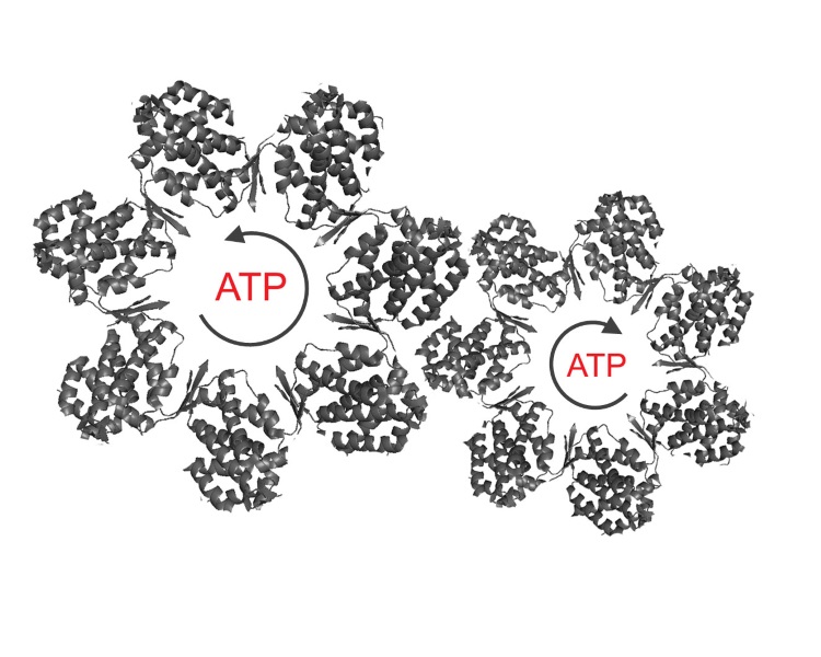
Scientific discussion meeting organised by Professor George Lorimer FRS, Professor Tom McLeish FRS and Professor Amnon Horovitz.
Many cellular processes are facilitated by multi-subunit, motor- proteins. These biological machines exploit allostery and the formation of distinct conformational states resulting from cycles of ligand binding and release, driven by the hydrolysis of ATP. We aim to discuss the latest experimental and computational approaches, so as to enhance our understanding of the structural changes underlying the operation of these molecular machines.
The schedule of talks and speaker biographies are available below.
Attending this event
This meeting has taken place.
Meeting papers will be published in a future issue of Philosophical Transactions B.
Enquiries: Contact the Scientific Programmes team
Organisers
Schedule
Chair
Professor George Lorimer FRS, University of Maryland, USA

Professor George Lorimer FRS, University of Maryland, USA
George Lorimer has been Professor of Biochemistry at the University of Maryland since 1997. In 1978 he joined the Central R&D department of the Du Pont Company. While there, he first worked on the mechanism of Rubisco, the enzyme that fixes CO2 in photosynthesis. He was elected a Fellow of the Royal Society in 1986 for his work on Rubisco. In 1989, using an unequivocally unfolded protein and the purified chaperonin proteins GroEL and GroES, his group was the first to demonstrate the ATP-dependent folding of Rubisco and many other proteins. In 1997, he was elected to the National Academy of Sciences for his work on chaperonin-assisted protein folding. George has since shown that GroEL can perform work on substrate protein during allosteric transitions. He has determined the crystal structure of the functional form, the symmetric GroEL:GroES2 ‘football’ and established that the GroEL rings operate as parallel-processing, iterative annealing molecular machines.
| 09:05 - 09:30 |
The nicotinic acetylcholine receptor a typical model of allosteric membrane protein
The concept of allosteric interaction (1) which was initially proposed to account for the inhibitory feedback mechanism mediated by bacterial regulatory enzymes contrasts with the classical mechanism of competitive, steric, interaction between ligands for a common site. Accordingly allosteric interactions are indirect interactions that take place between topographically distinct sites and are mediated by a discrete & reversible conformational change of the protein. 1. Changeux JP (1961) The feedback control mechanisms of biosynthetic L- threonine deaminase by L-isoleucine. Cold Spring Harb Symp Quant Biol 26:313–318 ; Gerhart JC, Pardee AB (1962] The enzymology of control by feedback inhibition. J Biol Chem 237:891–896 
Professor Jean-Pierre Changeux, Collège de France and Institut Pasteur, France

Professor Jean-Pierre Changeux, Collège de France and Institut Pasteur, FranceJean-Pierre Changeux PhD is International Faculty at the Kavli Institute for Brain and Mind University of California San Diego and Honorary Professor at the Collège de France and Institut Pasteur, Paris. Changeux PhD studies led to the discovery that chemical signals regulate the biological activity of proteins by acting at ‘allosteric’ sites distinct from the biologically active sites via a conformational change (1961-1965). He then proposed (1964,1966) that this type of regulation applies to receptor mechanisms engaged in the transmission of chemical signals in the nervous system and through his life-time work, validated this insight. His studies were initiated by the first identification of a neurotransmitter receptor: the nicotinic acetylcholine receptor together with Lee and Kasai (1970) and culminated by a contribution, with Corringer and Delarue to establishing the 3D structure and conformational transition of prokaryotic orthologs of nicotinic receptors by X-ray crystallography and molecular dynamics (2005-15). Changeux and his colleagues also deciphered the topology of allosteric modulatory sites for pharmacological ligands (1996-2011), thereby substantiating a novel strategy of drug design based on allosteric modulation. Moving to neuronal networks, Changeux, together with Courrège and Danchin (1973, 1976) formulated and experimentally tested the theory that long-term epigenesis of neuronal networks occurs by the activity-dependant selective stabilisation and elimination of developing synapses. Last, in particular with Dehaene, he proposed and tested models for defined cognitive tasks and their pharmacological modulation (1991-2015) in particular, a neuronal hypothesis for conscious processing, implicating a ‘global neuronal workspace’ composed of a brain-scale horizontal network of long axon neurons (1998-2015). His academic accolades include the Gairdner award (1978), the Wolf prize (1983), Médaille d'Or, Centre National de la Recherche Scientifique, Paris, (1992), the Goodman and Gilman Award in drug receptor pharmacology (1994), the Balzan Prize (2001), the US National Academy of Sciences Award in Neurosciences (2007), the Japanese Society for the Promotion of Science Award for Eminent Scientists,Tokyo (2012) and the Olav Thon prize Oslo (2016). |
|
|---|---|---|
| 09:40 - 10:05 |
Genetically tunable frustration controls allostery in an intrinsically disordered transcription factor
Intrinsically disordered proteins (IDPs) present a functional paradox because they lack stable tertiary structure, but nonetheless play a central role in signaling, utilizing a process known as allostery. Historically, allostery in structured proteins has been interpreted in terms of propagated structural changes that are induced by effector binding. Thus, it is not clear how IDPs, lacking such well-defined structures, can allosterically affect function. Here we show a mechanism by which an IDP can allosterically control function by simultaneously tuning transcriptional activation and repression, using a novel strategy that relies on the principle of energetic ‘frustration’. We demonstrate that human glucocorticoid receptor tunes this signaling in vivo by producing translational isoforms differing only in the length of the disordered region, which modulates the degree of frustration. We expect this frustration-based model of allostery will prove to be generally important in explaining signaling in other IDPs. 
Professor Vincent J. Hilser, Johns Hopkins University, USA

Professor Vincent J. Hilser, Johns Hopkins University, USAVincent J. Hilser has earned a BS degree in chemistry from St. John’s University (1987), and a PhD in Biochemistry from Johns Hopkins University (1995). From 1995 through 1997, he performed postdoctoral research in the lab of Ernesto Freire (John Hopkins University). In 1997, he accepted a position as an Assistant Professor in the Sealy Center for Structural Biology at the University of Texas Medical Branch in Galveston, Texas, ultimately rising to the rank of Professor and Director of the Center from 2005-2010. In 2010, he accepted a position in the Department of Biology at Johns Hopkins University, and has served as Chair of Biology since 2014. Dr Hilser’s research focuses on understanding the physical and energetic basis, as well as the functional consequences, of conformational heterogeneity in proteins and applying this understanding to the development of novel fold classification and protein design strategies and investigations into allosteric mechanism. |
|
| 10:35 - 11:05 |
New approaches for elucidating allosteric mechanisms and their application to chaperonins
Chaperonins are allosteric machines that consist of two back-to-back stacked heptameric rings with a cavity at each end where protein folding can take place. They assist protein folding by undergoing large conformational changes that are controlled by ATP binding and hydrolysis. The concerted Monod–Wyman–Changeux and sequential Koshland–Némethy–Filmer models of cooperativity are often used to describe such allosteric switching. In general, however, it has been impossible to distinguish between these different allosteric models using ensemble measurements of ligand binding in bulk protein solutions. In this talk, two approaches that break this impasse will be described: one that is kinetic and a second that is based on native mass spectrometry. Using these approaches, it was possible to show that the chaperonin GroEL from E. coli undergoes concerted intra-ring conformational changes whereas its eukaryotic homologue CCT/TRiC undergoes sequential intra-ring conformational changes. The impact of these different allosteric mechanisms on the folding functions of GroEL and CCT/TRiC will be discussed. 
Professor Amnon Horovitz, Weizmann Institute of Science, Israel

Professor Amnon Horovitz, Weizmann Institute of Science, IsraelAmnon Horovitz completed his undergraduate and graduate studies in biochemistry at the Hebrew University of Jerusalem. His thesis work on ‘Additivity in the effects of amino acid substitutions on protein-protein interactions’ was carried out under the supervision of Professors M. Rigbi and R. D. Levine. In 1989, he joined the laboratory of Professor Alan Fersht in Cambridge, England as a post-doctoral fellow where he worked on developing the double-mutant cycle method and applying it to study protein folding and stability. In 1991, he joined the faculty of the Department of Structural Biology at the Weizmann Institute of Science in Israel where he has been since. Amnon Horovitz was chair of the Department of Structural Biology from 2000 to 2006 and has been Full Professor since 2004. He has won several awards including the Hestrin Prize of the Israel Biochemical Society (1989) and the Zimmer Award of the University of Cincinnati (2008). He is currently President of the Israel Society for Biochemistry and Molecular Biology. |
|
| 11:15 - 11:45 |
Cooperative Dynamics of Neurotransmitter Transporters: Learning from Experiments and Computations
Recent years have seen a breakthrough in the elucidation of the structure and dynamics of sodium-coupled neurotransmitter transporters. These membrane proteins are essential regulators of neurotransmission in the brain, and their malfunction is implicated in several neurological disorders. We have now made significant progress in understanding the complex machinery of these secondary transporters, the way they undergo cooperative structural changes between outward-facing and inward-facing states for transporting their substrate and sodium ions, while they also permit for chloride channeling. We will present recent progress made in the elucidation of the mechanism of function of two major groups of transporters and their alteration by ligand binding and/or multimerization: Glutamate transporters, exemplified by the archaeal transporter GltPh which served as a useful model for understanding the dynamics of excitatory amino acid transporters (EAATs); and dopamine transporters as an important member of transporters sharing the LeuT fold. We will show how the multidomain structure or multimerization properties are essential to altering not only their conformational dynamics, but also the coupled membrane remodeling in the synapse, based on recent progresses made in both visualizing and modeling the molecular-to-cellular dynamics of these important transporters that regulate glutamatergic and dopaminergic signaling in the central nervous system. 
Professor Ivet Bahar, University of Pittsburgh, USA

Professor Ivet Bahar, University of Pittsburgh, USAIvet Bahar works at the interface between computational and life sciences, developing models and methods rooted in fundamental principles of physical sciences and engineering. She is specialised in biomolecular systems dynamics and developed several tools to facilitate the evaluation of collective dynamics for biomolecular systems. She is a Distinguished Professor in the Department of Computational & Systems Biology at the University of Pittsburgh, School of Medicine. She co-founded the Joint PhD Program in Computational Biology between the University of Pittsburgh and Carnegie Mellon University. She is a member of the European Molecular Biology Organization (EMBO). |
|
| 11:55 - 12:25 |
Kinetics and thermodynamics of protein assembly
The aim of my work is to understand the physicochemical principles that govern the highly complex process of protein assembly. This process may lead to fatal diseases, as in the case of Alzheimer's disease, but life also profits from it as many molecular processes within a cell are carried out by molecular machines that are built from a large number of proteins. All-atom molecular dynamics (MD) simulations of protein assembly in explicit solvent have been performed for over a decade, revealing valuable information about this phenomenon. The focus of my work lies on the analysis of MD simulations to elucidate the kinetics and thermodynamics of protein assembly processes. To this end, we developed kinetic transition networks showing the transitions between aggregates of different sizes and structural characteristics, allowing us to extract both the thermodynamics and kinetics of the assembly process. While the kinetic transition networks are based on conformational clustering, Markov state models (MSMs) use kinetic clustering for the identification of metastable states. The application of MSMs to protein assembly is highly desirable but challenging, as I will demonstrate in this talk for the aggregation of a small peptide. I will conclude my talk with a perspective on how the methods developed in my group can be applied to molecular machines in order to identify structural changes and kinetically relevant intermediates in their functional cycle. 
Professor Birgit Strodel, Jülich Research Centre, Germany

Professor Birgit Strodel, Jülich Research Centre, GermanyBirgit Strodel studied Chemistry at the universities of Düsseldorf (Germany) and North Carolina, Chapel Hill (USA). She received her PhD in Theoretical Chemistry from the University of Frankfurt/Main (Germany) in 2005 under the supervision of Professor Gerhard Stock. She then spent 2006–2008 as a post-doctoral research associate at the Chemistry Department at the University of Cambridge (UK), working with Professor David J. Wales. Since 2009 she had been head of the Computational Biochemistry Group at the Jülich Research Centre. In addition, she was appointed to a Professorship at Heinrich Heine University Düsseldorf in 2011. Her research primarily involves the study of protein-protein interactions and protein aggregation, for which she develops and applies simulation techniques to reveal the thermodynamics and kinetics of such processes. |
Chair
Professor Keith Willison, Imperial College London, UK

Professor Keith Willison, Imperial College London, UK
Professor Keith Willison is Professor of Chemical Biology in the Department of Chemistry, Imperial College London. He is a founder member of the Imperial College London Institute of Chemical Biology (http://www.imperial.ac.uk/chemical-biology) and the associated EPSRC Doctoral Training Centre (http://www.icb-cdt.co.uk/). He gained extensive experience of management and leadership roles in basic research, clinical research and medicine at the Institute of Cancer Research (1981-2011) where he was ICR Head of Laboratories (1995-2005) and a Trustee of the Royal Marsden Cancer Hospital (1995-2008). He has studied chaperone-assisted protein folding in eukaryotic cytoplasm; particularly the CCT folding machine. He takes a multidisciplinary approach to studying protein machines; in vivo genetics, enzyme kinetics, time resolved and single molecule spectroscopy, single-particle electron microscopy, X-ray crystallography and proteomics. His most notable scientific achievement has been the development of an energy landscape model for actin folding dynamics which explains the spring-like properties and symmetry-breaking nature of the actin filament.
| 13:30 - 14:00 |
Breaking symmetry via helix dipoles
The dipole moment of -helices (positive at the N-terminus, negative at the C-terminus is an important property that is rarely exploited in the transmission of allosteric information. Evidence will be presented here that the nucleotide binding sites of the different rings of the chaperonin GroEL communicate via the dipole moment of the 21-residue helix D (Gly88- Ala109). In the apo-state of GroEL the -amino group of Lys105 of helix D of one ring forms an inter-ring salt bridge with the negatively charged carbonyl oxygen of Ala109 of helix D of the other ring. Upon binding ATP the -phosphate interacts with the positively charged Gly88 at the N-terminal end of helix D, causing a ~2Å rigid body shift of the entire helix. This results in the breakage of the inert-ring Lys105-Ala109 salt bridges that are responsible for the negative cooperativity in ATP binding between the rings. The mutant K105A (i) removes these inter-ring salt bridges, (ii) abolishes negative cooperativity in ATP hydrolysis between the rings without altering the positive cooperativity within a ring or the fundamental rate of ATP hydrolysis determined from the pre-steady state burst kinetics, (iii) compromises the ability of the symmetric GroELK105A-GroES2 “football” complex to undergo breakage of symmetry. Absent the electron density associated with the side chains of K105, the crystal structure of apo-GroELK105A is virtually identical to that of apo-GroELWT. 
Professor George Lorimer FRS, University of Maryland, USA

Professor George Lorimer FRS, University of Maryland, USAGeorge Lorimer has been Professor of Biochemistry at the University of Maryland since 1997. In 1978 he joined the Central R&D department of the Du Pont Company. While there, he first worked on the mechanism of Rubisco, the enzyme that fixes CO2 in photosynthesis. He was elected a Fellow of the Royal Society in 1986 for his work on Rubisco. In 1989, using an unequivocally unfolded protein and the purified chaperonin proteins GroEL and GroES, his group was the first to demonstrate the ATP-dependent folding of Rubisco and many other proteins. In 1997, he was elected to the National Academy of Sciences for his work on chaperonin-assisted protein folding. George has since shown that GroEL can perform work on substrate protein during allosteric transitions. He has determined the crystal structure of the functional form, the symmetric GroEL:GroES2 ‘football’ and established that the GroEL rings operate as parallel-processing, iterative annealing molecular machines. |
|
|---|---|---|
| 14:15 - 14:45 |
Dynamic GroEL-GroES interaction revealed by high-speed atomic force microscopy
The GroEL−GroES chaperonin reaction system contains two rings of GroEL, seven ATPase sites in each ring, co-chaperonin GroES and substrate protein. Therefore, the reaction cycle of this system is too complicated to be analyzed by ensemble methods. Single-molecule fluorescence microscopy studies were recently performed and revealed a part of the reaction scheme with major appearance of the symmetric GroEL−(GroES)2 complexes. Nevertheless, the two rings of GroEL are closely positioned, so that it was difficult to know from which ring a detected fluorescence signal came. Here we used high-speed atomic force microscopy to visualize the dynamic GroEL−GroES interaction in the presence of ATP and unfoldable substrate protein, disulfide bond-reduced α-lactalbumin. The GroEL−GroES interaction was observed to branch into main and side pathways with probabilities of 2/3 and 1/3, respectively. In the main pathway, alternate binding and release of GroES occurs at the two rings. This alternate rhythm is made possible by two types of inter-ring communications; (i) the completion of ATP hydrolysis to ADP∙Pi at the new cis ring triggers Pi-release (and hence GroES release) from the opposite ring and (ii) the resulting asymmetric bullet-shaped structure retards ADP dissociation from the trans ring. This retardation contributes to providing an enough time for the substrate protein to be released from the trans ring but in turn possibly results in incomplete replacement of ADP with ATP at the trans ring. This incompletion is likely to deteriorate the inter-ring communication, resulting in the break of the alternate rhythm. 
Professor Toshio Ando, Kanazawa University, Japan

Professor Toshio Ando, Kanazawa University, JapanToshio Ando is a Biophysicist, studying at the Bio-AFM Frontier Research Center, Kanazawa University. He received his Dc. Sci. degree in physics from Waseda University. After working at UC San Francisco as a postdoctoral fellow and then an Assistant Research Biophysicist, he joined the faculty at Kanazawa University in 1986, where he is a Professor of Physics and Biophysics. He has been studying the molecular mechanism of proteins, while developing new tools. In 1993, he embarked on the development of high-speed atomic force microscopy and finally materialised this new microscopy in 2008. Currently, his research focuses on the nano-visualisation of protein molecules in dynamic action and on the development of high-speed scanning probe microscopy techniques. |
|
| 15:30 - 16:00 |
Division of labour among the subunits of a highly coordinated ring ATPase
As part of their infection cycle, many viruses must package their newly replicated genomes inside a protein capsid. Bacteriophage phi29 packages its 6.6 mm long double-stranded DNA using a pentameric ring nano motor that belongs to the ASCE (Additional Strand, Conserved E) superfamily of ATPases. A number of fundamental questions remain as to the coordination of the various subunits in these multimeric rings. The portal motor in bacteriophage phi29 is ideal to investigate these questions and is a remarkable machine that must overcome entropic, electrostatic, and DNA bending energies to package its genome to near-crystalline density inside the capsid. Using optical tweezers, the Bustamante group finds that this motor can work against loads of up to ~55 picoNewtons on average, making it one of the strongest molecular motors ever reported. The group establishes the force-velocity relationship of the motor. Interestingly, the packaging rate decreases as the prohead fills, indicating that an internal pressure builds up due to DNA compression attaining the value of ~6 MegaPascals at the end of the packaging. This pressure, Bustamante shows, is used as part of the mechanism of DNA injection in the next infection cycle. The group used high-resolution optical tweezers to characterize the steps and intersubunit coordination of the pentameric ring ATPase responsible for DNA packaging in bacteriophage Phi29. By using non-hydrolyzable ATP analogs and stabilizers of the ADP bound to the motor, they establish where DNA binding, hydrolysis, and phosphate and ADP release occur relative to translocation. Bustamante shows that while only 4 of the subunits translocate DNA, all 5 bind and hydrolyze ATP, suggesting that the fifth subunit fulfills a regulatory function. Finally, he shows that the motor not only can generate force but also torque. The group characterizes the role played by the special subunit in this process and identify this the symmetry-breaking mechanism. These results represent the most complete studies done to date on these widely distribute class of ring nano motors. 
Professor Carlos Bustamante, University of California, Berkeley, USA

Professor Carlos Bustamante, University of California, Berkeley, USACarlos Bustamante uses novel methods of single-molecule visualisation, such as scanning force microscopy, to study the structure and function of nucleoprotein assemblies. His laboratory is developing methods of single-molecule manipulation, such as optical and magnetic tweezers, to characterise the elasticity of DNA, to induce the mechanical unfolding of individual protein and RNA molecules, and to investigate the machine-like behaviour of molecular motors. Dr Bustamante is a professor of Molecular and Cell Biology, and Chemistry, and is the Raymond and Beverly Sackler Chair of Physics at the University of California, Berkeley. He has been a Howard Hughes Medical Institute Investigator since 2000. |
|
| 16:15 - 16:45 |
Symmetry, rigidity, and Allosteric Wiring Diagram in multi-domain proteins
I will introduce concepts describing the physical basis of allostery relying on propagation of excitations (phonons for example) in ordered solids. These ideas will be used to develop a Structural Perturbation Method (SPM) for identifying a network of residues that carry signals for allosteric transitions. Applications to bacterial chaperonin (GroEL) will be presented. In addition, the connection between the static SPM theory and the dynamics associated with allosteric transitions will be established. 
Professor Dave Thirumalai, University of Texas, USA

Professor Dave Thirumalai, University of Texas, USA |
Chair
Dr Paolo De Los Rios, EPFL, Switzerland

Dr Paolo De Los Rios, EPFL, Switzerland
Paolo De Los Rios earned a PhD in Statistical Physics at SISSA, Trieste (Italy) in 1993. After postdocs at the Max-Planck Institute for the Physics of Complex Systems (Dresden, Germany) and at the University of Fribourg, Switzerland, he was appointed Assistant Professor at the Ecole Polytechnique Fédérale de Lausanne, where he was promoted to Associate Professor in 2010. He is interested in understanding the function of proteins using tools from statistical physics (e.g. polymer physics, non-equilibrium stochastic processes) and in particular, how these tools can provide novel insights in the role of chaperones at protecting protein homeostasis. More recently, he has started using inverse methods in statistical physics to extract information from co-evolutionary ‘big data’, with the goal of reconstructing protein structures and protein interactions.
| 09:00 - 09:30 |
Allostery in Hsp70s: Intra-molecular pathways and domain dynamics
The 70 kDa heat shock proteins (Hsp70s) constitute the central hub of the cellular protein quality surveillance network. The versatility of Hsp70 chaperones is based on the interaction of their substrate binding domain (SBD) with short degenerative sequence motifs, found in most proteins on average every 30 to 40 residues. This binding is controlled by ATP binding and hydrolysis in the nucleotide binding domain (NBD) of Hsp70s, through an intricate allosteric mechanism. ATP binding increases polypeptide substrate association and dissociation rates by 100 and 1000-fold, respectively. In return, polypeptide substrate binding to Hsp70s stimulates the ATP hydrolysis rate in synergism with J-domain cochaperones. This synergistic stimulation of the ATP hydrolysis rate allows Hsp70s to efficiently trap substrates and is essential for all Hsp70 functions. I will report on extensive structure-function analysis and mutagenesis studies that delineate the allosteric mechanism of Hsp70s, elucidating signal transmission from NBD to SBD. In addition, data on the conformational dynamics of Hsp70s will be presented. Furthermore, we recently solved the crystal structure of the J-domain of E. coli DnaJ in complex with the E. coli Hsp70 DnaK in the ATP bound conformation. Our structure is consistent with a large body of biochemical and biophysical evidence and reveals the molecular mechanism of J-domain protein-mediated stimulation of the ATPase activity of Hsp70s.

Professor Matthias Mayer, Heidelberg University, Germany

Professor Matthias Mayer, Heidelberg University, GermanyMatthias P. Mayer completed his undergraduate and graduate studies at the University of Freiburg, Germany. His thesis work on ‘Desaturation reactions of the carotenoid biosynthesis’ were carried our under the supervision of Professor H. Kleinig. In 1990 he joined as postdoctoral research associate the lab of Professor C. D. Poulter in Salt Lake City, Utah, USA, where he worked on protein prenylation, and subsequently in 1992 the lab of Professor C. Georgopoulos in Geneva, Switzerland, where he worked on protein-protein interactions. In 1997 he moved back to Freiburg, Germany, joining Professor B. Bukau’s lab and working on Hsp70 chaperones. Matthias Mayer habilitated in 2002 and moved with B. Bukau to the Center for Molecular Biology Heidelberg University (ZMBH), where he became independent group leader in 2005. For his habilitation he won the Ernst Schering Prize of the German Society for Biochemistry and Molecular Biology. |
|
|---|---|---|
| 09:45 - 10:15 |
Can conformational dynamics and allostery be underlying mechanistic principle behind protein evolution?
The critical role of protein dynamics has become well recognized in various biological functions, including allosteric signaling and protein ligand recognition electron transfer. Likewise, in protein evolution, the classical view of the one sequence-one structure-one function paradigm is now being extended to a new view: an ensemble of conformations in equilibrium that can evolve new functions. Therefore, understanding inherent structural dynamics are crucial to obtain a more complete picture of protein evolution. A small local structural change due to a single mutation can lead to a large difference in conformational dynamics, even at quite distant residues due to allostery. We have recently analyzed evolution of different protein families including GFP proteins, beta-lactamase inhibitors, and nuclear receptors and observed that alteration of conformational dynamics through allosteric regulations leads to functional changes. Moreover, our site-specific dynamics-based metric reveals that enzymatic function is regulated by dynamically-coupled residues, which forms an allosteric communication network with the active sites. Disease causing mutations trigger a global loss in dynamic coupling, which disrupts the communication network ultimately inhibiting function. Analysis of over 200 missense mutations also shows that Gaucher disease (GD) mutations are abundant at dynamic allosteric residue coupling sites which we call DARC spots. Further tests using 75 human enzymes revealed that diseases emerging from DARC spot mutations are not isolated to GD; indeed, this phenomenon is observed across the proteome. 
Professor S. Banu Ozkan, Arizona State University, USA

Professor S. Banu Ozkan, Arizona State University, USAProfessor Ozkan is an Associate Professor of Physics at Arizona State University. She completed her PhD at Bogazici University in 2002. She worked as a Postdoctoral Fellow at University of Pittsburgh Medical School, and at University of California San Francisco. Her background is in computational biological physics with expertise that includes the prediction and design of protein structures as well as the modelling of protein dynamics and protein-ligand interactions. She has developed new methods for master-equation models of folding kinetics, lattice models, elastic networks, and all-atom physics-based computer simulations. These methods enable a better understanding of the principles of bio-molecular interactions at different length scales and the study of principles underlying protein folding and binding. Recently, she has developed a novel method called dynamic flexibility index, which quantifies the contribution of individual amino acid residues to functionally important dynamics. She has applied this method to understand the mechanistic insights about protein evolution, and disease development. |
|
| 11:00 - 11:30 |
Cryo-EM as a tool to explore the proteasome, its function and its ligands
The proteasome is essential in all eukaryotes for the highly regulated ATP dependent proteolysis of ubiquitin-tagged proteins, including key cell regulators. In eukaryotes its proteolytic core is formed by four hetero-heptameric rings arranged as an α(1 7)β(1 7)β(1 7)α(1 7) barrel shaped stack, which activity requires the binding of regulators at the outer surfaces of the α subunits. The 19S regulatory particle is the proteasome activator that recruits fully folded ubiquitinated proteins for degradation, associates with the core to form the fully active 26S proteasome and comprises a remarkable variety of functions, including ubiquitin receptors, deubiquitinases, ATP dependent unfoldase and translocase, and scaffolding. These are accomplished by at least 18 distinct canonical subunits, together with additional transiently bound proteins that further modulate the proteasome activity. Recently there was substantial progress in determining the 26S proteasome structure. However, a complete characterisation of how the proteasome components orchestrate the individual steps of substrate processing, from their recognition to the release of small peptides, is still missing. This information is required to fully understand the proteasome fundamental role and in order to optimise specific ligands as therapeutic drugs. In principle the final stages of substrate processing can be studied by the structural analysis of the proteasome core in the presence of substrate or product peptide mimetics. We investigated the use of cryo-EM and image processing for such studies, revealing advantages of their use for the overall study of protein-ligand interactions and leading to our contribution to the validation of the proteasome as a potential target for antimalarials. 
Dr Paula da Fonseca, MRC Laboratory of Molecular Biology, UK

Dr Paula da Fonseca, MRC Laboratory of Molecular Biology, UKDr Paula da Fonseca graduated in Biochemistry at the Faculty of Sciences, University of Lisbon, Portugal, and obtained her PhD in Biochemistry at the Imperial College London, UK. She held postdoctoral positions in London at the Imperial College School of Medicine and at the Institute of Cancer Research, before she moved to Cambridge, UK, to start her own research group at the MRC Laboratory of Molecular Biology. Her current work focuses on studying the structure and function of cell regulatory protein complexes, including the proteasome, primarily by electron microscope and image processing based methods. She recently investigated the use of high-resolution electron cryo-microscopy to study the mode by which inhibitory ligands bind to proteasome complexes from both humans and malarial parasites. Her work on the proteasome of malarial parasites has contributed to the identification of new compounds with antimalarial potential. |
|
| 11:45 - 12:15 |
Slow Brownian fluctuations for allosteric signalling without structural change
McLeish presents the results of a physics-biology collaboration in a foundational theory for how allostery can occur as a function of low frequency dynamics without a change in protein structure. Elastic inhomogeneities allow entropic ‘signalling at a distance’. Remarkably, many globular proteins display just this class of elastic structure, in particular those that support allosteric binding of substrates (long-range co-operative effects between the binding sites of small molecules). Through multi-scale modelling of global normal modes McLeish demonstrates negative co-operativity between the two cAMP ligands without change to the mean structure. Crucially, the value of the co-operativity is itself controlled by the interactions around a set of third allosteric ‘control sites’. The theory makes key experimental predictions, validated by analysis of variant proteins by a combination of structural biology and isothermal calorimetry. A quantitative description of allostery as a free energy landscape revealed a protein ‘design space’ that identified the key inter- and intramolecular regulatory parameters that frame CRP/FNR family allostery. Furthermore, by analysing naturally occurring CAP variants from diverse species, McLeish demonstrates an evolutionary selection pressure to conserve residues crucial for allosteric control. The methodology establishes the means to engineer allosteric mechanisms that are driven by low frequency dynamics. 
Professor Tom McLeish FRS, University of York, UK

Professor Tom McLeish FRS, University of York, UKTom McLeish, FRS, is Professor of Natural Philosophy in the Department of Physics at the University of York, England, the Centre for Medieval Studies and Humanities Research Centre. He has held previous academic posts at the universities of Cambridge, Sheffield, Leeds and Durham. His research in ‘soft matter and biological physics,’ draws on interdisciplinary collaborations to study relationships between molecular structure and material properties. He leads the UK ‘Physics of Life’ network, and holds a 5-year research fellowship focusing on the physics of protein signaling and the self-assembly of silk fibres. He has also pursued a programme of interdisciplinary research between the sciences and humanities, include the framing of science, theology, society and history, education and philosophy, leading to the recent books Faith and Wisdom in Science (OUP 2014), The Poetry and Music of Science (OUP 2019) and Soft Matter – A Very Short Introduction (OUP 2020). He co-leads the Ordered Universe project, a large interdisciplinary study of 13th century science. From 2008 to 2014 he served as Pro-Vice-Chancellor for Research at Durham University and was from 2015-2020 Chair of the Royal Society’s Education Committee. |
Chair
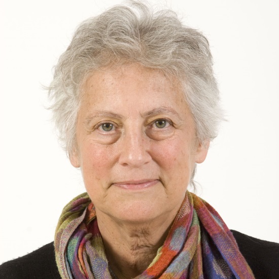
Professor Shoshana J. Wodak, VIB Structural Biology Research Center, Belgium

Professor Shoshana J. Wodak, VIB Structural Biology Research Center, Belgium
Shoshana J. Wodak obtained her PhD from Columbia University, USA. She was Professor at the Free University of Brussels and Group Leader at the European Bioinformatics Institute (EBI) UK. She subsequently held a Canada Research Chair and a professorship at the Departments of Biochemistry and of Molecular Genetics, at the University of Toronto. She is currently Professor and Group Leader at the Vlaamse Institute for Biotechnology (VIB), at the Free University of Brussels (VUB), in Brussels Belgium. Dr Wodak is a member of EMBO and a fellow of the ISCB.
Dr Wodak pioneered algorithms for protein-protein docking and for defining structural domains in proteins. She contributed key papers on the role of local interactions in stabilising the protein native state, on protein structure prediction, protein folding, and fold recognition. She also developed methods for computational protein design, for simulating protein interactions and conformational changes, and for analysing protein interactions networks and cellular pathways.
| 13:30 - 14:00 |
Time-resolved modelling of protein allosteric communication
Allostery represents a fundamental mechanism of biological regulation which is mediated via long-range communication between distant protein sites. While little is known about the underlying dynamical process, recent time-resolved infrared spectroscopy experiments on a photoswitchable PDZ domain (PDZ2S) have indicated that the allosteric transition occurs on multiple timescales. Employing extensive nonequilibrium molecular dynamics simulations, here a time-dependent picure of the allosteric communication in PDZ2S is developed. The simulations reveal that allostery amounts to the propagation of structural and dynamical changes that are genuinely nonlinear and can occur in a nonlocal fashion. A dynamic network model is constructed that illustrates the hierarchy and exceeding structural heterogeneity of the process. In compelling agreement with experiment, three physically distinct phases of the time evolution are identified, describing elastic response ( 0.1 ns), inelastic reorganization (∼ 100 ns) and structural relaxation ( 1μs). Issues such as the similarity to downhill folding as well as the interpretation of allosteric pathways are discussed. 
Professor Gerhard Stock, University of Freiburg, Germany

Professor Gerhard Stock, University of Freiburg, GermanyGerhard Stock studied Physics at the Technische Universität München and got his doctorate (1990) with Professor Wolfgang Domcke. He spent 1991 to 1992 at the University of California, Berkeley with Professor William H. Miller as a DFG postdoctoral fellow. Back at the TU München, he worked at the Department of Chemistry as a DFG Habilitation fellow and obtained his Habilitation in 1996. From 1997 to 2000 he was a Heisenberg Professor at the Department of Physics at the University of Freiburg, before he moved to the Goethe University in Frankfurt am Main where he held a full professorship (C4) in Theoretical Chemistry. Since 2009, he has held Chair of Theoretical Physics (successor of John Briggs) at the University of Freiburg. |
|
|---|---|---|
| 14:15 - 14:45 |
Exploring cooperativity of folding and binding in the tandem-repeat protein class
I will discuss the cooperative nature of protein folding and its relevance for function in the context of two contrasting systems. The first is the cks family of cell-cycle regulatory proteins. These proteins undergo three-dimensional domain swapping, in which two monomers exchange a part of their structure to form a highly intertwined dimer. Our work has explored how features of the cks architecture that are essential for domain swapping also control signal transduction between multiple binding partners that facilitate cks function as a hub for macromolecular complex assembly. The second is the tandem-repeat protein class such as ankyrin and tetratricopeptide repeats. Repeat proteins have strikingly simple, modular structures composed of small (30-50 residue) motifs repeated multiple times in tandem to produce elongated pseudo-one-dimensional spring-like architectures. We have been investigating how the folding cooperativity of repeat proteins can be manipulated by the insertion of long unstructured loops between adjacent repeats. In Nature such insertions, and the decoupling of the repeats that results, are likely to be important for the mechanics and the biological functions of these proteins. 
Dr Laura Itzhaki, University of Cambridge, UK

Dr Laura Itzhaki, University of Cambridge, UKDr Laura Itzhaki’s background is in protein folding and protein engineering. Her group’s research has helped to elucidate the folding-related phenomenon of three-dimensional domain swapping. They resolved the mechanism by which domain swapping occurs, delineated how this capability is encoded in the amino-acid sequence, and revealed the potential for domain swapping to regulate protein function and to drive disease-associated protein misfolding. Subsequent studies on several proteins have shown their findings to constitute universal principles of domain swapping. More recently their research has focused on understanding the rules governing the biophysics of a special protein class, so-called tandem-repeat proteins. These proteins have a simple and distinctive modular architecture, one consequence of which is that they can be subjected to gross perturbations (multi-site mutations, repeat deletions/insertions) yet remain correctly folded. This extraordinary malleability has enabled Dr Itzhaki’s group to resolve in unprecedented detail the multitude of inter-converting structures that comprise their energy landscapes and to apply methods originally designed for small proteins to investigate giant repeat arrays leading to unique insights. The simple, modular architecture of repeat proteins allows their properties to be engineered in a strikingly predictable way, and Dr Itzhaki’s group are currently exploiting this design-ability to dissect cellular mechanisms of folding and unfolding, and to engineer repeat proteins as a new therapeutics platform. |
|
| 15:30 - 16:00 |
Evolution of specificity for allosteric regulators: mechanisms and constraints
We have used ancestral protein reconstruction to identify the structural and genetic mechanisms by which specificity for allosteric effectors evolved in the steroid hormone receptor family of ligand-activated transcription factors. We find that dramatic shifts in ligand specificity -- and even a wholesale loss of allosteric control -- occurred numerous times during receptor evolution and were often mediated by a few mutations of large effect. The delicate energetics of allosteric regulation imposed strong constraints on the proteins' evolution, requiring rare permissive mutations to stabilize specific structural elements before changes in ligand regulation could occur. 
Professor Joseph Thornton, University of Chicago, USA

Professor Joseph Thornton, University of Chicago, USA |
