Links to external sources may no longer work as intended. The content may not represent the latest thinking in this area or the Society’s current position on the topic.
Mechanics of development
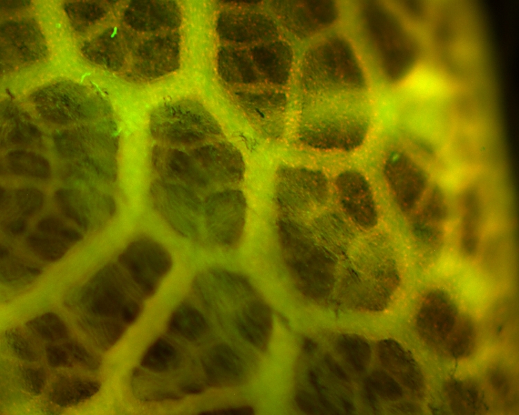
Theo Murphy international scientific meeting organised by Dr Niamh Nowlan, Professor Celeste Nelson and Professor Philippa Francis-West.
Mechanical forces are critical for embryonic development – yet very little is known about their roles. The meeting focused on the role of mechanobiology in multiple aspects of embryonic development. The meeting brought together multidisciplinary research groups in this exciting 'young' area of developmental biology for the first time, enabling formation of a network and sparking new collaborations.
The schedule of talks and speaker biographies can be found below. Recorded audio of some of the presentations is also available below. Meeting papers can be found in a themed issue of Philosophical Transactions B.
Attending this event
This meeting has taken place.
Enquiries: contact the Scientific Programmes Team
Organisers
Schedule
Chair
Professor Philippa Francis-West, King's College London, UK

Professor Philippa Francis-West, King's College London, UK
Philippa Francis-West is a Professor in Developmental and Cell Biology at the Centre for Craniofacial and Regenerative Biology, King’s College London. She earned a BA in Biochemistry at Cambridge University and carried out her PhD at the University of London. This was followed by post-doctoral position with Professors Lewis Wolpert, Paul Brickell and Cheryll Tickle at University College London. She established an independent research group at King’s College London in 1995 and has authored over 70 peer-reviewed publications. The group aims to understand the mechanisms of morphogenesis and cell differentiation within an embryo. The current research focus is on the Dchs1-Fat4-PCP-Hippo signaling pathway and the role of mechanics during cell differentiation. Her research has been funded by BBSRC, MRC together with Arthritis Research UK and Ageing Research charity funding.
Dr Niamh Nowlan, Imperial College London, UK

Dr Niamh Nowlan, Imperial College London, UK
Dr Niamh Nowlan is a Senior Lecturer in the Department of Bioengineering at Imperial College London. Following an undergraduate degree in Computer Engineering and a PhD in Biomechanical Engineering from Trinity College Dublin in Ireland, Dr Nowlan held research fellowships in Boston, USA and Barcelona, Spain. Dr Nowlan has received a number of awards for her research, including a Fulbright Award and the 2016 Bioengineering UK Early Career Scientist Award. She joined Imperial College in 2011, and now leads a research group of 7 full-time researchers. Dr Nowlan's group are working on a range of topics relating to foetal movements and skeletal development, funded by grants from the European Research Council and the Arthritis Research UK and Leverhulme Trust charities.
| 14:05 - 14:45 |
Active and passive forces in epithelial branching
The morphogenetic patterning that generates three-dimensional (3D) tissues requires dynamic concerted rearrangements of individual cells with respect to each other. The group has developed microfabrication- and lithographic tissue engineering-based approaches to investigate the mechanical forces and downstream signalling responsible for generating the airways of the lung and the milk ducts of the mammary gland. Professor Nelson will discuss how to combine these experimental techniques with computational models to uncover the physical forces that drive development of engineered tissues and tissues in vivo. Professor Nelson will also describe efforts to uncover and actuate the different physical mechanisms used to build the airways in lungs from birds, mammals, and reptiles. 
Professor Celeste Nelson, Princeton University, USA

Professor Celeste Nelson, Princeton University, USACeleste M Nelson is a Professor in the Departments of Chemical and Biological Engineering and Molecular Biology at Princeton University. She earned SB degrees in Chemical Engineering and Biology at MIT in 1998, a PhD in Biomedical Engineering from the Johns Hopkins University School of Medicine in 2003, followed by postdoctoral training in Life Sciences at Lawrence Berkeley National Laboratory until 2007. Her laboratory specializes in using engineered tissues and computational models to understand how mechanical forces direct developmental patterning events during tissue morphogenesis and during disease progression. She has authored more than 100 peer-reviewed publications. Dr Nelson’s contributions to the fields of tissue mechanics and morphogenesis have been recognized by a number of awards, including a Burroughs Wellcome Fund Career Award at the Scientific Interface (2007), a Packard Fellowship (2008), a Sloan Fellowship (2010), the MIT TR35 (2010), the Allan P. Colburn Award (2011), a Dreyfus Teacher-Scholar Award (2012), and a Faculty Scholars Award from the Howard Hughes Medical Institute (2016). |
|
|---|---|---|
| 14:45 - 15:30 |
Models and measurements of cortical folding
Cortical folding, or gyrification, is critical to human brain development and function. The folded shape of the human brain allows the cerebral cortex, the thin outer layer of neurons and their associated processes, to attain a large surface area (~1600 cm2) relative to brain volume (~1400 cm3). Abnormal cortical folding has been associated with severe cognitive and emotional disorders, including epilepsy, autism, and schizophrenia. Among different species the onset of cerebral cortical folding consistently takes place after the majority of cerebral cortical neurons have completed migration from their sites of origin (cell layers near the surface of the lateral ventricle) to the nascent cerebral cortex (the cortical plate). Despite decades of study the mechanical forces that lead to cortical folding remain incompletely understood. Leading hypotheses have focused on the roles of tangential growth, heterogeneities in the birth and migration of neurons, and internal tension in axons. Advances in the mathematical modelling of growth and morphogenesis, combined with new experimental data, promise to clarify the mechanical basis of cortical folding. Recent experimental studies have illuminated not only the fundamental cellular and molecular processes underlying cortical development, but also the stress state, mechanical properties, and spatiotemporal patterns of growth in the developing brain. The combination of mathematical modelling with these measurements allows us to evaluate hypothesized mechanisms objectively, and to ensure that they are consistent with physical law. Dr Bayly will review the neurobiology of cortical development, summarize pertinent experimental observations, and discuss the potential role of growth-induced instability in cortical folding. 
Dr Philip Bayly, University of Washington in St. Louis, USA

Dr Philip Bayly, University of Washington in St. Louis, USAPhilip V (Phil) Bayly is The Lilyan and E. Lisle Hughes Professor of Mechanical Engineering and Chair of the Department of Mechanical Engineering and Materials Science at Washington University in St. Louis. Dr Bayly earned an AB in Engineering Science from Dartmouth College, an MS in Engineering from Brown University, and a PhD in Mechanical Engineering from Duke University. Dr Bayly has been a member of the faculty at Washington University since 1993. His research involves the study of nonlinear dynamic phenomena in mechanical and biological systems, using imaging and image processing to understand the mechanics of cells and biological tissues. Dr Bayly’s research in brain biomechanics and flagellar oscillations has been funded by the National Science Foundation, the National Institutes of Health, and the Office of Naval Research. |
|
| 15:30 - 16:00 | Coffee |
Chair

Dr Niamh Nowlan, Imperial College London, UK

Dr Niamh Nowlan, Imperial College London, UK
Dr Niamh Nowlan is a Senior Lecturer in the Department of Bioengineering at Imperial College London. Following an undergraduate degree in Computer Engineering and a PhD in Biomechanical Engineering from Trinity College Dublin in Ireland, Dr Nowlan held research fellowships in Boston, USA and Barcelona, Spain. Dr Nowlan has received a number of awards for her research, including a Fulbright Award and the 2016 Bioengineering UK Early Career Scientist Award. She joined Imperial College in 2011, and now leads a research group of 7 full-time researchers. Dr Nowlan's group are working on a range of topics relating to foetal movements and skeletal development, funded by grants from the European Research Council and the Arthritis Research UK and Leverhulme Trust charities.
| 16:00 - 17:00 |
From mechanobiology to developmentally-inspired engineering
Professor Ingber will review work from his laboratory beginning with mechanistic studies relating to the biophysical basis of cell shape determination and tissue morphogenesis, and ending with demonstration of how the group has leveraged our understanding of the importance of mechanical forces as bioregulators to develop new medical devices and therapeutics. The work began with the hypothesis the mechanical forces are as important biological regulators as chemicals and genes, and that cells use tensegrity architecture to control their form and function. Testing this theory required development of new analytical tools, including magnetic cytometry, femtosecond laser nanosurgery, microcontact printing, and microfluidic culture systems. These studies led to confirmation of the critical role that physical forces play in developmental control, experimental confirmation of the cellular tensegrity model, and discovery that transmembrane integrins and cytoskeletal prestress mediate mechanotransduction and developmental control. Importantly, the group also leveraged these insights to develop new engineering innovations that are being translated into the clinic or commercial marketplace. Examples include: angiogenesis inhibitors for cancer therapy identified based on cell shape changes; ‘shrink wrap’-like polymer gels that trigger tissue and organ formation by physically inducing mesenchymal condensation; mechanotherapeutics that prevent pulmonary edema by inhibiting integrin-mediated mechanochemical signal transduction; shear stress-targeted drug delivery systems that are directed to sites of vascular occlusion and dissolve blood clots without systemic toxicity; and microfluidic cell culture devices lined by living human cells, known as ‘Organs-on-Chips’, which recapitulate organ-level structure and functions by providing appropriate mechanical cues as a way to replace animal testing for drug development, mechanistic discovery, and personalized medicine. 
Professor Donald Ingber, Wyss Institute at Harvard University, USA

Professor Donald Ingber, Wyss Institute at Harvard University, USADonald E Ingber, MD, PhD is the Founding Director of the Wyss Institute for Biologically Inspired Engineering at Harvard University, Judah Folkman Professor of Vascular Biology at Harvard Medical School and Boston Children's Hospital, and Professor of Bioengineering at the Harvard School of Engineering and Applied Sciences. He received his BA, MA, MPhil, MD and PhD from Yale University. Dr. Ingber has made major contributions to mechanobiology, cell biology, bioengineering, tissue engineering, nanobiotechnology, angiogenesis, cancer, medical device development, and translational medicine. He has authored more than 425 publications and 150 patents that have been licensed to multiple companies, founded 5 startups, presented more than 450 presentations world-wide, and received numerous honours in a broad range of disciplines, including membership of the National Academy of Medicine, National Academy of Inventors, American Institute for Medical and Biological Engineering, and the American Academy of Arts and Sciences. |
|---|
Chair
Professor Andrew Pitsillides, The Royal Veterinary College, UK

Professor Andrew Pitsillides, The Royal Veterinary College, UK
Andrew Pitsillides is Professor of Skeletal Dynamics in the department of Comparative Biomedical Sciences at The Royal Veterinary College, London. His research explores skeletal mechanobiology across diverse biological settings; from the role of embryo movement in the emergence of skeletal form, to how bones and joints respond to functional/ traumatic load in growth, adulthood and ageing.

Dr Chrissy Hammond, University of Bristol, UK

Dr Chrissy Hammond, University of Bristol, UK
Dr Chrissy Hammond (CLH) is currently an Arthritis Research UK Fellow, with a proleptic appointment at the University of Bristol. She has worked with animal models of musculoskeletal development for over a decade, holding independent fellowships in the UK and the Netherlands. In 2011 she established a group at Bristol. Her group makes use of the genetic amenability and excellent high resolution imaging options of zebrafish along with computational modelling to unpick the relative contributions of biomechanics and genetics on joint development and homeostasis.
| 08:30 - 09:00 |
Uncovering the mechanistic basis of biomechanical input controlling skeletal development: exploring the interplay with Wnt signalling at the joint
Embryo movement is essential to the formation of a functional skeleton. Using mouse and chick models, the group has previously shown that mechanical forces influence gene regulation and tissue patterning, particularly at developing joints. However, there remains a lack of knowledge of the molecular mechanisms that underpin the influence of mechanical signals. Wnt signalling is required during skeletal development and is altered under reduced mechanical stimulation. To explore Wnt signalling as a mediator of mechanical input, the expression of Wnt ligand and Fzd receptor genes in the developing skeletal rudiments was profiled. Canonical Wnt activity restricted to the developing joint is reduced under immobilization while over-expression of activated b-catenin or the Wnt antagonist Sfrp3 following electroporation of chick embryo limbs, supports the proposed role for Wnt signalling in mechanoresponsive joint patterning. Two key findings advance our understanding of the interplay between Wnt signalling and mechanical stimuli: firstly, the loss of canonical Wnt activity at the joint shows reciprocal, co-ordinated regulation of Wnt and BMP pathways under mechanical influence. Secondly, this occurs simultaneously with increased expression of several Wnt pathway component genes in a territory peripheral to the joint, identifying the importance of mechanical stimulation on a population of potential joint progenitor cells. 
Professor Paula Murphy, Trinity College Dublin, the University of Dublin, Ireland

Professor Paula Murphy, Trinity College Dublin, the University of Dublin, IrelandPaula Murphy is Professor in Developmental Biology, in the School of Natural Sciences, Trinity College Dublin, the University of Dublin. She is a developmental biologist interested in the mechanisms that generate spatially organized patterns of tissue differentiation in the developing embryo. A science graduate of Trinity College Dublin, specialising in Genetics, she carried out doctoral research at the University of Edinburgh (MRC Human Genetics Unit) under the supervision of Prof RE Hill on Hox genes. Two Postdoctoral Research Fellowships from the European Molecular Biology Organisation and the Human Frontiers Science Programme provided research experience at the University of Rome (La Sapienza) (1991 – 1993) and the Ecole Normale Superieur, Paris (1993 – 1995), working on muscle and peripheral nervous system development. Following research positions in Oslo and Edinburgh she returned to take up her current post at Trinity College in 2001. In 2008 she was elected Fellow of Trinity College Dublin. |
|
|---|---|---|
| 09:00 - 09:30 |
New players and concepts in muscoskeletal biomechanics
Muscle spindles and Golgi tendon organs (GTOs), proprioceptive mechanoreceptors located inside striated muscles and in myotendinous junctions, respectively, are components of the stretch reflex circuitry that control muscle activity. Using genetic mouse models, the group demonstrates the involvement of proprioception in regulating spine alignment and spontaneous realignment of fractured bones, dubbed natural reduction. Failure of the mechanism that maintains posture may result in spinal deformity as in adolescent idiopathic scoliosis. The group shows that null mutants for Runx3 transcription factor, which lack connectivity between proprioceptors and spinal cord, developed peripubertal scoliosis not preceded by vertebral dysplasia or muscle asymmetry. Deletion of Runx3 in the peripheral nervous system or specifically in peripheral sensory neurons, or of enhancer elements driving Runx3 expression in proprioceptive neurons, induced a similar phenotype. Egr3 knockout mice, lacking spindles but not GTOs, displayed a less severe phenotype, suggesting that both receptor types are required for this regulatory mechanism. Fracture repair involves restoration of bone morphology. Comparison among mice of different ages revealed, surprisingly, that three-month-old mice exhibited more rapid and effective natural reduction than newborns. Fractured bones of Runx3-null mutants failed to realign properly. Blocking Runx3 expression in peripheral nervous system, but not in limb mesenchyme, recapitulated the null phenotype, as did inactivation of muscles flanking the fracture site. Egr3 knockout mice displayed a less severe phenotype, suggesting that both receptor types, as well as muscle contraction, are required for this regulatory mechanism. Overall, these findings uncover physiological roles for proprioception in non-autonomous regulation of skeletal integrity and repair. 
Professor Elazar Zelzer, Weizmann Institute of Science, Israel

Professor Elazar Zelzer, Weizmann Institute of Science, IsraelProfessor Zelzer received his MSc in immunology from Ben-Gurion University, Beer-Sheva in 1994 and his PhD in molecular genetics in 1999 from the Weizmann Institute of Science. He conducted his postdoctoral studies in developmental biology, focusing on bone angiogenesis, at Professor Bjorn Olsen’s lab, Harvard Medical School. Professor Zelzer established his lab at the Weizmann Institute in 2004. The main goal of his lab is to decipher the fundamental mechanisms of the development of a complex, integrated system in order to expose the underlying biological principles. Using the limb musculoskeleton and its vasculature as a model system, the group have investigated subjects such as growth coordination and scaling, tissue attachment, cross-tissue regulatory interactions and mechanotransduction. |
|
| 09:30 - 10:00 |
The role of fetal movements in shaping the developing human skeleton
Mechanical stimulation generated by fetal kicking and movements is known to be important for prenatal musculoskeletal development, and there are a number of human conditions that emphasise the link between abnormal fetal movements and delayed or impaired skeletal development. The most common of these is developmental dysplasia of the hip (DDH), a relatively common joint shape abnormality (clinical incidence 1.3 in 1000), the risk of which is strongly associated with restricted fetal movement, such as fetal breech position. The group is using computational modelling approaches (finite element analysis and musculoskeletal modelling), combined with human fetal imaging data to try to understand how the biomechanical stimulation (stresses and strains) caused by fetal movements evolve in the prenatal developing hip joint. Furthermore, the group models a range of intra-uterine conditions and situations that increase the risk of DDH, such as fetal breech position and oligohydramnios (reduced amniotic fluid), in order to be able to understand how a range of factors affecting movement may impact on the developing hip joint. 
Dr Niamh Nowlan, Imperial College London, UK

Dr Niamh Nowlan, Imperial College London, UKDr Niamh Nowlan is a Senior Lecturer in the Department of Bioengineering at Imperial College London. Following an undergraduate degree in Computer Engineering and a PhD in Biomechanical Engineering from Trinity College Dublin in Ireland, Dr Nowlan held research fellowships in Boston, USA and Barcelona, Spain. Dr Nowlan has received a number of awards for her research, including a Fulbright Award and the 2016 Bioengineering UK Early Career Scientist Award. She joined Imperial College in 2011, and now leads a research group of 7 full-time researchers. Dr Nowlan's group are working on a range of topics relating to foetal movements and skeletal development, funded by grants from the European Research Council and the Arthritis Research UK and Leverhulme Trust charities. |
|
| 10:00 - 10:30 |
Mechanobiology of embryonic tendon development and regeneration
Tendons transmit muscle-generated forces to bones to enable skeletal movements and stabilize joint structures. Proper development and maintenance of these extracellular matrix-rich tissues is critical to their demanding physical roles throughout the body. Abnormal tendon formation during embryogenesis is associated with frequently occurring musculoskeletal deformities such as congenital tallipes equino varus. Furthermore, tendons injured postnatally fail to recapitulate development during healing, and instead heal with aberrant matrix composition and organization, resulting in reduced functionality and greater susceptibility to re-injury. The group is interested in understanding how tendon mechanical properties elaborate during normal embryonic development to inform the prevention of tendon-related musculoskeletal birth defects and the enhancement of regenerative postnatal healing. This talk will focus specifically on the novel approaches the group has utilized to characterize the mechanical properties of tendons during embryonic development, and crosslinking mechanisms that have been identified to be critical to this process. Furthermore, the talk will discuss the development of engineered hydrogel systems that mimic the embryonic tendon mechanical microenvironment, enabling study of how dynamic changes in tissue stiffness continuously influence cell behaviours (differentiation, extracellular matrix synthesis, etc.) during development. Finally, the talk will discuss the role of physical movements (eg, kicking) in regulating tendon formation during embryonic development. These findings provide new insights into the crucial roles of mechanics and tissue mechanical properties in new tendon formation. 
Professor Catherine K Kuo, University of Rochester, USA

Professor Catherine K Kuo, University of Rochester, USACatherine K Kuo is an Associate Professor in the Department of Biomedical Engineering, the Department of Orthopaedics, and the Center for Musculoskeletal Research at the University of Rochester. She received her BSE in Materials Science and Engineering and PhD in Biomaterials and Macromolecular Science and Engineering from the University of Michigan, and completed her Postdoctoral Fellowship in the Cartilage Biology and Orthopaedics Branch of NIAMS at the NIH. Her research focuses on elucidating mechanically and biochemically driven mechanisms of embryonic tendon development and healing, and using these findings to inform novel stem cell-based tissue engineering and regenerative medicine strategies. Professor Kuo is the recipient of numerous accolades for her research, including the Go:Life Award for Innovation in Research (2015), Stem Cell Research and Therapy Emerging Investigator Award (2015), NSF CAREER Award (2013), and March of Dimes Basil O'Connor Starter Scholar Research Award (2011). Her research has been continuously funded by the National Institutes of Health (NIH), Department of Defense (DoD), National Science Foundation (NSF), March of Dimes Foundation, and Biogen Idec. She serves on the editorial review board for the Journal of Orthopaedic Research and the advisory council for the International Society of Ligaments and Tendons. She is Chair of the ORS Tendon Section Research Committee, ORS Program Committee Regenerative Medicine Topic Co-chair, and ORS Ambassador for the US Northeast. |
|
| 10:30 - 11:00 | Coffee |
Chair

Professor Celeste Nelson, Princeton University, USA

Professor Celeste Nelson, Princeton University, USA
Celeste M Nelson is a Professor in the Departments of Chemical and Biological Engineering and Molecular Biology at Princeton University. She earned SB degrees in Chemical Engineering and Biology at MIT in 1998, a PhD in Biomedical Engineering from the Johns Hopkins University School of Medicine in 2003, followed by postdoctoral training in Life Sciences at Lawrence Berkeley National Laboratory until 2007. Her laboratory specializes in using engineered tissues and computational models to understand how mechanical forces direct developmental patterning events during tissue morphogenesis and during disease progression. She has authored more than 100 peer-reviewed publications. Dr Nelson’s contributions to the fields of tissue mechanics and morphogenesis have been recognized by a number of awards, including a Burroughs Wellcome Fund Career Award at the Scientific Interface (2007), a Packard Fellowship (2008), a Sloan Fellowship (2010), the MIT TR35 (2010), the Allan P. Colburn Award (2011), a Dreyfus Teacher-Scholar Award (2012), and a Faculty Scholars Award from the Howard Hughes Medical Institute (2016).

Dr Chrissy Hammond, University of Bristol, UK

Dr Chrissy Hammond, University of Bristol, UK
Dr Chrissy Hammond (CLH) is currently an Arthritis Research UK Fellow, with a proleptic appointment at the University of Bristol. She has worked with animal models of musculoskeletal development for over a decade, holding independent fellowships in the UK and the Netherlands. In 2011 she established a group at Bristol. Her group makes use of the genetic amenability and excellent high resolution imaging options of zebrafish along with computational modelling to unpick the relative contributions of biomechanics and genetics on joint development and homeostasis.
| 11:00 - 11:45 |
Notochord mechanics shape the vertebrate axis
The notochord is a conserved axial structure that serves as a hydrostatic scaffold for embryonic axis elongation and, later on, for proper spine assembly. The vertebrate notochord consists of a core of large vacuolated cells surrounded by an epithelial sheath that is encased by an elaborate extracellular matrix. Each vacuolated cell contains a single fluid filled vacuole that occupies most of the cellular volume. These cells are arranged in a stereotypical staircase pattern within the notochord. The group investigated the origin of this pattern and found that it can be achieved purely by physical principles. Moreover, the group is able to model the arrangement of vacuolated cells within the notochord using a physical model composed of silicone tubes and water absorbing beads, suggesting that the notochord is a self-organizing structure. Furthermore, it was found that the biological structure and the physical model can be accurately described by the theory developed for cylindrical foams. In spite of their large size and being under constant mechanical stress, vacuolated cells possess a remarkable structural integrity. This is in part due to the presence of numerous caveolae, omega shaped cell surface invaginations, at the plasma membrane of vacuolated cells, which buffer mechanical tension. In zebrafish that lack caveolae, vacuolated cells collapse under the strain of axial bending during locomotion. Then, release of nucleotides from collapsed vacuolated cell leads to the invasion and trans-differentiation of sheath cells into new vacuolated cells. This regenerative response restores the mechanical properties of the notochord, thus allowing normal spine development. 
Associate Professor Michel Bagnat, Duke University, USA

Associate Professor Michel Bagnat, Duke University, USAMichel was born in Rio Negro, Northern Patagonia, Argentina. He did his undergrad at the Universidad Autónoma de Madrid in Spain. For his graduate studies, he went to the EMBL in Heidelberg, Germany and the Max Planck Institute in Dresden, Germany to study protein and lipid sorting in budding yeast. He then went to UCSF as a postdoc to learn about zebrafish genetics and development. He started his lab in the Department of cell Biology at Duke University in 2008 and was promoted to Associate Professor in 2016. The Bagnat lab uses zebrafish to study the reciprocal relationship between the development of organs and the physiological function they perform. |
|
|---|---|---|
| 11:45 - 12:30 |
Haemodynamic regulation of heart valve development
Recent studies using a variety of animal models support a central role for flow and fluid forces in the development of valves in the heart, blood vessels and lymphatic vessels. An outstanding question in this field is how information sensed by endothelial cells that are directly subject to fluid forces is converted to instructions to underlying cells that participate most directly in formation of valves. To address this question the group has investigated the role of the transcription factor Krupple-like Factor 2 (KLF2) in heart valve development. KLF2 expression is highly upregulated by fluid shear forces in endothelial cells in vitro and its expression in vivo corresponds closely to predicted endothelial fluid shear forces as well. The group finds that loss of endocardial KLF2 in mouse embryos results in mid-gestation death due to an inability to remodel the cardiac cushion to a mature valve as the embryo grows. Loss of KLF2 is accompanied by cell non-autonomous changes in mesenchymal cell proliferation and condensation, two key steps in valve development. Loss of KLF2 results in virtually complete loss of the secreted protein Wnt9b as well as a severe loss of canonical WNT signalling in the sub-endocardial mesenchymal cells that participate in valve formation. Endocardial loss of Wnt9b recapitulates the Klf2 deficient phenotype in mice and studies in zebrafish embryos performed in collaboration with the Vermot lab demonstrate that Wnt9b is strongly regulated by both fluid shear forces and klf2a in vivo. These studies provide one answer to the question of how the information generated by fluid forces at the blood-endothelial interface is transmitted to cells not in contact with blood to orchestrate the creation of heart valves. 
Professor Mark Kahn, University of Pennsylvania, USA

Professor Mark Kahn, University of Pennsylvania, USAMark L Kahn received his BA and MD from Brown University. He completed his internal medicine residency at Oregon Health Sciences University, completed his medical staff fellowship at NIH’s National Heart, Lung and Blood Institute and his clinical and post-doctoral fellowships at UCSF. Mark is a practicing cardiologist in the Division of Cardiovascular Medicine at Penn. He has won many prestigious awards including the Judah Folkman Award in Vascular Biology from the North American Vascular Biology Organization (NAVBO) and has recently been awarded the Cooper-McClure Chair in Medicine. Mark has a full time laboratory that studies cardiovascular development and function. Their interest in the CCM pathway began with studies of the HEG receptor in mouse and fish development, and have extended to understanding CCM signalling and disease pathogenesis using a mouse model and human patient samples. His lab recently identified a molecular pathway by which CCM signalling regulates MEKK3-ERK5 signalling and the expression of KLF2 and KLF4 transcription factors in the endothelium during cardiac development (Developmental Cell, 2015). The lab’s most recently published studies used a mouse model of CCM disease and analysis of human CCM lesions to demonstrate that loss of CCM negative regulation of MEKK3-KLF2/4 signalling also underlies CCM pathogenesis (Nature, 2016). These new findings extend these studies to identify an upstream mechanism of MEKK3 signalling that appears to play an important role in the human disease. |
Chair

Professor Philippa Francis-West, King's College London, UK

Professor Philippa Francis-West, King's College London, UK
Philippa Francis-West is a Professor in Developmental and Cell Biology at the Centre for Craniofacial and Regenerative Biology, King’s College London. She earned a BA in Biochemistry at Cambridge University and carried out her PhD at the University of London. This was followed by post-doctoral position with Professors Lewis Wolpert, Paul Brickell and Cheryll Tickle at University College London. She established an independent research group at King’s College London in 1995 and has authored over 70 peer-reviewed publications. The group aims to understand the mechanisms of morphogenesis and cell differentiation within an embryo. The current research focus is on the Dchs1-Fat4-PCP-Hippo signaling pathway and the role of mechanics during cell differentiation. Her research has been funded by BBSRC, MRC together with Arthritis Research UK and Ageing Research charity funding.

Professor Andrew Pitsillides, The Royal Veterinary College, UK

Professor Andrew Pitsillides, The Royal Veterinary College, UK
Andrew Pitsillides is Professor of Skeletal Dynamics in the department of Comparative Biomedical Sciences at The Royal Veterinary College, London. His research explores skeletal mechanobiology across diverse biological settings; from the role of embryo movement in the emergence of skeletal form, to how bones and joints respond to functional/ traumatic load in growth, adulthood and ageing.
| 13:30 - 14:15 |
A mechanical coupling checkpoint required for mechanotransduction in endothelial cells
The Tzima lab investigates the role of mechanotransduction in regulating cardiovascular function in health and disease. We focus on endothelial mechanosensing and how fluid shear stress affects vessel and cardiac morphogenesis and function. Over the last decade, we identified and systematically characterised one of the most comprehensive models of endothelial mechanotransduction available to date. Using state-of-the-art approaches we showed that PECAM is a part of a junctional mechanosensory complex that senses and transduces mechanical force that ultimately regulates flow-mediated vascular remodelling. PECAM does not act alone; it crosstalks with integrins, another established mechanotransduction site, to collectively determine downstream responses. Recent work from our group, identified the signalling adaptor protein Src homologous and collagen protein (Shc) as a major player in endothelial mechanotransduction. We showed that Shc is tyrosine phosphorylated in response to mechanical force and binds to both the junctional mechanosensory complex and integrins. We now present data showing that Shc tyrosine phosphorylation provides a mechanical coupling checkpoint that allows certain signals to proceed, while blocking others in endothelial cells. 
Professor Ellie Tzima, University of Oxford, UK

Professor Ellie Tzima, University of Oxford, UKEllie was an AHA-funded postdoctoral fellow in Martin Schwartz’s lab at The Scripps Research Institute working on endothelial mechanisms of shear stress sensing and later a Senior Research Associate in Paul Schimmel’s lab working on the protein translation machinery in cardiovascular function. In 2005, she was recruited at the University of North Carolina at Chapel Hill as an Assistant Professor and was promoted to Associate Professor with Tenure in 2011. In 2015, funded by a Wellcome Senior Fellowship, she joined Cardiovascular Medicine at the Radcliffe Department of Medicine where she is currently Professor of Cardiovascular Mechanotransduction. Ellie held NIH R01 grants, served as Director of Graduate Studies, and was on the Editorial Board of Circulation Research and ATVB. She was a recipient of an American Heart Association Established Investigator Award, Ellison Medical Foundation Scholar in Aging and a Charter member of the NIH Study section Vascular Cell Molecular Biology. The lab focuses on the role of mechanotransduction in regulating cardiovascular function in health and disease. |
|
|---|---|---|
| 14:15 - 15:00 |
Tissue mechanics and mechanical forces during cardiovascular development
Mechanical forces are fundamental to cardiovascular development and physiology. The interactions between mechanical forces and endothelial cells are mediated by mechanotransduction feedback loops. The group is interested in understanding how hemodynamic forces modulate cardiovascular function and morphogenesis. Overall, their recent work is unravelling the biological links between mechanical forces, mechanotransduction and endothelial cell responses. Here the talk will discuss the recent work aiming at addressing how mechanical forces are spreading into the embryonic heart at each contraction and how these forces are sensed to modulate cardiac valve development. 
Dr Julien Vermot, Institut de génétique et de biologie moléculaire et cellulaire, France

Dr Julien Vermot, Institut de génétique et de biologie moléculaire et cellulaire, FranceJulien Vermot graduated from the University of Strasbourg. After a post training in the US, Julien Vermot established his own lab at the IGBMC of Strasbourg. He explores the roles of mechanical forces in the process of morphogenesis and their impact on the genetic program. |
|
| 15:00 - 15:30 | Coffee |
Chair

Professor Celeste Nelson, Princeton University, USA

Professor Celeste Nelson, Princeton University, USA
Celeste M Nelson is a Professor in the Departments of Chemical and Biological Engineering and Molecular Biology at Princeton University. She earned SB degrees in Chemical Engineering and Biology at MIT in 1998, a PhD in Biomedical Engineering from the Johns Hopkins University School of Medicine in 2003, followed by postdoctoral training in Life Sciences at Lawrence Berkeley National Laboratory until 2007. Her laboratory specializes in using engineered tissues and computational models to understand how mechanical forces direct developmental patterning events during tissue morphogenesis and during disease progression. She has authored more than 100 peer-reviewed publications. Dr Nelson’s contributions to the fields of tissue mechanics and morphogenesis have been recognized by a number of awards, including a Burroughs Wellcome Fund Career Award at the Scientific Interface (2007), a Packard Fellowship (2008), a Sloan Fellowship (2010), the MIT TR35 (2010), the Allan P. Colburn Award (2011), a Dreyfus Teacher-Scholar Award (2012), and a Faculty Scholars Award from the Howard Hughes Medical Institute (2016).
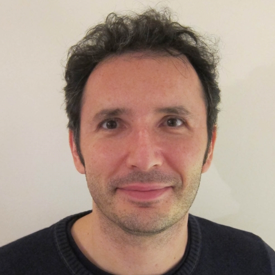
Dr Nic Tapon, The Francis Crick Institute, UK

Dr Nic Tapon, The Francis Crick Institute, UK
Dr Tapon was born in Poitiers, France. He received his BSc in Biochemistry from Imperial College in 1993. He carried out his PhD research in Alan Hall’s lab at University College (1993 – 1997), working on small GTPases. Following this he was a postdoctoral fellow in Iswar Hariharan’s lab at Massachusetts General Hospital Cancer Center, USA (1998 – 2001), working on the growth of fruit flies. Dr Tapon was a staff scientist (CNRS) in Pierre Leopold’s lab at the University of Nice, France from 2002 until 2003, and junior group leader at the Cancer Research UK London Research Institute from 2003 to 2009. He has been senior group leader at the Cancer Research UK London Research Institute since 2009, moving to the Francis Crick Institute in August 2016. His lab is currently interested in growth control and tissue architecture.
| 15:30 - 16:15 |
Tissue dynamics in development and repair
Actomyosin contractility is a key regulator of tissue dynamics. During development, tissue dynamics, such as cell intercalations and oriented cell divisions, are critical for shaping tissues and organs. However, less is known about how tissues regulate their dynamics during tissue homeostasis and repair, to maintain their shape after development. In this talk, Dr Mao will discuss how tissues respond to mechanical perturbations, such as stretching or wounding, by altering their actomyosin contractile structures, to change tissue dynamics, and thus preserve tissue shape and patterning. 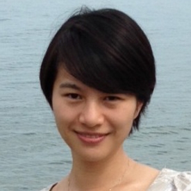
Dr Yanlan Mao, University College London, UK

Dr Yanlan Mao, University College London, UKYanlan Mao is a Group Leader at the MRC Laboratory for Molecular Cell Biology, University College London. After receiving her BA in Natural Sciences at Cambridge University, she completed her PhD with Matthew Freeman at the MRC LMB in Cambridge on Drosophila cell signaling and epithelial patterning. During her postdoc with Nic Tapon at the CRUK London Research Institute (now Francis Crick Institute), she became interested in tissue mechanics and computational modeling approaches. She now holds a UCL Excellence Fellowship and a MRC Career Development Award Fellowship to pursue her interests in the mechanical regulation of tissue growth and regeneration. |
|
|---|---|---|
| 16:15 - 17:00 |
Blood flow and heart formation
Heart formation involves a complex progression of finely orchestrated biological and biophysical processes. Blood flow dynamics, which is established early during cardiac development, when the heart is a linear tube without valves or chambers, is indispensable for proper heart formation. Cardiac looping, septation and valve formation all happen under blood flow, and, in fact, perturbations in blood flow dynamics during tubular heart stages lead to structural heart malformations. Blood flow mechanics, sensed by cardiac cells, act as a feedback loop regulating heart development. Feedback from blood flow mechanical stimuli is needed so that cardiovascular development addresses the increasing requirements of the embryo, while ensuring proper cardiac function. However, the discovery of the mechanisms by which developing cells sense and respond to blood flow mechanical stimuli is only starting to emerge. Quantification of both mechanical stimuli and biological responses is needed to determine the magnitude of the response to blood flow, affected regulatory processes, and possible implications to congenital heart disease. This talk will discuss these results in quantifying blood flow stimuli and biological responses, as well as implications on heart malformation. 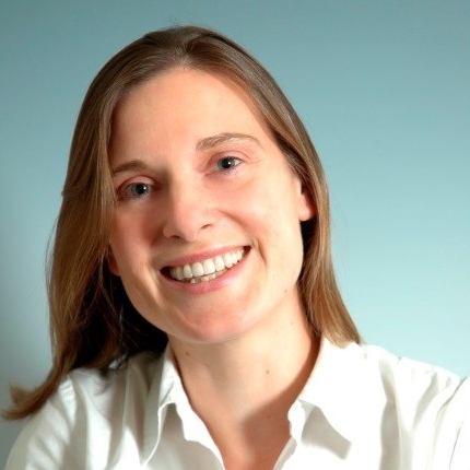
Professor Sandra Rugonyi, Oregon Health & Science University, USA

Professor Sandra Rugonyi, Oregon Health & Science University, USASandra Rugonyi is Professor of Biomedical Engineering at the Oregon Health & Science University (OHSU) School of Medicine, in Portland, Oregon, USA. In 1995, she earned her degree in Nuclear Engineering from Instituto Balseiro (Balseiro Institute), Argentina, one of the most prestigious research centers in Latin America. In 1999, she earned her MS in Mechanical Engineering from the Massachusetts Institute of Technology (MIT), and went on to complete her PhD in 2001, also in Mechanical Engineering at MIT. From 2001 – 2005, Dr Rugonyi held a postdoctoral fellowship at OHSU, after which she became Faculty of Biomedical Engineering at OHSU. In 2017, she was promoted to the rank of Professor. Dr Rugonyi's main research interest is in the study of the cardiovascular system, and the development of cardiovascular mathematical and computational models. Her current research projects involve investigating the effects of blood flow conditions on cardiovascular tissues during development and beyond. |
Chair

Professor Philippa Francis-West, King's College London, UK

Professor Philippa Francis-West, King's College London, UK
Philippa Francis-West is a Professor in Developmental and Cell Biology at the Centre for Craniofacial and Regenerative Biology, King’s College London. She earned a BA in Biochemistry at Cambridge University and carried out her PhD at the University of London. This was followed by post-doctoral position with Professors Lewis Wolpert, Paul Brickell and Cheryll Tickle at University College London. She established an independent research group at King’s College London in 1995 and has authored over 70 peer-reviewed publications. The group aims to understand the mechanisms of morphogenesis and cell differentiation within an embryo. The current research focus is on the Dchs1-Fat4-PCP-Hippo signaling pathway and the role of mechanics during cell differentiation. Her research has been funded by BBSRC, MRC together with Arthritis Research UK and Ageing Research charity funding.

Dr Nic Tapon, The Francis Crick Institute, UK

Dr Nic Tapon, The Francis Crick Institute, UK
Dr Tapon was born in Poitiers, France. He received his BSc in Biochemistry from Imperial College in 1993. He carried out his PhD research in Alan Hall’s lab at University College (1993 – 1997), working on small GTPases. Following this he was a postdoctoral fellow in Iswar Hariharan’s lab at Massachusetts General Hospital Cancer Center, USA (1998 – 2001), working on the growth of fruit flies. Dr Tapon was a staff scientist (CNRS) in Pierre Leopold’s lab at the University of Nice, France from 2002 until 2003, and junior group leader at the Cancer Research UK London Research Institute from 2003 to 2009. He has been senior group leader at the Cancer Research UK London Research Institute since 2009, moving to the Francis Crick Institute in August 2016. His lab is currently interested in growth control and tissue architecture.
| 09:00 - 09:45 |
Organoid development by design
Over the past years, organoids have stepped into the limelight as unique in vitro models for studying organ development, function and disease, owing to the previously unmatched fidelity with which they approximate real organs. Organoids form through poorly understood morphogenetic processes in which initially homogeneous aggregates of stem cells spontaneously self-organize within three-dimensional extracellular matrices (ECM). Yet, the absence of any predefined patterning influences such as morphogen gradients or mechanical cues results in an extensive heterogeneity. Moreover, the current mismatch in shape, size and lifespan between native organs and their in vitro counterparts severely hinders their applicability. In this talk, Professor Lutolf will discuss some of the ongoing efforts in developing programmable organoids that are assembled by combining bioengineering approaches with insights from developmental biology. Specifically, using intestinal organoids (‘mini-guts’) as a model system, Professor Lutolf will show for example how it is possible to overcome the stochasticity in self-organization by controlling cell fate decisions through localized changes in ECM mechanics. The convergence of bioengineering and cellular self-organization may be broadly applicable to attain more physiological organoid sizes, shapes and function. 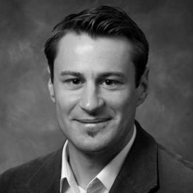
Professor Matthias Lutolf, École polytechnique fédérale de Lausanne, Switzerland

Professor Matthias Lutolf, École polytechnique fédérale de Lausanne, SwitzerlandMatthias Lutolf is the director of the Institute of Bioengineering at EPFL, Switzerland. He was trained as a Materials Engineer at ETH Zurich where he also carried out his PhD studies. After a Postdoc in stem cell biology at Stanford University, Lutolf started up his independent group at EPFL in with a European Young Investigator (EURYI) award. His research is focused on the development of novel technologies for improved stem cell and organoid culture. |
|---|---|
| 09:45 - 10:30 |
Physical organogenesis of the gut
The intestine is our body’s longest and most anisotropic organ. In this talk, Dr Chevalier will present the physical factors (physiological static and dynamic forces, biomechanical properties) that drive anisotropic gut growth during embryonic development. 
Dr Nicolas R Chevalier, Laboratoire Matière et Systèmes Complexes; CNRS - Université Paris Diderot, France

Dr Nicolas R Chevalier, Laboratoire Matière et Systèmes Complexes; CNRS - Université Paris Diderot, FranceDr Chevalier grew up in France and Austria and studied physics (MSc) at Ecole Polytechnique Fédérale de Lausanne (EPFL, Switzerland) and Moscow State University (Russia). His initial investigations pertained to crystal growth, thin films and colloids. He obtained his PhD in the field of biomineralization in 2010 from Pierre and Marie Curie University, France. He became passionate about embryonic development when he joined V Fleury’s group as a postdoc in 2013 at Laboratoire Systèmes Complexes (MSC). He has been a CNRS staff scientist at MSC, University Paris Diderot since 2016. Dr Chevalier develops a physical perspective on how organs develop in an embryo, with a particular focus on biomechanics, gut development, motility, neural crest cell migration and enteric nervous system development. |
| 10:30 - 11:00 | Coffee |
Chair

Professor Philippa Francis-West, King's College London, UK

Professor Philippa Francis-West, King's College London, UK
Philippa Francis-West is a Professor in Developmental and Cell Biology at the Centre for Craniofacial and Regenerative Biology, King’s College London. She earned a BA in Biochemistry at Cambridge University and carried out her PhD at the University of London. This was followed by post-doctoral position with Professors Lewis Wolpert, Paul Brickell and Cheryll Tickle at University College London. She established an independent research group at King’s College London in 1995 and has authored over 70 peer-reviewed publications. The group aims to understand the mechanisms of morphogenesis and cell differentiation within an embryo. The current research focus is on the Dchs1-Fat4-PCP-Hippo signaling pathway and the role of mechanics during cell differentiation. Her research has been funded by BBSRC, MRC together with Arthritis Research UK and Ageing Research charity funding.
| 11:00 - 12:00 |
Mechanobiology of mammalian epidermis
The ability to reconstitute human epidermis in culture has afforded the opportunity to examine stem cell-niche interactions at single cell resolution. Professor Watt will describe the responses of human epidermal stem cells to different topographical cues, ranging from the 1 to 100 micron scale. Professor Watt will describe the signal transduction pathways that lead to the onset of terminal differentiation and show how different cues trigger differentiation via different pathways. 
Professor Fiona Watt FMedSci FRS, King's College London, UK

Professor Fiona Watt FMedSci FRS, King's College London, UKFiona Watt obtained her first degree from Cambridge University and her DPhil, in cell biology, from the University of Oxford. She was a postdoc at MIT, where she first began studying differentiation and tissue organisation in mammalian epidermis. She established her first research group at the Kennedy Institute for Rheumatology and then spent 20 years at the CRUK London Research Institute (now part of the Francis Crick Institute). She helped to establish the CRUK Cambridge Research Institute and the Wellcome Trust Centre for Stem Cell Research and in 2012 she moved to King's College London to found the Centre for Stem Cells and Regenerative Medicine. Fiona Watt is a Fellow of the Royal Society, a Fellow of the Academy of Medical Sciences and an Honorary Foreign Member of the American Academy of Arts and Sciences. Her awards include the American Society for Cell Biology (ASCB) Women in Cell Biology Senior Award, the Hunterian Society Medal and the FEBS/EMBO Women in Science Award. In 2016 she was awarded Doctor Honoris Causa of the Universidad Autonoma de Madrid. |
|
|---|---|---|
| 12:00 - 12:30 | Summary of discussions and closing remarks |
