Links to external sources may no longer work as intended. The content may not represent the latest thinking in this area or the Society’s current position on the topic.
Single cell ecology
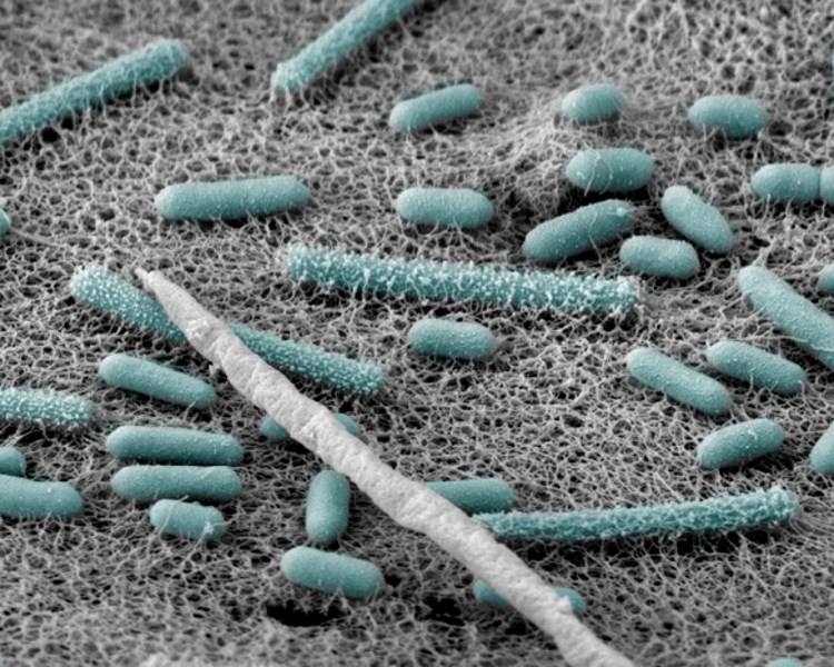
Scientific discussion meeting organised by Professor Thomas Richards, Dr Ramon Massana and Professor Neil Hall
Interdisciplinary meeting to explore the use of single cell technologies to understand the function, diversity and interactions of microbes. By bringing together physicists who manipulate cells, microbiologists who seek to understand the nature of microbial communities and genomicists who are developing new approaches to study individual cells we will achieve a greater understanding of the potential of this new field.
The speaker biographies abstracts are below. Recorded audio of the presentations will be available on this page after the meeting has taken place. Meeting papers will be published in Philosophical Transactions B.
Enquiries: Contact the Scientific Programmes team.
Organisers
Schedule
Chair
Professor Tom Richards, University of Exeter, UK

Professor Tom Richards, University of Exeter, UK
Tom Richards is an evolutionary biologist who works on the rise and diversification of the eukaryotes. His work focuses on the use of environmental DNA methods to understand the diversity of eukaryotic microbes in natural environments and how they fit on to the tree of life. He also works on trying to understand the evolutionary processes that have driven the diversification of the eukaryotic form such as endosymbiosis and horizontal gene transfer.
| 09:05 - 09:30 |
Cooperative growth and community assembly at the microbe scale
Bacterial cooperation, whereby cells secrete compounds that can facilitate the growth of neighbouring cells, has been extensively studied through the lens of evolutionary biology. However, the environmental implications of cooperation and the ecological scenarios under which it takes place remain much less understood. In this talk Otto Cordero will discuss the conditions under which cooperation emerges in microbial populations that degrade complex organic particles in the ocean. Otto will show that organisms that privatise their enzymes by tethering them to their membranes are more likely to engage in cooperative interactions by forming cell-aggregates that enables them to efficiently uptake of the solubilised organic matter. By contrast, when organisms secrete highly active enzymes dynamics quickly turn competitive, and the efficiency of carbon uptake drops. Otto will back up this results with theory, data from individual based models and experiments. Finally, he will discuss the genomic underpinnings and potential role of social cheaters in the natural environment, based on a study with hundreds of micro-scale particle colonisation experiments in natural seawater. 
Professor Otto X Cordero, MIT, USA

Professor Otto X Cordero, MIT, USAOtto X Cordero received a BS in Computer and Electrical Engineering from the Polytechnic University of Ecuador; an MSc in Artificial Intelligence from Utrecht University; and a PhD in Theoretical Biology also from Utrecht University. His main research focus is the ecology and evolution of natural microbial collectives. The Cordero lab is interested in understanding how social and ecological interactions at micro-scales impact the global productivity, stability and evolutionary dynamics of microbial ecosystems. |
|
|---|---|---|
| 09:45 - 10:15 |
A single cell view on host-virus interactions in the ocean
Investigating host-pathogen interactions at the whole-population level tends to mask cell-to-cell variability, hampering the ability to discern between diverse phenotypes. To overcome this limitation, the Vardi group applied single-cell dual RNA-seq to study the dynamic transcriptomes of a cosmopolitan bloom-forming alga, Emiliania huxleyi, and its specific large DNA virus EhV. Gene expression was profiled for multiple genomes (nuclear, plastid, mitochondrial and viral) using the massively parallel RNA single-cell sequencing method for cells sorted from infected cultures during a time course. Striking heterogeneity in the overall viral transcription was observed among individual cells from all samples, with cells exhibiting either high or very low viral transcript numbers, but very few at intermediate levels. The cells with high viral expression formed subpopulations characterised by clusters of strongly co-expressed viral genes. By re-ordering cells based on their transcriptional pattern, the group defined a continuum infection states along a pseudo-temporal scale. As the infection progressed, the viral genes were successively expressed in five partially overlapping kinetic classes that end with constitutive expression of the virion structural protein genes. Majority of cells with high viral gene expression showed a selective shutoff of host nucleus-encoded genes and minor fraction exhibited differential gene expression relate to cell fate regulation, lipid biosynthesis, and flagellated resistant phenotype. Vardi’s group further applied this scRNAseq approach to track viral infection dynamics during natural bloom of E. huxleyi which enabled a first single cell view on host and virus arms race at sea. 
Professor Assaf Vardi, Weizmann Institute of Science, Israel

Professor Assaf Vardi, Weizmann Institute of Science, IsraelAssaf Vardi’s laboratory aims to explore the cellular mechanisms involved in sensing and acclimation to diverse environmental stress conditions during algal bloom dynamics in the ocean. Their research utilises the recent advances in the field of chemical ecology, combined with single cell imaging and transcriptomic approaches to understand cell-cell interactions at the micro(be)-scale. The group studies key biotic interactions (host-virus, host-bacteria, predator-prey and allelopathy) that regulate the fate of algal blooms, in order to discover novel signalling and metabolic pathways employed during these interactions. Newly identified genes and metabolites induced during specific host-pathogen interactions are used as functional biomarkers to assess the ecological impact of microbial interactions in structuring microbial food webs and potentially global nutrient cycles. |
|
| 10:30 - 11:00 | Coffee | |
| 11:00 - 11:30 |
Single cell genomics
An exciting emerging area revolves around the use of microfluidic tools for single-cell genomic analysis. Stephen Quake’s group has been using microfluidic devices for both gene expression analysis and for genome sequencing from single cells. In the case of gene expression analysis, it has become routine to analyse hundreds of genes per cell on hundreds to thousands of single cells per experiment. This has led to many new insights into the heterogeneity of cell populations in human tissues, especially in the areas of cancer and stem cell biology. These devices make it possible to perform “reverse tissue engineering” by dissecting complex tissues into their component cell populations, and they are also used to analyse rare cells such as circulating tumour cells or minor populations within a tissue. The group has also used single-cell genome sequencing to analyse the genetic properties of microbes that cannot be grown in culture – the largest component of biological diversity on the planet – as well as to study the recombination potential of humans by characterising the diversity of novel genomes found in the sperm of an individual. Stephen expects that single cell genome sequencing will become a valuable tool in understanding genetic diversity in many different contexts. 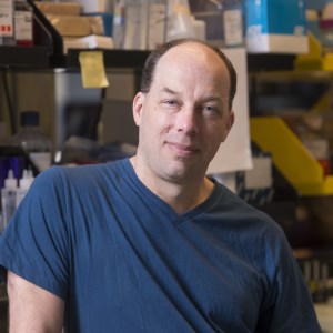
Professor Stephen Quake, Stanford University, USA

Professor Stephen Quake, Stanford University, USAStephen Quake is the Lee Otterson Professor of Bioengineering and Professor of Applied Physics at Stanford University and is co-President of the Chan Zuckerberg Biohub. He received a doctorate in Theoretical Physics from the University of Oxford in 1994. He began his faculty career at the California Institute of Technology in 1996, later becoming the Everhart Professor of Applied Physics and Physics. He joined Stanford in 2005 to help found and lead Stanford’s new Bioengineering department, cultivating it to nearly two dozen faculty members. He was simultaneously an Investigator of the Howard Hughes Medical Institute from 2006-2016. Stephen has invented many measurement tools for biology, including new DNA sequencing technologies that have enabled rapid analysis of the human genome and microfluidic automation. Stephen is also well known for his work in the first non-invasive prenatal test for Down syndrome and other aneuploidies. His test is rapidly replacing risky invasive approaches such as amniocentesis, and millions of women each year now benefit from this approach. Honours include the Human Frontiers of Science Nakasone Prize, the MIT-Lemelson Prize, the Raymond and Beverly Sackler International Prize in Biophysics, the American Society for Microbiology Promega Biotechnology Research Award, the Royal Society of Chemistry Publishing Pioneer of Miniaturization Award, and the NIH Director’s Pioneer Award. He is an elected fellow of the National Academy of Sciences, the National Academy of Engineering, the National Academy of Medicine, the American Academy of Arts and Sciences, the National Academy of Inventors, the American Institute for Medical and Biological Engineering and the American Physical Society. |
|
| 11:45 - 12:15 |
Single cell analysis with droplet microfluidics
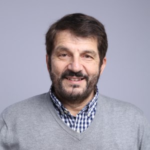
Professor David Weitz, Harvard University, USA

Professor David Weitz, Harvard University, USADavid Weitz received his PhD in physics from Harvard University and then joined Exxon Research and Engineering Company, where he worked for nearly 18 years. He then became a professor of physics at the University of Pennsylvania and moved to Harvard at the end of the last millennium as professor of physics and applied physics. He leads a group studying soft matter science with a focus on materials science, biophysics and microfluidics. Several startup companies have come from his lab to commercialize research concepts. |
Chair
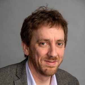
Professor Neil Hall, Earlham Institute, UK

Professor Neil Hall, Earlham Institute, UK
Neil Hall has been working in genomics for over 15 years. He has previously led research groups at the Sanger Institute, The Institute for Genomic Research, and The University of Liverpool. His research focusses on comparative and evolutionary genomics in pathogens (particularly parasitic protists) to understand the molecular basis of important phenotypes such as virulence and host specificity. His group also apply genomics to the analysis of microbial communities in order to understand how they may influence health or respond to changing environments.
| 13:30 - 14:00 |
The Malaria Cell Atlas and using scRNA-seq to examine developmental decisions made by single-celled parasites
Single-cell RNA sequencing is revolutionising our understanding of malaria parasites. The Lawniczak group has used a combination of Smart-seq2 and 10X to characterise the transcriptional networks underpinning the developmental trajectories of parasites. Here Mara presents thousands of single-cell transcriptomes covering the entire life cycle of the rodent malaria parasite Plasmodium berghei, both in the mosquito and in the mammalian host. The group uses these data to identify how modules of genes are used throughout the life cycle, the variability in their expression, and how they are putatively regulated. They have developed an open-access interactive interface to view the complete dataset, enabling researchers to explore the behaviour of any gene within single parasites across the entire lifecycle. Finally, Mara will present work that uses both multiplexed gene knock-out screens and scRNA-seq to examine one of the most critical decisions a parasite makes: whether or not to become sexually committed. This work demonstrates how scRNA-seq can be used to profile the entire life-cycle of a single-celled organism, revealing novel insights into its genome. 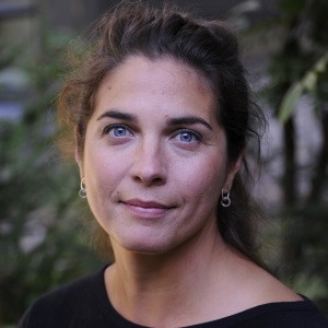
Dr Mara Lawniczak, Wellcome Sanger Institute, UK

Dr Mara Lawniczak, Wellcome Sanger Institute, UKMara Lawniczak is an evolutionary geneticist interested in understanding what makes some mosquitoes better than others at transmitting malaria, and what makes some parasites better at getting transmitted. She also likes trying out new technologies that enhance our understanding of genomes. Mara obtained her PhD in population biology from UC Davis in 2004. She became a postdoc at Imperial College London in 2007 with Fotis Kafatos and George Christophides. In 2012 Mara was awarded an MRC Career Development Award, and in 2014 she joined the Wellcome Sanger Institute as a group leader. |
|
|---|---|---|
| 14:15 - 14:45 |
Cell Atlas Technologies and the maternal-foetal interface
Genomics has undergone a resolution revolution, such that the nucleic acid content of individual cells is now sequenced routinely in high-throughput mode. Thus we can define the cellular composition of tissues in an unbiased and comprehensive way using single cell transcriptomics, revealing the molecular fingerprint of cell states and their predicted signalling circuits in tissues across development and disease. Sarah Teichmann will illustrate this with the maternal-foetal interface in the first trimester placenta/decidua. 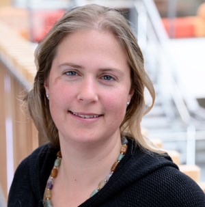
Dr Sarah Teichmann FMedSci, Wellcome Sanger Institute, UK

Dr Sarah Teichmann FMedSci, Wellcome Sanger Institute, UKSarah Teichmann is interested in global principles of regulation of gene expression and protein complexes, specifically in the context of immunity. Sarah did her PhD at the MRC Laboratory of Molecular Biology, Cambridge, UK and was a Beit Memorial Fellow at University College London. She started her group at the MRC Laboratory of Molecular Biology in 2001. In 2013, she moved to the Wellcome Trust Genome Campus in Hinxton/Cambridge, jointly with the EMBL-European Bioinformatics Institute and the Wellcome Trust Sanger Institute (WTSI). In January 2016 she became Head of the Cellular Genetics Programme at the WTSI. Sarah is an EMBO member and a Fellow of the Academy of Medical Sciences, and her work has been recognised by a number of prizes, including the Lister Prize, Biochemical Society Colworth Medal, Royal Society Crick Lecture, EMBO Gold Medal and the Mary Lyon Medal. |
|
| 15:00 - 15:30 | Tea | |
| 15:30 - 16:00 |
Single-cell responses to environmental cues
Clonal microbial populations feature cell-to-cell differences in physiological parameters such as gene expression, growth rate, and resistance to stress. This phenotypic heterogeneity is at the basis of fundamental biological processes such as membrane transport, stem cell differentiation, and tolerance to antimicrobial compounds. Therefore, it is paramount understanding how changes in the environment affect the phenotypic heterogeneity within a clonal microbial population. Stefano will illustrate how embryonic stem cells respond to physical environmental cues such as transient confinement into narrow grooves. He will show that this mechanical response is heterogeneous. Remarkably, the nuclei of some embryonic stem cells display a unique material property that is they are auxetic; exhibiting a cross-sectional expansion when stretched and a cross-sectional contraction when compressed. He will then talk about the different strategies that individual bacteria can exploit to escape antibiotics and illustrate current efforts to understand how the environment predetermines the outcome of antibiotic treatment. 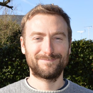
Dr Stefano Pagliara, University of Exeter, UK

Dr Stefano Pagliara, University of Exeter, UKThe aim of Stefano Pagliara research is to devise approaches for microbe isolation, growth, and characterisation, thus allowing to understand how microbial cells interact with their natural environment both as a community and as individual cells. Stefano Pagliara obtained his PhD in Nanoscience from the University of Salento (Italy) in 2010, where he introduced a new microfluidic technology embedding organic light polarisers. Afterwards, he joined the Cavendish Laboratory at the University of Cambridge where he advanced the understanding of molecular transport across biological membranes and discovered that the nuclei of embryonic stem cells can be auxetic. Since 2017 he has been a Senior Lecturer at the University of Exeter where his research group investigates the molecular mechanisms underlying drug uptake and tolerance to antibiotics in bacteria. |
|
| 16:15 - 18:00 | Poster session |
Chair
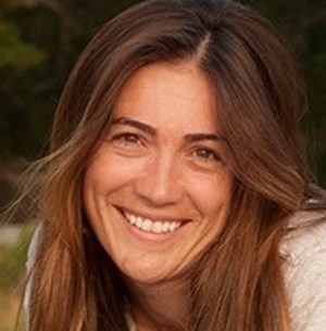
Dr Alyson Santoro, University of California, Santa Barbara, USA

Dr Alyson Santoro, University of California, Santa Barbara, USA
Dr Alyson Santoro is an Assistant Professor in the Department of Ecology, Evolution and Marine Biology at the University of California, Santa Barbara. Alyson’s research focuses on microbes involved in nutrient cycling in the ocean, especially of the element nitrogen. She is interested in cultivating new microbes and discovering novel ways of tracking their activity. This research combines laboratory experiments with field observations, and to date has used genomics, transcriptomics, proteomics and stable isotope geochemistry as tools to uncover the activity of microbes in the mesopelagic ocean. A particular focus of the lab is the marine archaea, a largely uncultured group of microbes. Findings from their recent research include the discovery that archaea in the ocean can make the greenhouse gas nitrous oxide, and that some marine archaea have exceptionally small genomes. Alyson received her PhD in Environmental Engineering at Stanford University and completed a postdoctoral fellowship at Woods Hole Oceanographic Institution.
| 09:00 - 09:30 |
Short-range interactions govern cellular dynamics in microbial multi-genotype systems
Many microorganisms live in communities that are spatially structured, for example in biofilms. Such communities exhibit activities and functions that are shaped by metabolic interactions between the community member. Interactions are expected to mainly occur between cells that are close in space. As a consequence, the nature and strength of the interactions that will occur will depend on the spatial arrangement of different types of microbial cells. In turn, the spatial arrangement is expected to be shaped by metabolic interactions, which determine regions where a given cell type grows well. The Ackermann group’s goal here is to better understand this interplay between the spatial arrangement of different types of microbial cells and the interactions that arise between them. Working with synthetic consortia of different E. coli strains with well-defined metabolic interactions, Martin can quantify the spatial range over which interactions occur and understand the consequences for the spatial self-organisation of these multi-genotype systems. The goal of this work is to contribute to identifying general principles that govern how different types of microorganisms organise in space, and how this spatial self-organisation shapes the activities and functions of microbial systems. 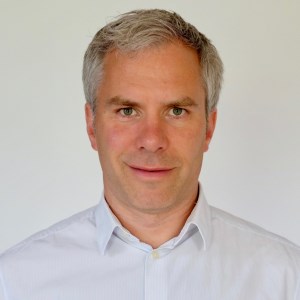
Professor Martin Ackermann, ETH Zurich and Eawag, Switzerland

Professor Martin Ackermann, ETH Zurich and Eawag, SwitzerlandMartin Ackermann is a Professor at ETH Zürich. His group consists of about 15 PhD students and postdocs with backgrounds in microbiology, evolutionary biology, physics and computer science. The goal of the group is to work on general principles of how bacteria interact with each other and with their environment. A first interest of the group is bacterial individuality, that is, differences in behaviour and properties between genetically identical cells. The group works on how individual cells that specialise in different tasks can interact with each other and engage in the division of labour. A second interest is how bacteria cope with dynamic environments — how cellular decisions of individual bacteria are influenced by past events as well as by stochastic processes. A third interest is on biological systems that are composed of several interacting genotypes; the group is interested in how new functionality at the level of microbial consortia emerges based on the properties of individuals and their interactions. |
|
|---|---|---|
| 09:45 - 10:15 |
Virtual fluidic channels: from functional single cell rheology to tissue mechanics
The mechanical properties of cells have long been established as a sensitive and label-free biomarker. While mechanical cell assays have been traditionally limited to low throughput or small sample size, the introduction of real-time deformability cytometry (RT-DC) increased analysis rates to up to 1,000 cells per second. RT-DC has demonstrated its relevance in basic and fundamental life science research, e.g. by observing the activation of immune cells and describing the membrane dynamics of Malaria pathogenesis. However, linking immune cell activation to underlying tissue alterations has not been possible so far. Here, the concept of virtual fluidic channels is introduced to bridge the gap between single cell rheology and tissue mechanics. Virtual channels can be created in almost any microfluidic geometry and can be tailored dynamically towards hydrodynamic stress distributions sufficient to probe the rheology of arbitrary cell sizes. Using spheroids as a tissue model, results from virtual channel measurements indicate that the Young’s modulus of single cells exceeds the one of spheroids and that their elasticity increases with size. The availability of a high-throughput assay for mechanical spheroid characterization might lead to a better understanding of tissue rheology and help to study the interplay of virus infiltration and tissue regeneration. 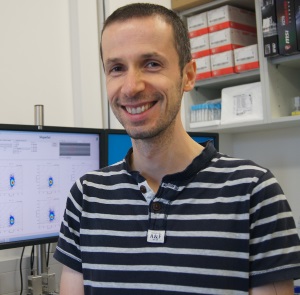
Dr Oliver Otto, University of Greifswald, Germany

Dr Oliver Otto, University of Greifswald, GermanyThe research group of Oliver Otto develops and applies microfluidic technologies for the label-free characterization of suspended and adherent cells. Specifically, he is interested in the role of mechanical properties for homeostasis in the cardiovascular system. Oliver received his PhD in Physics from the University of Cambridge, UK, where he investigated single molecule dynamics at the Cavendish Laboratory. In 2012, he joined the Technical University of Dresden, Germany, as a postdoctoral researcher where he was working on the translation of high-throughput screening into the field of cell mechanics. His method, real-time deformability cytometry, allows for the first time the measurement of cell mechanical properties with the throughput of a flow cytometer. Since 2016 Oliver has been an independent group leader at the Centre of Innovation Competence – Humoral Immune Reactions in Cardiovascular Diseases at the University of Greifswald, Germany. He leads a group of interdisciplinary researchers combining expertise in Biology, Engineering as well as Physics. |
|
| 10:30 - 11:00 | Coffee | |
| 11:00 - 10:30 |
Studying uncultivated diversity via single-cell approaches – from microorganisms to viruses
The bacterial and archaeal tree of life has undergone significant expansion, chiefly from candidate phyla obtained through genome-resolved metagenomics and, at smaller scale, via single-cell sequencing efforts. Following this path, viral diversity is being uncovered at a rapid pace. Tanja Woyke discusses how the combination of flow cytometry followed by genome amplification and sequencing can provide a means to cataloguing uncultivated microbial and viral diversity. Focusing on Nanoarchaeota symbionts attached to their hosts, she illustrates that this approach further allows the assignment of novel putative host associations, facilitating the exploration of cell-cell interactions and fine-scale genomic diversity. 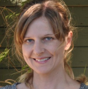
Dr Tanja Woyke, DOE Joint Genome Institute, USA

Dr Tanja Woyke, DOE Joint Genome Institute, USAAfter studying the mechanism of action of antifungal natural products and their derivatives during her PhD in Microbiology at the Eberhard Karls University of Tübingen, Germany, Dr Tanja Woyke pursued her postdoctoral research at the JGI in 2004. The main objective of her research was the symbiont community of a gutless oligochaete for which she deciphered function and host-symbiont interplay using metagenomics. Taking on a Research Scientist position in 2007, she switched gears from metagenomics to single-cell genomics, which is now her primary interest and passion. Tanja stepped into the microbial genomics program lead position in 2009. She now holds a Staff Scientist position at the JGI, an Adjust Scientist position at the Bigelow Laboratory for Ocean Sciences and an Adjunct Associate Professor at the UC Merced School of Natural Sciences. |
|
| 11:45 - 12:15 |
Interrogating marine microbes for their activity and growth: an overview and recent developments
Recent developments in community and single-cell genomic approaches have provided an unprecedented amount of information on the ecology of microbes in the aquatic environment. However, linkages between each specific microbe’s identity and their in situ level of activity (be it growth, division, or just metabolic activity) are much more difficult to obtain. One of the ultimate goals of marine microbial ecology would be integrate three levels: the genomic (including identity) one, the activity/growth one, and the morphology/visualisation, and all this for as many individual cells as possible, as a means to understand how each environmental characteristic determines the types of different microbes in nature and their activity or growth, alongside with information on morphology and cell-to-cell associations. Recent reviews have stressed ways for capturing the activity level of different genomic entities, but often one of the three legs, that of visualization, has not been well covered by the available methods. A review of current methodologies that have been applied to marine microbes, particularly prokaryotes, will be presented combined with a discussion of the difficulties in identifying and categorizing activity and growth, in doing so at with the minimal manipulation of the environment, and the level of within-population single-cell variability in activity that occurs in natural marine environments. 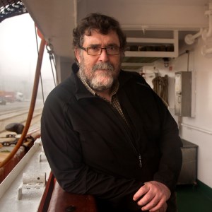
Dr Josep M Gasol, Institut de Ciències del Mar, CSIC, Spain

Dr Josep M Gasol, Institut de Ciències del Mar, CSIC, SpainDr Josep M Gasol is a Research Professor in Barcelona, interested in planktonic microbe abundance and activity, and their ecosystem effects, with emphasis on microorganism community structure (size, functional and taxonomical structure), and how the physical and biological factors shape it. He approaches these question by empirical analysis of data bases, oceanographic cruise, mesocosm and microcosm experiments, and by the combined use of image analysis and flow cytometry and metabolic fluorescent probes. Josep started his career working on the microbes of one of the most interesting lakes in the world, Lake Cisó. He was then a postdoc in Montreal where instead of focussing on one lake, he tried to study as many lakes as possible, and then started to focus on the Ocean. Josep is very involved with the Microbial Observatory of Blanes Bay, where he coordinates the long term research, was the Microbiology coordinator of the Malaspina-2010 circumnavigation, and he has recently edited the last edition of the Microbial Ecology of the Ocean book. |
Chair
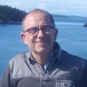
Dr Ramon Massana Institute of Marine Sciences, CSIC, Spain

Dr Ramon Massana Institute of Marine Sciences, CSIC, Spain
Ramon Massana is a Research Scientist at the ICM-CSIC with a wide experience in marine microbial ecology. His broad research line is the study of the phylogenetic and functional diversity of marine protists, especially those species that still remain uncultured. He conducts this exploration by applying simultaneously several tools, such as sequencing of environmental rDNA genes, metagenomics, single cell genomics, specific FISH counts and experiments. He is particularly interested in opening the black box of heterotrophic flagellates and exploring the role of these unpigmented protists in marine food webs as bacterial grazers and nutrient remineralisers. He is the coordinator of the EU Innovative Training Network SINGEK conceived to promote the use of single cell genomics to address critical ecological and evolutionary questions in microbial eukaryotes.
| 13:30 - 14:00 |
Ocean microbiome datafication
Data-rich, molecular analyses are becoming important sources of knowledge about the composition and activities of microorganisms in diverse environments. Yet, today only a small fraction of these meta-omics data (short DNA, RNA and peptide sequences) can be accurately assigned to specific lineages and metabolic pathways, due to the lack of adequate reference genome databases. To address this challenge, the Stepanauskas group sequenced 20,000 individual bacteria, archaea and viral particles from the tropical and subtropical epipelagic using an unbiased, Big Data approach. This GORG-Tropics database provides some of the first insights into the global pangenomes, infections, biogeography and sizes of many of the bacterial and archaeal lineages that dominate surface ocean. The group shows that GORG-Tropics enables accurate taxonomic and functional assignments of the majority of meta-omics data from this global environment. Intriguingly, the group found that every sequenced cell was genomically unique, and that a large fraction of the global bacterioplankton coding potential was present in a single drop of seawater. GORG-Tropics expands the capacity of oceanography’s cyber-infrastructure and enhances the interpretation of diverse marine omics studies. It also demonstrates that relevant genomic representation of complex microbiomes is an achievable goal and may be applied in other environments. 
Dr Ramunas Stepanauskas, Bigelow Laboratory for Ocean Sciences, USA

Dr Ramunas Stepanauskas, Bigelow Laboratory for Ocean Sciences, USADr Ramunas Stepanauskas obtained his PhD in ecology from Lund University, Sweden. He was a postdoctoral associate at the University of Georgia in 2000-2004. He then went on to become an assistant research scientist at Savannah River Ecology Laboratory at the University of Georgia. In 2005, Ramunas became a senior research scientist at the Bigelow Laboratory for Ocean Sciences. In 2009, he became a founding director of Bigelow Laboratory Single Cell Genomics Center, having established the first high throughput, shared user facility for microbial single cell genomics. He has also held a research faculty appointment at Colby College since 2009. |
|
|---|---|---|
| 14:15 - 14:45 |
How do we define the role of eukaryotic cells in complex microbial ecosystems?
One challenge associated with developing a functional understanding of microbial ecosystems is the magnitude of the diversity encompassed by the word “microbial”. Most ecology is biased towards a few macroscopic plants and animals. In contrast, microbial ecosystems are made up of more than ten-times the diversity of eukaryotes alone, in addition to bacteria, archaea, and viruses. Microbial ecology is also heavily biased, towards bacteria (and sometimes archaea). So we are faced with a need to develop a fuller picture of these networks, but to do so both a quantitative and qualitative problem, because the diversity is so great that neither the technical approaches nor the basic biology required to understand one group is readily translatable to others. For microbial eukaryotes there are paths forward to solving many technical challenges though single cell methods, and advances in both genomics and transcriptomics will be discussed. But this still leaves a thornier conceptual problem of how such data will (or will not) reveal ecological roles of microbial eukaryotes because translating genomics to ecological function is largely restricted to metabolism. For most microbial eukaryotes, complex cellular structures and resulting behaviours are much more significant, but we lack strong conceptual frameworks to derive these traits from genomic data alone. 
Professor Patrick Keeling, University of British Columbia, Canada

Professor Patrick Keeling, University of British Columbia, CanadaPatrick Keeling is a Canadian evolutionary microbiologist and Professor in Botany at the University of British Columbia. He grew up in rural Ontario, and completed a Bachelors at the University of Western Ontario in Genetics and a PhD in Biochemistry at Dalhousie, followed by postdoctoral research at the University of Melbourne and Indiana University. He began his current position as a professor at UBC in Vancouver in 1999. |
|
| 15:00 - 15:30 | Tea | |
| 15:30 - 16:00 |
Exposing uncultured marine eukaryotes and their ecological interactions through single cell genomics
For decades and longer scientists have described diverse unicellular eukaryotic organisms in aquatic ecosystems. The introduction of molecular approaches to ocean ecology has expanded our view of the diversity of marine protists and their importance in these ecosystems, yet many key taxa remain uncultured. Hence it has been difficult to examine their evolutionary relationships beyond what is possible with marker gene analyses. Likewise, it has not been possible to characterise their nuclear and organellar genomes. Moreover, lack of cultures has obviated deeper exploration of their interactions with other biological entities in the sea. In 2005 the Worden group began using at-sea flow cytometry to sort wild protists in order to generate both targeted (population) metagenomes and eukaryotic single-cell metagenomes. In addition to providing insights into the evolution and distribution of wild phytoplankton species these studies are increasingly revealing co-associations between the targeted eukaryote and other entities. Here, Alexandra will discuss a suite of findings from studies in the tropical Atlantic and eastern North Pacific Oceans. Her long term goal is to use these approaches to facilitate research that goes deeper into the biology of key uncultured groups and their ecological interactions. Collectively, these types of studies are critical for establishing a baseline against which future changes in microbial community structure can be assessed and for understanding the diversity and evolution of marine protists. 
Professor Alexandra Worden, Monterey Bay Aquarium Research Institute (MBARI), USA

Professor Alexandra Worden, Monterey Bay Aquarium Research Institute (MBARI), USAProfessor Worden leads a molecular ecology research group at the Monterey Bay Aquarium Research Institute, a non-profit organization focused on the intersection of oceanographic science and technology development. Her research focuses on photosynthetic organisms and integrates across genomics, evolutionary biology and ecology to explore microbial roles in carbon dioxide uptake and fate. Her group develops methods and technologies for systems biology and sea-going studies of unicellular eukaryotes, and for quantify their contributions to global primary production, activities in the deep sea and physical interactions. An underlying principle for her research is that organisms must be studied at habitat scales relevant to their adaptive strategies to determine how their metabolism and activities influence larger-scale ecosystem dynamics. She considers this principle essential for understanding how biological communities and global carbon dioxide uptake by algae will transition during climate change. Worden is an elected Fellow of the American Academy of Microbiology and a Gordon & Betty Moore Foundation Marine Investigator. |
|
| 16:15 - 17:00 | Panel discussion/overview |
