Links to external sources may no longer work as intended. The content may not represent the latest thinking in this area or the Society’s current position on the topic.
Contemporary morphogenesis
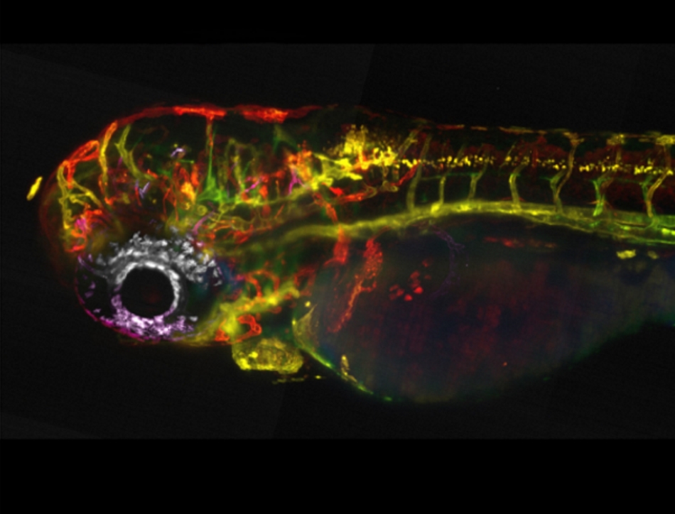
Scientific discussion meeting organised by Dr Kyra Anne Campbell, Dr Emily Noel, Dr Alexander Fletcher and Dr Natalia Bulgakova.
Recent advances have revolutionised the field of developmental biology, heralding a new era of state-of-the art super-resolution live imaging, precision analyses, and à la carte genetic engineering. As a result, this fast-moving field now impacts on almost every other biological discipline. This meeting showcased the cutting edge of research in developmental biology, with a focus on emerging talent.
Recorded audio of the presentations will be available on this page under the abstract of each talk. An accompanying journal issue for this meeting was published in Philosophical Transactions of the Royal Society B.
Enquiries: contact the Scientific Programmes team
Organisers
Schedule
Chair
Dr Natalia Bulgakova, Bateson Centre, University of Sheffield, UK

Dr Natalia Bulgakova, Bateson Centre, University of Sheffield, UK
Natalia Bulgakova has been a lecturer at the Department of Biomedical Science, University of Sheffield, since October 2015. Natalia’s research focuses on cell-cell adhesion, which is the mechanism that connects individual cells in our body together, currently funded by the BBSRC and Leverhulme Trust. She studies the regulation and roles of cell-cell adhesion during development and tissue homeostasis using Drosophila as a model. Natalia’s research greatly benefits from her passion for developing new computational approaches for automated image analysis. These approaches allow the extraction of quantitative, high-quality information from microscopy data to better understand cellular mechanisms of morphogenesis.
After studying Natural Sciences at the Novosibirsk State University, Russia, Natalia moved in 2004 to Germany to do her PhD with Prof Elisabeth Knust, first at the University of Düsseldorf and later at the Max-Planck Institute of Molecular Cell Biology and Genetics, Dresden. During this time, she explored the roles of an apical-basal polarity regulator, Stardust, in Drosophila retinal morphogenesis and prevention of light-induced retinal degeneration. For her postdoctoral training Natalia moved in 2009 to the lab of Professor Nick Brown at the University of Cambridge, UK, and focused on the regulation of cell-cell adhesion by the microtubule cytoskeleton in Drosophila epithelia, which sparked her interest in cell-cell adhesion and inspired her current research focus.
| 08:05 - 08:30 |
Role of sidekick, a novel resident protein at tricellular adherens junctions, in morphogenesis
Epithelia change shape extensively during embryo development and this requires the remodeling of the contacts between cells, which include adherens and occluding junctions. Whereas there is much knowledge about how bicellular contacts are remodeled, less is known about tricellular contacts, the epithelial cell’s “corners”. Cell behaviours important to shape tissues such as polarised cell intercalation, cell division and cell extrusion require the remodelling of cell-cell contacts and pose a conformational problem at tricellular junctions that has started to be addressed. Also there is evidence that tricellular vertices may be important for tension sensing and force transmission during epithelial remodelling. Until recently, resident proteins at tricellular contacts were known for occluding junctions, both in vertebrates (angulins and tricellulins) and in invertebrates (Gliotactin, Anakonda, M6), but not for adherens junctions. We have now identified the adhesion molecule Sidekick, known before for its role in synaptogenesis in the visual system, as the first known component of tricellular adherens junctions (tAJs) in Drosophila epithelia. The identification of Sidekick gives an opportunity to study the role of tricellular adherens junctions in morphogenesis. Clonal analysis showed that two cells, rather than three cells, contributing Sdk are sufficient for tAJs localization. Super-resolution imaging using structured illumination reveals that Sdk proteins form string-like structures at vertices, suggesting an adhesion plaque there. Postulating that Sdk may have a role in epithelia where adherens junctions are actively remodeled, we analyzed the phenotype of sdk null mutant embryos during Drosophila axis extension, using quantitative methods. We find that apical cell shapes are abnormal in sdk mutants, suggesting a defect in tissue remodeling during convergence and extension. Moreover, adhesion at apical vertices is compromised in rearranging cells, with apical tears in the cortex forming and persisting throughout axis extension, especially at the centers of rosettes. Finally, we show that polarized cell intercalation is decreased in sdk mutants. Mathematical modeling of the cell behaviours supports the notion that the T1 transitions of polarized cell intercalation are delayed in sdk mutants, in particular in rosettes. We propose that this delay, in combination with a change in the mechanical properties of the converging and extending tissue, causes the abnormal apical cell shapes in sdk mutant embryos. 
Dr Bénédicte Sanson, University of Cambridge, UK

Dr Bénédicte Sanson, University of Cambridge, UKAfter a PhD in Paris on the molecular mechanisms of mRNA processing in phage, Bénédicte Sanson switched to Drosophila developmental genetics for a postdoc in Cambridge at the MRC-LMB, to work on Wingless (the fly Wnt-1 homologue) signaling and gradient formation. Next, she started a lab supported by a Wellcome Trust Career Development Award in the Department of Genetics at the University of Cambridge, to study morphogenetic mechanisms in developing epithelia. Now a University Reader in the department of Physiology, Development and Neuroscience, she investigates the cellular and physical mechanisms underlying cell behaviours during developmental morphogenesis, supported by a Wellcome Trust Investigator Award. Approaches used by her group include genetics, live imaging, quantitative analysis of cell and tissue behaviours and mathematical modeling: www.pdn.cam.ac.uk/directory/benedicte-sanson.
|
|
|---|---|---|
| 08:30 - 08:40 | Discussion | |
| 08:40 - 09:10 |
Folding tissues across length scales: cell-based origami
Throughout the lifespan of an organism, tissues are remodeled to sculpt organs and organisms and to maintain tissue integrity and homeostasis. Apical constriction is a ubiquitous cell shape change of epithelial tissues that promotes epithelia folding and cell/tissue invagination in a variety of contexts. Apical constriction promotes tissue bending by changing the shape of constituent cells from a columnar-shape to a wedge-shape. Drosophila gastrulation is one of the classic examples of apical constriction, where cells constrict to fold the primitive epithelial sheet and internalize cells that will give rise to internal organs. Studies of Drosophila gastrulation have illustrated the intricate spatial and temporal organization of the proteins that drive apical constriction. Apical constriction of presumptive mesoderm cells occurs via repeated contractile pulses, specifically via the pulsed accumulation of myosin II motors. Contractile pulses are stabilised to promote incremental apical constriction, similar to a ratchet. Furthermore, the cytoskeleton exhibits clear spatial organization. The upstream signals that regulate myosin II activity exhibit are polarized to the center of the apical domain (medioapical). Actin turnover regulated by the microtubule cytoskeleton is important to maintain medioapical actomyosin connected to peripheral adherens junctions. Finally, apical actomyosin fibers connect between cells to form a supracellular cytoskeletal network. Intercellular connections in this cytoskeletal network are guided by tissue geometry and result in anisotropic tension that promotes a furrow shape. 
Adam Martin, Massachusetts Institute for Technology, USA

Adam Martin, Massachusetts Institute for Technology, USAAdam Martin received his PhD from UC Berkeley where he combined biochemistry, genetics, live imaging, and quantitative image analysis to demonstrate important roles for Arp2/3 complex mediated actin assembly and myosin motor activity to generate force during endoctyosis. As a postdoctoral fellow at Princeton University he extended his expertise to Drosophila gastrulation where live imaging of cell shape changes can be combined with genetics, mechanical manipulations, and computational analysis to study cellular forces that underlie tissue morphogenesis. Here, he discovered that apical constriction during Drosophila gastrulation occurs incrementally, via a ratchet-like contraction of an actin-myosin network. Pulsatile and incremental cell shape changes have now been identified in many other developmental processes that drive tissue morphogenesis. He started his own lab at MIT in January of 2011, where members rely heavily on imaging and quantitative analysis to analyze the spatial organization and dynamics of the cellular activities that transmit forces from the molecular to tissue scale. He is a recipient of the NIH Pathway to Independence Award and a Thomas D. and Virginia W. Cabot Career Development Assistant Professor of Biology. He was promoted to Associate Professor with Tenure in July 2018.
|
|
| 09:10 - 09:20 | Discussion | |
| 09:20 - 09:50 | Coffee | |
| 09:50 - 10:20 |
AP-DV embryo patterning synergy in cell shape change and tissue morphogenesis
Morphogenesis is a process by which the embryo is reshaped into the final form of a developed animal. Tissue morphogenesis is under the control of genes which expression follows precise and instructive patterns that can extend from the anterior to the posterior (AP) or from the dorsal to the ventral (DV) axis of the embryo. While much work has been done in understanding how AP and DV patterning independently control morphogenesis, little is known on how cross-patterning functions. We use the Drosophila embryo as a model system and focus on the process of tissue folding, process that is vital for the animal since folding defects can impair neurulation in vertebrates and gastrulation in all animals which are organized into the three germ layers. Past work has shown that an acto-myosin meshwork spanning the apical-medial side of prospective mesoderm cells and under the control of the embryo DV patterning plays a key role in mesoderm invagination. Nevertheless, experimental evidence and theoretical simulations have argued that apical constriction per se is not sufficient for invagination. In our lab we have uncovered a cell junctional lateral network under the control of both AP and DV patterning. This contractile network generates tension along the apical-basal axis and within the tissue plane 10-15 µm inside the mesoderm epithelium initiating lateral cell intercalation. Lateral forces in mesoderm cells seem to play a multivalent role both driving mesoderm extension and invagination. Finally, by implementing 4D multi-view light sheet imaging, infra-red femtosecond ablation to perturb the cytoskeleton and optogenetics to synthetically control tissue morphology, this work shines new light on the origin and functions of a novel mechanism responsible for simultaneous tissue elongation and folding. 
Dr Matteo Rauzi, University Côte d'Azur, CNRS, France

Dr Matteo Rauzi, University Côte d'Azur, CNRS, FranceMatteo Rauzi, with a university degree in engineering and a PhD in developmental biology, joined the EMBL Heidelberg in 2011. Since 2016 he has been a group leader at the University Côte d’Azur, France. The Rauzi team is focused in better understanding the fundamental mechanisms controlling and driving epithelial morphogenesis during embryo development. More specifically the team studies the dynamic properties of the cytoskeleton coupled to junctions and the forces necessary for cell shape changes and epithelial tissue remodeling. The aim is to provide new understanding of morphogenesis from within the cell to the embryo scale. The projects developed in the lab gather people from different backgrounds (biology, informatics, physics, and engineering) to generate an interdisciplinary and synergistic group in an international environment. |
|
| 10:20 - 10:30 | Discussion | |
| 10:30 - 11:00 |
Epithelial cell reintegration: ins and outs
Epithelial tissues form chemical and mechanical barriers in animal bodies, and must therefore maintain their integrity to function. This is a particular challenge during development, when new cells are being added to the tissue. Work in a number of systems shows that one answer to this challenge is cell reintegration: epithelial cells can be born protruding from the sheet, then reincorporate into it. The Bergstralh lab is working to understand this process. A previous study demonstrated that reintegration in the Drosophila follicular epithelium relies in part on Neuroglian (Nrg) a homophilic adhesion molecule that promotes axonal growth and pathfinding. Current research demonstrates that Nrg coordinates with another neuronal adhesion molecule, Fasciclin 3 (Fas3), and with the juxtamembrane spectrin-based cytoskeleton to drive reintegration. These proteins are likely to provide a traction force for the reintegrating cell, analogous to their function in the nervous system. Accumulating evidence suggests that this assembly is evolutionarily conserved, and also acts to maintain tissue integrity in proliferating vertebrate epithelia. 
Dr Dan Bergstralh, University of Rochester, USA

Dr Dan Bergstralh, University of Rochester, USADan Bergstralh is an Assistant Professor of Biology at the University of Rochester (New York), where his lab was established in 2016. Dan earned his undergraduate degree at the University of Maryland, then undertook his PhD research in Jenny Ting’s lab at the University of North Carolina, where he studied cancer chemotherapy and innate immunity. Dan performed a short post-doc working on DNA repair in Jeff Sekelsky’s lab, then moved to the UK to work with Daniel St Johnston and his group in the Gurdon Institute at the University of Cambridge. During that time, Dan became interested in understanding how cell division works within the context of an epithelial sheet. How do epithelial cells determine the direction in which they divide? What happens when that mechanism fails? These questions are the basis of continuing work in the Bergstralh lab. |
|
| 11:00 - 11:10 | Discussion | |
| 11:10 - 12:10 | Lunch |
Chair
Dr Emily Noel, Bateson Centre, University of Sheffield, UK

Dr Emily Noel, Bateson Centre, University of Sheffield, UK
Emily Noël became a Research Fellow in the Department of Biomedical Science at the University of Sheffield in 2015, and has held a British Heart Foundation Intermediate Basic Science Research Fellowship since 2017. Emily’s research seeks to understand the mechanisms which drive heart development during embryogenesis, using zebrafish as a model organism. Her current work investigates how cells interact with their neighbours and local environment to promote the morphogenetic processes which allow the heart to form the correct shape and size. Emily studied Biochemistry at the University of Warwick, before taking up a PhD position with Dr Elke Ober at the National Institute for Medical Research in 2004. In 2009 she moved to the lab of Prof Jeroen Bakkers at the Hubrecht Institute in Utrecht, where her postdoctoral research investigated the establishment of embryonic left-right asymmetry and the links between embryonic laterality and heart development.
| 12:10 - 12:40 |
Patterning morphogenesis through the planar cell polarity pathway
How cells assemble into precise spatial patterns from undifferentiated progenitors is a fundamental but still poorly understood question in developmental biology and tissue engineering. Using the mouse embryonic skin as a model system, which is decorated with regularly spaced, globally polarized hair follicles (HFs) that arise through self-organized epidermal-dermal signaling and planar polarized morphogenesis, the Devenport lab has established methods to perform long-term live imaging of epidermal development to capture the individual and collective cell behaviors that drive polarized morphogenesis of mammalian hair follicles. Using cell tracking methods to monitor the behaviors of every cell within developing hair placodes over the course of polarization, this live imaging approach revealed an unanticipated and novel pattern of collective cell movements that generates both morphological and cell fate asymmetry of developing follicles. The spatial patterning of hair follicle progenitors through Wnt and Shh pathways establish a morphogenetic program of collective cell motion. Moreover, this morphogenetic program displays unanticipated robustness, being able to withstand perturbations to spatial patterning though feedback between cell fate specification and cell motility. 
Dr Danelle Devenport, Princeton University, USA

Dr Danelle Devenport, Princeton University, USADanelle Devenport is an Associate Professor of Molecular Biology at Princeton University, where her research focuses on the development of tissue patterning and its maintenance during growth and regeneration. She pioneered the mouse epidermis as a model system to investigate the planar cell polarity pathway in an expansive and highly regenerative organ. Her work showed how epidermal stem cells erase and restore cell polarity when they divide, and led to the discovery of a new mechanism for collective cell motion during hair placode morphogenesis. She was previously a postdoctoral fellow with Elaine Fuchs at The Rockefeller University, and trained with Nick Brown as a Wellcome Trust graduate student at the University of Cambridge. She is the recipient of the NIH Pathway to Independence Award, Searle Scholars Award, and Vallee Scholar Award. |
|
|---|---|---|
| 12:40 - 12:50 | Discussion | |
| 12:50 - 13:20 |
Revealing functional interactions between PAR proteins and the cytoskeleton in C. elegans zygote
C. elegans zygote polarity relies on the asymmetric distribution of key polarity effectors, the PAR proteins, which in turn drive the zygotes’ asymmetric cell division leading to the germline and somatic cell precursors. Zygote polarisation is triggered by the sperm-donated centrosome via two semi-redundant pathways. First, the centrosome reorganises the cortical acto-myosin meshwork, inducing a cortical flow away from the newly defined posterior pole. This flow transports a subset of PAR proteins to the anterior half of the zygote. Second, centrosomal-microtubules (MT) induce the membrane loading of another set of PAR proteins at the posterior. Anteriorly and posteriorly localised PARs mutually antagonise each other, further ensuring their asymmetric distribution. The existence of these two pathways confers robustness, but also makes it hard to tease these mechanisms apart and has impeded the identification of regulatory components for the MT pathway. We reasoned that knock-down of MT-pathway regulators in a mutant strain where the acto-myosin flow is perturbed, should lead to strong polarity defects and lethality that are not observed when knocked-down in a wild-type strain. Using this strategy we are identifying MT-pathway candidates and their characterisation is revealing new roles for microtubules in zygote polarity. I will discuss the unexpected role of a chromokinesin in cell polarity establishment. 
Dr Josana Rodriguez, Newcastle University, UK

Dr Josana Rodriguez, Newcastle University, UKJosana Rodriguez obtained her BA in Biochemistry and Molecular Biology from the ‘Universidad Autonoma de Madrid’ (Spain). She obtained her PhD in 2005 for her research project with Paola Bovolenta (Cajal Institute, Madrid) and Christine Holt (University of Cambridge), studying axon guidance in the central nervous system. She was awarded a Human Frontiers Science Program and Herschel Smith postdoctoral fellowships to conduct in the laboratory of Julie Ahringer (The Gurdon Institute, Cambridge) large-scale genetic screens identifying regulators of cell polarity. In 2014 she started her laboratory at the University of Newcastle. Her research currently focuses on understanding the molecular mechanisms underlying asymmetric cell division. She exploits the outcomes of her genetic screens in combination with high-end phenotypic analyses of cell polarity mutants (i.e. super-resolution imaging). Using this strategy, she has recently uncovered a mechanism that can confer plasticity to the signalling of PAR proteins (conserved effectors of cell polarity).
|
|
| 13:20 - 13:30 | Discussion | |
| 13:30 - 14:00 | Tea | |
| 14:00 - 14:30 |
Left-right asymmetry of the heart : forming the right loop
Left-right partitioning of the heart underlies the double blood circulation. Impairment of left-right patterning leads to heterotaxy, including severe cardiac malformations. Asymmetric heart morphogenesis is initiated in the embryo, by the rightward looping of the cardiac tube, which determines cardiac chamber alignment. Whereas the molecular cascade breaking the symmetry has been well characterised, how Nodal signalling is sensed by precursor cells to generate asymmetric organogenesis remains unknown. Heart looping has been previously analysed simply as a direction. The associated 3D shape changes have now been reconstructed and quantified in the mouse. In combination with cell labelling and computer simulations, a model of heart looping has been proposed, centred on the buckling of the tube growing between fixed poles, which functions as a random asymmetry generator. Sequential and opposite left-right asymmetries have been identified at the poles, which bias the buckling, thus leading to a helical shape. Manipulating Nodal signalling in time and space shows that it is not involved in the buckling, but that it is required transiently in heart precursors, to amplify and coordinate asymmetries at the heart tube poles and thus generate a robust helical shape. Laterality defects are often partially penetrant, and thus it is a challenge to correlate embryonic anomalies with specific congenital defects. A multimodality imaging pipeline has been developed to phenotype laterality defects at multiple stages and at multiple scales within a single individual. These tools and model provide a novel framework to analyse the origin of complex congenital heart defects. 
Dr Sigolène Meilhac, Institut Imagine, Institut Pasteur, France

Dr Sigolène Meilhac, Institut Imagine, Institut Pasteur, FranceSigolène Meilhac is a developmental biologist and INSERM Research Director, leading the team of Heart Morphogenesis jointly affiliated to the Institut Pasteur and Institut Imagine in Paris, France. She obtained a PhD under the supervision of M. Buckingham (Institut Pasteur) in 2003 and carried out post-doctoral work at the Gurdon Institute of Cambridge (UK). The work of her team now aims at deciphering the mechanisms of heart growth and left-right asymmetry in the mouse embryo. Using interdisciplinary approaches in quantitative image analyses and computer modelling, the team addresses shape changes in 3 dimensions. Within the campus of the Hospital Necker, the team interacts closely with paediatric cardiologists and human geneticists to investigate the emergence of specific congenital heart defects. Sigolène Meilhac was the recipient of the Pasteur Vallery-Radot prize in 2018. |
|
| 14:30 - 14:40 | Discussion | |
| 14:40 - 15:10 |
Mechanics and mechanisms of tube formation
We study the dynamic behaviour of epithelial sheets of cells during organ formation, in particular during the formation of tubular organs, using the formation of the tubes of the salivary glands in the Drosophila embryo as our main model system. These tubes form through a process of budding, and we have recently uncovered that cell behaviours across the tissue primordium, the placode, are highly patterned during the initial formation of the tube from a flat epithelial sheet. Within the apical domain, isotropic constriction near the invagination point combines with polarised cell intercalation away from the invagination point. In 3D, this was due to strong wedging of cells near the pit, as well as tilting towards it, and interleaving of cells across the tissue. I will discuss these findings in the light of the analysis of mutants that fail proper tube formation and that allow us to dissect the contribution of different behaviours as well as their potential mechanical interplay. We know that apical constriction in the placodal cells is driven by an apical medial acto-myosin network that depends on an intact longitudinal microtubule cytoskeleton. This microtubule network becomes acentrosomal concomitant with apical constriction, and we can now show that this change in organisation is driven by a two-pronged mechanism, combining loss of nucleation capacity at centrosomes with release of controsomal microtubules through severing followed by selective stabilisation of new free minus ends within the apical domain. 
Dr Katja Röper, Medical Research Council Laboratory of Molecular Biology, UK

Dr Katja Röper, Medical Research Council Laboratory of Molecular Biology, UKAfter a degree in Biochemistry at the Free University of Berlin, Katja Röper undertook her PhD at the Ruprecht-Karls University of Heidelberg, Germany, studying membrane traffic in polarised epithelial cells as well as asymmetric divisions in the mouse neuroepithelium. During her postdoc at the Gurdon Institute in Cambridge, she started working with Drosophila melanogaster as a model system and studied the role of cytoskeletal crosslinkers in tissue morphogenesis. She started her lab at the University of Cambridge, and since 2011 is a group leader at the MRC-Laboratory of Molecular Biology in Cambridge, UK. Using advanced imaging, genetic and quantitative approaches her lab investigates how tubular organs are built from epithelial sheets of cells, and in particular the contribution of cytoskeletal control and cell-cell adhesion to these processes.
|
|
| 15:10 - 15:20 | Discussion | |
| 15:20 - 17:15 | Poster Session |
Chair
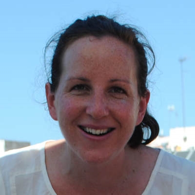
Dr Kyra Campbell, Bateson Centre, University of Sheffield, UK

Dr Kyra Campbell, Bateson Centre, University of Sheffield, UK
I have long been fascinated by the question of how cells assemble into functional tissues at both the subcellular and intercellular levels. After studying Natural Sciences at the University of Cambridge and being fired up by my final year course in Developmental Biology, I stayed on to do my PhD with Helen Skaer. In close collaboration with Elisabeth Knust, I explored how cell polarity is established and maintained as cells undergo the extensive remodelling that underlies tissue morphogenesis. For my postdoctoral training, I moved to the lab of Jordi Casanova in Barcelona, and focused on developing a novel model system for studying the mechanisms underlying cell plasticity during development, and in collaboration with Eduard Batlle’s lab at the IRB, in tumourigenesis. In 2017 I activated a Sir Henry Dale Fellowship that I was awarded by the Wellcome Trust/the Royal Society, and started my group in the University of Sheffield. In addition to continuing our exciting work on EMT and MET during development, we are collaborating closely with the lab of Andreu Casali, IRB LLeida, with whom we recently developed the first model for metastatic cancer in adult Drosophila. Our future work will focus on unravelling the cellular and molecular mechanisms that underlie epithelial cell plasticity during development, and in metastatic cancer progression.
| 08:00 - 08:30 |
Cellular mechanisms of organ self-assembly in vivo
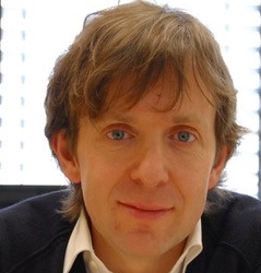
Professor Darren Gilmour, University of Zurich, Switzerland

Professor Darren Gilmour, University of Zurich, SwitzerlandPhD in Martin Evans’ lab at the Gurdon Institute, Cambridge. Post-doc in the lab of Janni Nüsslein-Volhard at Max Planck Institute for Developmental Biology, Tübingen. Group leader at the European Molecular Biology Laboratory (EMBL), Heidelberg. Since 2017, group leader at the Institute of Molecular Life Sciences (IMLS), University of Zurich. |
|
|---|---|---|
| 08:30 - 08:40 | Discussion | |
| 08:40 - 09:10 |
Dynamics of EMT and cell migration in Xenopus Neural Crest cells
Epithelial-mesenchymal transition (EMT) and cell migration are essential for numerous normal and pathological processes such as embryo development and cancer progression. Neural crest cells are multipotent embryonic stem cells whose EMT and migration rely on an array proto-oncogenes (e.g. Twist, Snail1/2, Ets1). In Xenopus, the early steps of neural crest development recapitulate what is observed in many carcinoma including an E-to-N cadherin switch followed by invasion of the local extracellular matrix. Neural crest cells express proteinases of the ADAM and MMP families and respond to factors that control tropism and homing of cancer cells such as CXCL12 and Semaphorins. Thus, Xenopus neural crest cells are an excellent model to study EMT and migration in a physiological context. Recently, Eric Theveneau’s group focused on two metalloproteinases (MMP14 and 28) that are known for their role in cancer invasion and wound healing, respectively. While most attention has been focused on the role of MMP14 and 28 in matrix remodeling during these processes, ongoing work on Xenopus neural crest cells by the Theveneau lab unraveled new functions for these enzymes, identifying them as early players during the EMT process and regulating the coordination between adjacent cell populations during morphogenesis. 
Dr Eric Theveneau, Center for Integrative Biology, CNRS/Université Paul Sabatier, France

Dr Eric Theveneau, Center for Integrative Biology, CNRS/Université Paul Sabatier, FranceEric Theveneau studied biology at Université Pierre et Marie Curie in Paris where he obtained a PhD in Cell and Developmental Biology in 2006 for investigating the transcriptional regulation of EMT in chick cephalic Neural Crest cells. He undertook his post-doctoral training at University College London with Professor Roberto Mayor (2007-2013). There, he studied mechanisms of cell cooperation. His work highlighted the ability of mesenchymal cells to undergo collective migration. Since then, Eric Theveneau has been recruited as a staff scientist by the French Centre for Scientific Research (CNRS) and appointed as a group leader in the Centre for Integrative Biology at Université Paul Sabatier in Toulouse, France, in 2014. His group currently works on elucidating in vivo functions of Matrix Metalloproteinases during EMT and cell migration.
|
|
| 09:10 - 09:20 | Discussion | |
| 09:20 - 09:50 | Coffee | |
| 09:50 - 10:20 |
Deciphering epithelial cell movements during embryogenesis
During early post-implantation embryogenesis of the mouse, the epiblast contributes the majority of the cells of the fetus but it is another tissue, the anterior visceral endoderm (AVE), that is responsible for imparting axial pattern upon the epiblast. AVE cells show a stereotypic unidirectional migration that is essential for correct orientation of the anterior-posterior axis. We do not understand how cells within epithelia such as the visceral endoderm show directed migration, how surrounding cells within intact epithelia accommodate the movement of a subset and the relative contributions of cell shape changes, regional differences in proliferation rates, oriented division etc. to such migration. Furthermore, we do not understand the extent to which movements within epithelia is coordinated with cell movements in abutting tissues such as the epiblast. To address these questions, we have used lightsheet microscopy to capture multi-dimensional image volumes of developing mouse embryos during AVE migration. To quantitatively analyse cell movements in the visceral endoderm, we have developed machine learning based approaches for the automated detection of cell boundaries and division events. I will discuss the new insights into cellular behaviour during AVE migration this approach reveals and a previously unknown movement within the epiblast that occurs in coordination with AVE migration. 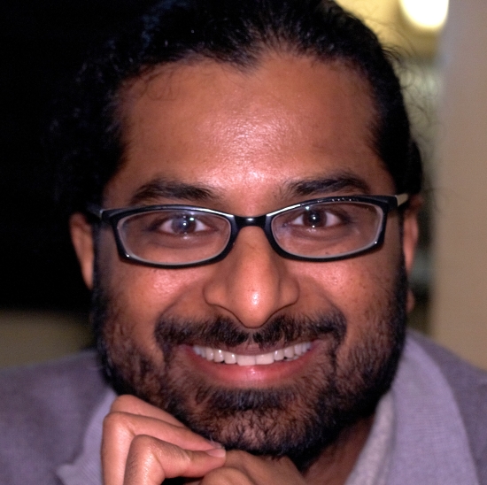
Professor Shankar Srinivas, University of Oxford, UK

Professor Shankar Srinivas, University of Oxford, UKShankar completed a BSc in Nizam College in Hyderabad, India. He then joined the group of Frank Costantini in Columbia University, New York, where he received a PhD for work on the molecular genetics of kidney development. Following this, he moved to the NIMR in Mill Hill, London, where he worked as a HFSPO fellow in the groups of Rosa Beddington and Jim Smith on how the anterior-posterior axis is established. Here, he developed time-lapse microscopy approaches to study early post-implantation mouse embryos. In 2004 Shankar started his independent group at the University of Oxford as a Wellcome Trust Career Development Fellow and as Zeitlyn Fellow and Tutor in Medicine at Jesus College. In 2016 he became Professor of Developmental Biology. He is currently a Wellcome Senior Investigator. The research in Shankar’s group continues to focus on understanding how the coordinated cell movements that shape the early mammalian embryo are controlled.
|
|
| 10:20 - 10:30 | Discussion | |
| 10:30 - 11:00 |
Signaling mechanisms that coordinate individual cell movements for epithelial migration
The collective migration of cells within an epithelial sheet underlies tissue remodeling events associated with morphogenesis, wound repair, and the spread of many cancers. Yet little is known about how each cell coordinates its movements with those of its neighbors. Studying the rotational migration of the follicular epithelium in the Drosophila egg chamber, the Horne-Badovinac lab has identified two planar signaling systems that operate along leading-trailing cell-cell interfaces to coordinate individual cell movements for collective motility. In the first signaling system, the giant cadherin Fat2 localizes to the trailing edge of each cell and sends an attractive signal to the receptor tyrosine phosphatase Lar at the leading edge of the cell behind. In the second signaling system, the transmembrane semaphorin, Sema-5c, localizes to the leading edge of each cell and sends a repulsive signal to the Plexin A receptor at the trailing edge of the cell ahead. In this talk, Dr. Horne-Badovinac will introduce both signaling systems as well as her lab’s efforts to understand how these local signals are propagated to polarize the entire epithelium for directed migration. 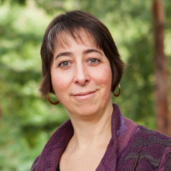
Dr Sally Horne-Badovinac, University of Chicago, USA

Dr Sally Horne-Badovinac, University of Chicago, USASally Horne-Badovinac is an Associate Professor in the Department of Molecular Genetics and Cell Biology at the University of Chicago. Her work seeks to understand how collective cell migration and reciprocal interactions between cells and their extracellular matrix sculpt an organ’s shape during development. Studying a dramatic epithelial migration that shapes the Drosophila egg, her lab has made fundamental contributions to our understanding of the signaling mechanisms that coordinate individual cell movements for epithelial motility, and how migrating epithelial cells build structured extracellular matrices to direct organ morphogenesis. Sally performed her doctoral training at the University of California San Francisco, where she was a National Sciences Foundation Graduate Research Fellow, and her postdoctoral training at University of California Berkeley, where she was a Jane Coffin Childs Postdoctoral Fellow. She has been at the University of Chicago since 2008.
|
|
| 11:00 - 11:10 | Discussion | |
| 11:10 - 12:10 | Lunch |
Chair
Dr Alexander Fletcher, Bateson Centre, University of Sheffield, UK

Dr Alexander Fletcher, Bateson Centre, University of Sheffield, UK
Alexander Fletcher has been a Vice-Chancellor’s Fellow in the School of Mathematics and Statistics at the University of Sheffield since 2015. He uses mathematical modelling to study how cell-level processes contribute to tissue-level dynamics in health and disease, with particular application to epithelial patterning and morphogenesis. To support this work he is a lead developer of Chaste, the first open source simulation package for off-lattice and multiscale computational models of cell populations. Before moving to Sheffield, he was a postdoctoral researcher at the University of Oxford, based in the Wolfson Centre for Mathematical Biology. He has an undergraduate degree in mathematics from the University of Cambridge and a DPhil in mathematics from the University of Oxford.
| 12:10 - 12:40 |
Guts and gastrulation: the lineages & dynamics driving the morphogenesis of the gut endoderm in the mouse embryo
Gastrulation is a paradigm for the coupling of cell fate specification and tissue morphogenesis. In mammals, gastrulation transforms an embryo comprising two tissue layers (epiblast and visceral endoderm), into one comprising three tissue layers (epiblast, mesoderm and gut endoderm). The hallmark morphogenetic event of gastrulation is an epithelial-to-mesenchymal transition (EMT), which occurs at, and defines, a structure called the primitive streak. At the primitive streak epiblast cells lose pluripotency and undergo an EMT and begin to move away as they concomitantly acquire a mesoderm or definitive endoderm fate. Having left the vicinity of the primitive streak, cells specified as definitive endoderm will intercalate into the overlying visceral endoderm epithelium to generate the gut endoderm, the precursor tissue of the respiratory and digestive tracts and associated organs. Using various contemporary approaches, including imaging and transcriptomics, coupled with the analysis of mutants, we are systematically investigating the sequential steps leading to the formation of the gut endoderm, towards developing a mechanistic understanding of this process. I will highlight some open questions and overview some of our recent work. 
Dr Anna-Katerina Hadjantonakis, Sloan Kettering Institute, Memorial Sloan Kettering Cancer Center

Dr Anna-Katerina Hadjantonakis, Sloan Kettering Institute, Memorial Sloan Kettering Cancer CenterDevelopmental biologist Anna-Katerina (Kat) Hadjantonakis studies cell lineage commitment, tissue patterning, and morphogenesis in mouse embryos and in vitro stem cell models. The overarching goal of her work is to understand how single cells reproducibly build complex tissues; from molecular circuits to cellular states and behaviors, to tissue-level architecture. She is a Member of the Sloan Kettering Institute of the Memorial Sloan Kettering Cancer Center, and a Professor at Cornell University, New York City, USA. She received a BSc in Biochemistry in 1990, and PhD in Molecular Genetics in 1994, from Imperial College London. From 1996 to 2003 she undertook postdoctoral work, first at the Samuel Lunenfeld Research Institute, in Toronto, Canada, and thereafter at Columbia University, in New York, USA. She established her independent research group at the Sloan Kettering Institute in 2004: www.mskcc.org/research/ski/labs/anna-katerina-hadjantonakis
|
|
|---|---|---|
| 12:40 - 12:50 | Discussion | |
| 12:50 - 13:20 |
Coordination of patterning and growth in the spinal cord
As the spinal cord grows during embryonic development, an elaborate pattern of molecularly distinct neuronal precursor cells forms along the DV axis. This pattern depends both on the dynamics of a morphogen-regulated gene regulatory network, and on tissue growth. We study how these processes are coordinated. Our data revealed that during mouse and chick development the gene expression pattern changes but does not scale with the overall tissue size. These changes in the pattern are sequentially controlled by distinct mechanisms. Initially, neural progenitors integrate signaling from opposing morphogen gradients to determine their identity by using a mechanism equivalent to maximum likelihood decoding. This strategy allows accurate assignment of position along the patterning axis and can account for the observed precision and shifts of pattern. During the subsequent developmental phase, cell-type specific regulation of differentiation rate, but not proliferation, elaborates the pattern. 
Dr Anna Kicheva, Institute of Science and Technology (IST), Austria

Dr Anna Kicheva, Institute of Science and Technology (IST), AustriaAnna Kicheva has been an Assistant Professor at IST Austria since November 2015. She studies the control of tissue growth and pattern formation during development. The research aim of her group is to unravel the feedbacks between cell fate specification, tissue growth and morphogen signaling in the developing spinal cord. Their approach combines biophysics, mouse genetics, and ex vivo assays. Anna started working on spinal cord development during her postdoc with James Briscoe at the NIMR (currently The Crick) in London. Before that she did her PhD with Marcos Gonzalez-Gaitan at the MPI-CBG in Dresden and the University of Geneva, studying morphogen gradient formation in the Drosophila wing disc.
|
|
| 13:20 - 13:30 | Discussion | |
| 13:30 - 14:00 | Tea | |
| 14:00 - 14:30 |
Getting in shape: in vivo and in silico studies of tissue mechanics in growth control
Tissue folding is a fundamental process that shapes epithelia into complex 3D organs. The initial positioning of folds is the foundation for the emergence of correct tissue morphology. Mechanisms forming individual folds have been studied, but the precise positioning of folds in complex, multi-folded epithelia is less understood. In this talk, a novel computational model of morphogenesis will be presented. The model encompasses local differential growth and tissue mechanics, to investigate tissue fold positioning. The Drosophila wing disc is used as the model system, as there is spatial-temporal heterogeneity in its planar growth rates. This differential growth, especially at the early stages of development, is the main driver for fold positioning. Increased apical layer stiffness and confinement by the basement membrane drive fold formation, but influence positioning to a lesser degree. The model successfully predicts the in vivo morphology of overgrowth clones and wingless mutants via perturbations solely on planar differential growth in silico. 
Dr Yanlan Mao, University College London, UK

Dr Yanlan Mao, University College London, UKYanlan Mao is a Group Leader at the MRC Laboratory for Molecular Cell Biology, University College London. After receiving her BA in Natural Sciences at Cambridge University, she completed her PhD with Matthew Freeman at the MRC LMB in Cambridge on Drosophila cell signaling and epithelial patterning. During her postdoc with Nic Tapon at the CRUK London Research Institute (now Francis Crick Institute), she became interested in tissue mechanics and computational modeling approaches. She now holds a UCL Excellence Fellowship and a MRC Career Development Award Fellowship to pursue her interests in the mechanical regulation of tissue growth and regeneration. |
|
| 14:30 - 14:40 | Discussion | |
| 14:40 - 16:00 | Panel discussion/Overview (future directions) |
