Open, reproducible hardware for microscopy

Theo Murphy meeting organised by Dr Richard Bowman, Dr Caroline Müllenbroich, Dr Benedict Diederich, Dr Julieta Arancio, Dr Sanli Faez and Professor Gail McConnell.
Reproducible experiments in microscopy require well-understood instruments that can be replicated independently. Open Source Hardware, the practice of sharing complete designs under an open license can make instrument development more reproducible, more accessible, and reduce duplicated effort. This meeting brought together researchers, manufacturers, and others involved in open microscopy to discuss recent developments, best practice, and goals for the future.
The meeting papers have been published in Philosophical Transactions of the Royal Society A.
The organisers would like to acknowledge the contribution of Dr Kirti Prakash, who has since stepped down from the organising committee, for helping to set up this meeting.
Attending this event
The meeting has taken place.
Follow-up online event
There will be a follow-up online meeting on 30 May 2023 at 2.30pm BST that aims to keep the conversation going after the main 'Open, reproducible hardware for microscopy' meeting.
Organisers
Schedule
Chair
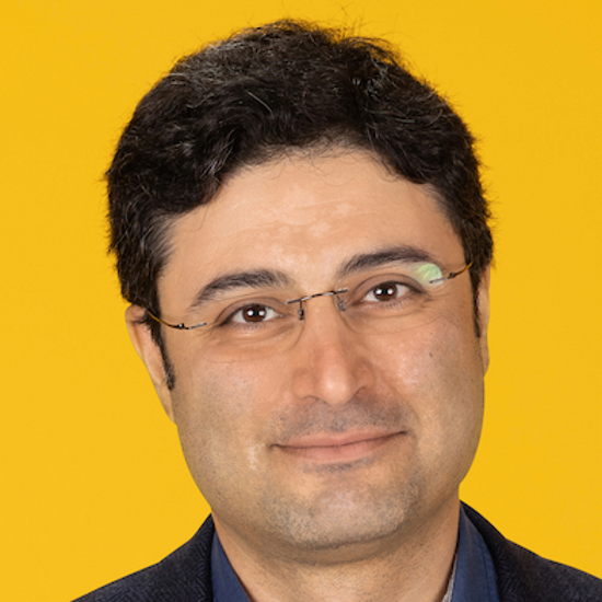
Dr Sanli Faez, Utrecht University, the Netherlands

Dr Sanli Faez, Utrecht University, the Netherlands
Sanli Faez is a professor (UD) at the Physics department of Utrecht University, a member of KNAW De Jonge Akademie, and a fellow at the Centre for Unusual Collaborations. He studied Physics at the Sharif University of Technology in Iran, and received his master’s degree in Nanotechnology from the University of Twente. His doctorate research at the University of Amsterdam was on wave propagation in strongly scattering media and Anderson localization. After post-doctoral research at the Max Planck Institute for the Science of Light and Leiden Institute of Physics, focused on single-molecule optics and (cryogenic) microscopy, he became the principal investigator for the research direction nanoElectroPhotonics at the Nanophotonics section of the Debye Institute for Nanomaterials at Utrecht University. He has hosted and produced several podcasts about open science.
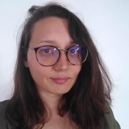
Dr Julieta Arancio, University of Bath, UK

Dr Julieta Arancio, University of Bath, UK
Dr Julieta Arancio is a postdoctoral researcher at the Centre for Science, Technology and Society, Drexel University (USA), and an associated researcher at CENIT-UNSAM (Argentina). She holds a degree in environmental science from Universidad de Buenos Aires and a PhD in science and technology studies from Universidad Nacional de Quilmes. Julieta has followed the open science hardware movement since 2017, looking at its potential contributions to more equitable science. Her current work, funded by the Alfred P Sloan Foundation, explores how open hardware is enabling new research activities and agendas. She has written academic papers and policy briefs on the topic, having presented her research at venues such as University of Cambridge (UK) and MIT (US), and teaching seminars at Universidad Nacional de Rosario (Argentina) and TU Berlin (Germany). Beyond academia, Julieta co-founded the Open Science Hardware network in Latin America (reGOSH) and the mentorship program Open Hardware Makers.
|
Session 1: Open hardware reaches further
Sharing hardware openly both enables more people and labs to access it, but through encouraging replication it makes science more reproducible. In this session, we will hear from speakers who work with or develop open projects, that have achieved a much wider reach because of that openness. The discussion will touch on the benefits of openness applied to experimental hardware, particularly as applied to reducing the inequalities entrenched in modern scientific practice. We will discuss issues around how to make projects open, barriers we often face to doing so, and tools that can help. |
|
| 09:00-09:05 |
Welcome by the lead organiser
|
| 09:05-09:45 |
Panel 1
Dr Jenny Molloy - Collaborations on open hardware for biotechnology in the global South Biotechnology contributes enormously to sustainable development and yet many researchers are unable to participate in biotechnology research due to lack of reagents, equipment and infrastructure. In this talk Dr Molloy will describe the work her research group, the Open Bioeconomy Lab, has undertaken in distributed manufacturing of hardware and enzymes, how open sharing has enabled their resources to reach over 40 countries, some of the barriers they have encountered and potential solutions to ensure that future open hardware projects achieve global reach. Professor Dan Fletcher - Mobile phone microscopy Optical microscopy remains a central tool for basic research and a key technology for disease diagnosis. While new microscopy techniques continue to expand the information that can be collected from microscopic samples, the availability of even basic microscopy equipment remains limited outside of well-funded research and clinical centres. Lack of access to microscopes and skilled users is not only a barrier to research but also to essential healthcare, such as the diagnosis of infectious diseases. Mobile phones, with their ever advancing camera and computational capabilities, offer opportunities for creating powerful, portable, and affordable microscopy systems that combine imaging with on-board image processing. The author will briefly describe ongoing work that combines mobile phones with both hardware and software engineering to enable multifunctional mobile microscopes. 
Dr Jenny Molloy, University of Cambridge, UK

Dr Jenny Molloy, University of Cambridge, UKDr Jenny Molloy is a Senior Research Associate at the University of Cambridge building technologies for an open, globally inclusive and equitable bioeconomy through the Open Bioeconomy Lab. She develops biomanufacturing tools and technologies that are sustainable by design and deployable in low-resource contexts. Her group also investigates the most effective ways that their research can make a positive impact in developing and emerging economies, particularly the effect of open licensing and collaborative development with end users. Jenny was a member of the World Economic Forum Global Future Council on Synthetic Biology, chairs an independent multi-sectoral working group on Local Production and Diagnostics in Low and Middle Income Countries and has co-founded four social enterprises and non-profits supporting the development and deployment of open source tools for science. Professor Manu Prakash, Stanford University, USA
Professor Manu Prakash, Stanford University, USABiography will be available soon. 
Professor Dan Fletcher, University of California, Berkeley, USA

Professor Dan Fletcher, University of California, Berkeley, USADr Dan Fletcher is a Professor of Bioengineering and Biophysics at UC Berkeley and the Pernendu Chatterjee Chair of Engineering Biological Systems. He is also a Chan-Zuckerberg Biohub Investigator and currently serves as Director of the Blum Centre for Developing Economies at UC Berkeley. Dr Fletcher and his laboratory develop optical instruments and study fundamental mechanisms of cellular organization, with applications to infectious diseases, immunology, and cancer biology. His research has been recognized with an NSF CAREER Award, a Tech Award from the San Jose Tech Museum, and a “Best of What’s New” citation by Popular Science magazine. Dr Fletcher received a BS from Princeton University, a DPhil from Oxford University where he was a Rhodes Scholar, and a PhD from Stanford University as an NSF Graduate Research Fellow. 
Dr Catherine Mkindi, Ifakara Health Institute, Tanzania

Dr Catherine Mkindi, Ifakara Health Institute, TanzaniaCatherine Mkindi is a Senior Research Scientist at Ifakara Health Institute, Tanzania since 2012. After earning her first and second degrees in Veterinary Medicine from Sokoine University, Tanzania, she enrolled in PhD studies in Medical-Biological research at the University of Basel, Switzerland, to further explore her passion in infectious diseases research. Dr Mkindi has an extensive research experience in malaria vaccine clinical trials in which they evaluate immune responses after participants vaccination with candidate malaria vaccines, followed by assessment of vaccine efficacy by human malaria control infection (CHMI) using microscopy and molecular methods. Recently, Dr Mkindi has been involved in clinical evaluation of the open flexure microscope, an automated, simple but robust microscope, that can be built and maintained by local biomedical engineers in the country and that has a potential to be used in limited resource settings in developing countries. This technology will not only help in assessing malaria vaccine effectiveness in a large population including rural settings but also revolutionize routine malaria diagnosis in lower-level remote health clinics with equipment and expert technicians’ constraints. |
| 09:45-10:25 |
Panel discussion – Q&A
|
| 10:25-10:40 |
Introduction to the unconference format
|
| 10:40-11:10 |
Break
|
| 11:10-12:10 |
Unconference session 1 following on from the panel discussion
|
| 12:10-12:30 |
Plenary feedback and discussion
|
| 12:30-13:30 |
Lunch
|
| 18:30-20:00 |
Dinner
|
Chair
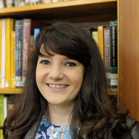
Dr Caroline Müllenbroich, University of Glasgow, UK

Dr Caroline Müllenbroich, University of Glasgow, UK
Dr Caroline Müllenbroich is a lecturer at the School of Physics and Astronomy at the University of Glasgow. She studied physics at the University of Heidelberg, Germany and obtained her PhD at the Institute of Photonics, University of Strathclyde, Glasgow in 2012. She then joined the European Laboratory for Nonlinear Spectroscopy (LENS) in Florence, Italy working on light-sheet microscopes for structural imaging of clarified mouse brains and functional imaging in Zebrafish. From 2016 on, she was a researcher with the Italian National Institute of Optics, National Research Council and focused her activities on the application of non-diffractive Bessel beams for the high-fidelity interrogation of neuronal structure and function. She obtained a Marie Curie Fellowship and a position at Glasgow University where her group now works on deep cardiac imaging, remote focusing and light-sheet microscopy. She is an advocate for increased inclusion and diversity in our the STEM (Science, Technology, Engineering, Maths and Medicine) community.
|
Session 2: Faster, more reproducible instrument development
Many cutting-edge techniques are now being shared under open licenses. This enables us to build on each other's work without the need for licensing deals, which is good for reproducibility and also for the pace of innovation. This session will let us hear from speakers developing novel technologies and sharing them openly. |
|
| 13:30-14:10 |
Panel 2
Professor Paul French - openScopes: an open, modular platform for microscopy and high content analysis Professor French and his collaborators are developing open multidimensional fluorescence imaging instrumentation, including high content analysis (HCA), super-resolved microscopy, quantitative phase imaging and optical projection tomography. For fluorescence microscopy, they are developing a modular open-source microscopy platform based on openFrame, a low-cost, modular, microscope stand that can be used for low-cost and sustainable instruments in lower resource settings or for rapidly prototyping of advanced microscopy concepts. For open HCA and slide scanning, the scientists have developed novel optical autofocus modules including a long-range (~200 micron) implementation utilising machine learning. This can be combined with easySTORM for cost-effective single molecule localisation microscopy (SMLM) including automated multiwell plate SMLM for super-resolved HCA. They have also developed a polarization differential phase contrast microscopy (pDPC) module providing single-shot quantitative phase imaging that we are applying to label-free single cell segmentation and tracking for long time-base assays. Dr Johannes Hohlbein - Open microscopy in the life sciences: sharing is caring Together with other colleagues enthusiastic about open science, Dr Hohlbein recently published a comment on the past, presence and future potential of open microscopy. Here, the author will iterate on some of the key points with a focus on single-molecule localisation microscopy (SMLM). SMLM allows monitoring molecular interactions in live cells and other complex samples. A few years ago, the scientists developed the miCube microscopy framework to increase the general accessibility and affordability of SMLM. They were intrigued to see how quickly other groups came up with new ideas and improvements in addition to our own developments including new algorithms for fast data analysis, addition of adaptive optics for localising proteins in turbid media and a scheme for spectrally resolved SMLM. These few examples demonstrate the huge potential of open microscopy in enabling interdisciplinary science and lowering the threshold for researchers to find the best solutions for their scientific imaging challenges. Dr Ulrike Boehm - Open hardware dissemination challenges and how to go about it The open hardware movement in science is gaining more and more traction. More and more developers openly share information about their tools with the scientific community. Still, the dissemination of these tools also needs to gain more traction. This presentation will focus on the reasons behind the dissemination challenges and suggests solutions for how to go about it. The solutions provided were direct learnings from two successfully realized open hardware projects at HHMI Janelia Research Campus, namely Karel Svoboda’s Mesoscope and Eric Betzig’s Lattice Lightsheet Microscope. Professor Paul French, Imperial College London, UK
Professor Paul French, Imperial College London, UK
Professor Paul French was awarded the BSc Degree in Physics in 1983 and the PhD degree (for work on femtosecond dye lasers) in 1987 from Imperial College London. In 1988 he was a visiting professor at the University of New Mexico working on femtosecond dye lasers and in 1989 he was awarded a Royal Society University Research Fellow at Imperial, where he joined the academic staff in 1994. From 1990 to 1991 he worked on ultrafast all optical switching in optical fibres at AT&T Bell Laboratories, Holmdel, NJ. He is currently a Professor of Physics at Imperial College London and is Head of the Photonics Group. His research has evolved from ultrafast dye and solid-state laser physics to biomedical optics. Today his group develops and applies multidimensional fluorescence imaging technology for molecular cell biology, drug discovery and clinical diagnosis with a strong emphasis on fluorescence lifetime imaging (FLIM) using microscopy, endoscopy and tomography. Paul French is a Fellow of the Institute of Physics, the European Physical Society and the Optical Society of America and holds a Royal Society Wolfson Research Merit Award.

Dr Johannes Hohlbein, Wageningen University & Research, the Netherlands

Dr Johannes Hohlbein, Wageningen University & Research, the NetherlandsDr Johannes Hohlbein studied Medical Physics at the MLU Halle-Wittenberg (Germany). In 2008 he obtained his PhD in Physics working on single-molecule detection in nanoscale confinement at the MPI of Microstructure Physics (Halle, Germany). He then joined the ‘Gene Machines’ group of Professor Kapanidis at the University of Oxford (UK) to work on DNA polymerases and developing assays for DNA sequencing. In 2012 he accepted a position as an Assistant Professor in the Laboratory of Biophysics at Wageningen University & Research (The Netherlands). Since then, his lab has been working on studying DNA-protein interactions in vitro and advancing single-molecule detection schemes. In 2018, Dr Hohlbein obtained tenure followed by promotion to Associate Professor. The current research in the lab is focussed on exploring DNA-RNA-protein interactions in live bacteria and performing functional food imaging using super-resolution microscopy and related techniques. 
Dr Ulrike Boehm, Carl Zeiss AG, Germany

Dr Ulrike Boehm, Carl Zeiss AG, GermanyUlrike is a physicist, optical scientist and data scientist passionate about community building/engagement, outreach, and teaching. She has over ten years of experience designing, building and running advanced optical systems, analysing (microscopy) data and developing image acquisition and analysis workflows. She studied physics at the Technical University of Munich and did her diploma at the Max Planck Institute of Biochemistry. Followed by her PhD studies in Göttingen at the Max Planck Institute for Biophysical Chemistry, she spent time in the US at the National Institutes of Health & the HHMI Janelia Research Campus, where she designed and built new tools for electron microscopy and light microscopy. Now she is part of the Corporate Research and Technology team at ZEISS in Oberkochen, Germany. At ZEISS, she works on the latest optical trends for imaging, microscopy, and optical metrology. In addition, Ulrike is a huge advocate for open science/education and women/diversity in science. 
Dr Lin Wang, Central Laser Facility, Rutherford Appleton Laboratory, Science and Technology Facilities Council, UK Research and Innovation

Dr Lin Wang, Central Laser Facility, Rutherford Appleton Laboratory, Science and Technology Facilities Council, UK Research and InnovationLin Wang is a Senior Scientist at the Central Laser Facility, Science and Technology Facilities Council, UKRI. He obtained his PhD in Electrical and Electronic Engineering from the University of Nottingham in 2010, and subsequently joined the University of Sheffield as a post-doctoral research fellow until 2015. He serves as an Honorary Associate Professor at the University of Nottingham, and holds Honorary Professorships at Xidian University and Jiangsu University of Technology in China, respectively. His research interest lies at the technology development end of the biomedical imaging spectrum. He has a strong interest in developing novel super-resolution microscopy. He also enjoys working with biologists, chemists and material scientists to explore how optical microscopy may help them address challenging questions and problems. |
| 14:10-14:40 |
Panel discussion - Q&A
|
| 14:40-15:10 |
Poster pitches
|
| 15:10-15:40 |
Break
|
| 15:40-16:25 |
Unconference session 2
|
| 16:25-16:45 |
Plenary feedback and discussion
|
| 16:45-18:15 |
Poster/tabletop presentations
|
| 18:30-20:00 |
Dinner
|
Chair
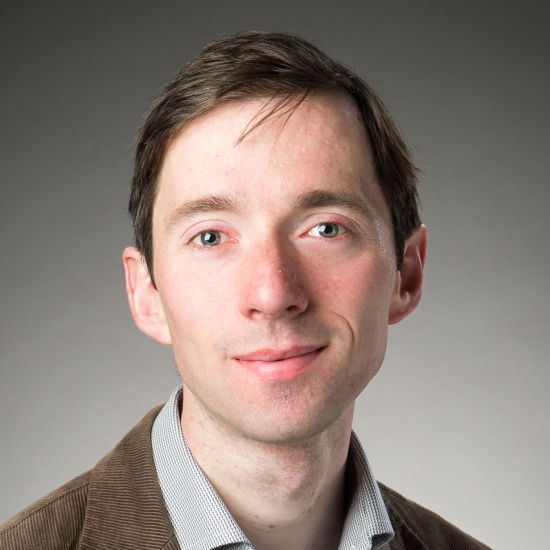
Dr Richard Bowman, University of Glasgow, UK

Dr Richard Bowman, University of Glasgow, UK
Dr Richard Bowman is a Royal Society University Research Fellow and Reader at the University of Glasgow, specialising in optical instrumentation and laboratory automation. Over the last six years he has led the OpenFlexure Project, centred on open source designs for a laboratory microscope suitable for diagnostics, research, and teaching. It has been reproduced thousands of times by a diverse community in over 40 countries around the world, including research labs in many disciplines, pathology research and teaching, community groups looking at environmental samples, and companies using it as a basis for new products. He is a strong advocate of openness as a means to improve both the reproducibility and accessibility of modern scientific apparatus, and very much hopes this meeting helps to forge links between the microscopy and open hardware communities.
|
Session 3: Interoperability and reproducibility for experiments
Any design can be shared, but when interoperability, openness, and reproducibility are included in a design from the start, it often has great results. We will have a discussion led by panellists who are experienced in working openly, and in designing interfaces between different systems. The utopia of a lab where different experiments can be designed and implemented by connecting standard components in a seamless and rapid way is still some way off, but we will discuss how we can achieve this using open tools. |
|
| 09:00-09:40 |
Panel 3
Dr Gemma Cairns - The Development of M4All: MultiModal Modular Microscopy for All M4All is a fully open-source and 3D-printable optics system which can be used to build low-cost microscopes combining different imaging modalities. M4All was primarily developed to address imaging challenges when investigating macrophage-pathogen interactions. Streptococcus pneumoniae is the most common cause of community acquired pneumonia. Mitochondrial reactive oxygen species (mROS) have been shown as critical microbicidal factors employed by alveolar macrophages in the clearance of internalised bacteria. Understanding and quantifying interactions with mROS could provide a future target for pharmacologically enhancing host responses to S. pneumoniae as an alternative to conventional antimicrobials, to address the increasing issue of antibiotic resistance. This is especially important in some developing countries in Africa and Asia where prevalence of invasive pneumococcal diseases is much higher. The short half-life of mROS (from 10-9 s to a few seconds) and the time frame of the overall response of alveolar macrophages to S. pneumoniae (16+ hours) poses challenges with detecting short lived interactions over long time frames. Advanced microscopy hardware for live cell microscopy can be expensive, leading to issues with accessibility. Furthermore, imaging over long periods of time introduces challenges with photobleaching and phototoxicity. Low-cost microscopes have the potential to widen accessibility to these techniques. With a focus on stability for timelapse imaging studies within incubators, M4All was developed as modular CAD files which are printed monolithically to build up the desired optomechanical system. M4All is also compatible with the OpenFlexure microscope stage. We have developed three microscopes using M4All in combination with the OpenFlexure microscope: a dual channel fluorescence microscope, a single channel fluorescence and computational phase contrast microscope, and a single channel brightfield incubator microscope. Here I present initial imaging results of macrophages and discuss how M4All could enable wider accessibility to advanced technologies by combining low-cost 3D-printable microscopes with computational microscopy techniques. Dr Fernan Federici - How open source hardware projects have enabled our capabilities for education, research, and outreach. The need to address inequalities in education, scientific research, and technological development has brought open science and open source technology to the forefront. The use of open protocols, public domain reagents, and open hardware is leading to more inclusive, efficient, and multidisciplinary research models. Collaborative networks such as ReClone, JOGL, iGEM, and GOSH, which openly share these resources online, are also enabling the emergence of more decentralized models for biotechnology. In case of Dr Federici and his group of scientists, engaging with these communities has expanded their research opportunities and facilitated the development of new teaching resources. For instance, they were able to use well-documented open hardware projects, such as OpenFlexure, OpenLabTools, and FlyPi, to create microscope prototypes for various applications, ranging from yeast monitoring during fermentation processes to time-lapse fluorescence experiments with optogenetic control. During the COVID-19 lockdown, they assembled portable minilabs for teaching hands-on molecular biology practicals at home. In this presentation, Dr Federici will share their experiences working with open source hardware projects in his lab and within the context of the local reGOSH-CYTED Latin American network. Furthermore, he will discuss the limitations they are facing in expanding these open frameworks in our region and share insights into how the plan to address these challenges. Dr David Baddeley - Python-microscopy and pyoptic - open source software for microscope control and design Python-microscopy is a suite of software tools for the control of open-source microscopes, for simulating microscopic imaging, and for image data analysis. Dr Baddeley will introduce python-microscopy, describe some of the innovations they have made for handling high data-rate streams and extended imaging durations, and discuss how to interface with custom hardware. He will also describe the use of pyoptic2 – a python-based optical-CAD package – in conjunction with python-microscopy and 3D printing for the simulation and rapid-prototyping of custom optical systems. Dr Rifka Vlijm - Going Live! Technological developments for live cell (STED) super-resolution imaging With the development of super-resolution microscopy, and great progress in labelling techniques, direct visualization of cellular structures in living cells now is possible at resolutions of 30-40nm. Commercial Stimulated Emission Depletion (STED) microscopes now ensure access to this technique to researchers not specialised in optics. One important limitation which hinders a broad application to cell biological questions is the low throughput due to the manual steps required for optimal results still. Capturing ‘rare’ events, such as for example a specific cell division stage, with only manual structure selection and image parameter selection becomes near impossible. To overcome these limitations, Dr Vlijm and her group have managed to fully automate our data acquisition, and thereby improved the throughput by two orders of magnitude. They have shown the value of these improvements by detecting cells in a specific cell division stage without the typical required chemical or genetic modifications necessary to increase the occurrence. In combination with full incubator conditions on the microscope the scientists have furthermore developed a protocol for live cell STED which still enables normal cell proliferation. 
Dr Gemma Cairns, Beatson Institute, UK

Dr Gemma Cairns, Beatson Institute, UKDr Gemma Cairns is a Postdoctoral Research Scientist at the CRUK Beatson Institute in the Leukocyte Dynamics group. After gaining an interest in optics and microscopy during her undergraduate physics degree at Heriot-Watt University, she pursued a PhD through the OPTIMA EPSRC and MRC CDT in Optical Medical Imaging. Working between the NanoBioPhotonics group at the University of Strathclyde and the Centre for Inflammation Research at the University of Edinburgh, she developed M4All, an open-source, 3D-printable and modular optics system, to address imaging challenges when investigating macrophage host defence responses to Streptococcus pneumoniae. Now working in a cancer immunology research group, she hopes to bring her multidisciplinary background and expertise in microscopy to help investigate the role of neutrophils in breast cancer metastasis. Dr David Baddeley, University of Auckland, New Zealand
Dr David Baddeley, University of Auckland, New ZealandFollowing under-graduate and MSc level training in Physics at the University of Auckland, David did his doctorate at the University of Heidelberg in Germany. During this time he started his association with optical microscopy and super-resolution imaging. This was followed by a Postdoc in the Department of Physiology at Auckland, and a stint as an Assistant Professor in the Cell Biology Department at Yale. He is now back in Auckland, at the Auckland Bioengineering institute. He has published across a wide range of different microscopy techniques including confocal, structured illumination 4Pi, single-molecule switching (STORM/PALM/PAINT, etc), and STED. 
Dr Rifka Vlijm, The University of Groningen, The Netherlands

Dr Rifka Vlijm, The University of Groningen, The NetherlandsRifka Vlijm is a Biophysicist who studied the physical properties of DNA and nucleosome assembly at the single molecule level using techniques such as magnetic tweezers and (high-speed) AFM (Technical University Delft, Netherlands). Driven by her findings, she joined the lab of Professor Stefan Hell as a Postdoc to work on (live-cell) Stimulated Emission Depletion (STED) super-resolution microscopy (DKFZ & Max Planck Institute for Medical Research, Heidelberg, Germany). In 2019 she became a group leader at the University of Groningen (Netherlands). Her group uses STED microscopy to study protein structures in fixed and living cells. The focus of her research is on the structural changes of, for example, the DNA and kinetochore during cell division. The recent developments in her lab are focused on turning STED microscopy into a non-invasive high-throughput technique suitable for live-cell imaging at resolutions of 30-40nm. 
Niamh Burke, University College Dublin, Ireland

Niamh Burke, University College Dublin, IrelandNiamh Burke is a PhD student and Anatomy Demonstrator at University College Dublin (UCD), Ireland and a Postgraduate Representative with the Microscopy Society of Ireland. During her undergraduate degree in Physiology in UCD, Niamh completed a short project under the supervision of Dr Mark Pickering. It was here where she developed a strong interest in building simple, low-cost, open-source lab tools, from remote aquarium monitoring systems to 3D printed centrifuges and simple light-sheet microscopes. Taking a brief break from the world of open hardware, Niamh then worked as a Research Assistant in the Cancer Biology and Therapeutics Group in the UCD Conway Institute under Professor William Gallagher. Following this, Niamh returned to the Pickering Lab for her PhD. Niamh's PhD focuses on designing open and accessible tools to measure microplastic pollution in the sea. As part of this she has designed the EnderScope - a low-cost, 3D printer based microscope for automatic scanning of filtered seawater samples for detection of microplastics. The philosophy behind the EnderScope is that by making this tool low-cost, open-source and easy for the user to build and reproduce, it can be used as a truly accessible and scalable solution to the global problem of microplastic pollution. 
Teja Potocnik, University of Cambridge, UK

Teja Potocnik, University of Cambridge, UKTeja is a PhD student at the University of Cambridge in the Low-Dimensional Electronics Group. She started her PhD in 2020 after studying Material Science and Engineering at the University of Manchester. As part of her PhD she is currently developing passivation and encapsulation strategies for 2D material devices, as well as automated protocols for high throughput nanomaterial characterisation. |
| 09:40-10:20 |
Panel Discussion
|
| 10:20-10:50 |
Break
|
| 10:50-11:50 |
Unconference session 3
|
| 11:50-12:20 |
Unconference recap
|
| 12:20-13:20 |
Lunch
|
Chair
Dr Benedict Diederich, Leibniz Institute of Photonic Technology, Germany
Dr Benedict Diederich, Leibniz Institute of Photonic Technology, Germany
After doing an apprenticeship as an electrician Benedict Diederich started studying electrical engineering at the University for Applied Science Cologne. A specialisation in optics and an internship at Nikon Microscopy Japan pointed him to the interdisciplinary field of microscopy. After working for Zeiss he started his PhD in the Heintzmann Lab at the Leibniz IPHT Jena, where he focusses on bringing cutting edge research to everybody by relying on tailored image processing and low-cost optical setups. Part of his PhD program took place at the Photonics Center at the Boston University in the Tian Lab. A recent contribution was the open-source optical toolbox UC2 (You-See-Too) which tries to democratise science by making cutting-edge affordable and available to everyone, everywhere.
|
Session 4: Making open, reproducible hardware a reality
Open hardware is already possible in microscopy, and is increasingly being seen as good practice. If we're to make open hardware more widespread, we need to understand how to recognise this practice in ways that are picked up by academic metrics, and how to produce instruments commercially without exclusive licenses. Our panellists represent a variety of different stakeholders from the scientific ecosystem, including suppliers, funders, publishers, and a legal expert. The discussion will start focused on questions around how we can help open hardware become more widely practised, including how it's published, purchased, and funded. |
|
| 13:20-14:20 |
Panel 4
Dr Rita Strack - An editor’s perspective on open microscopy Innovations in both hardware and software have propelled the advancement of microscopy in the life sciences. From an editorial perspective, they have seen an important movement in open science that has made a huge impact in terms of technology dissemination, uptake, and development. Dr Strack will discuss trends they at Nature Methods have seen in open microscopy and how these fit into a larger landscape of both technology development and improving reporting and reproducibility in microscopy across biology. Dr Brian Mehl - Thorlabs Imaging Research Group: How to accelerate advanced imaging technologies to the wider research community At Thorlabs, researchers transform the world by identifying, enabling, and accelerating key photonics technologies. The imaging research group implements this mission statement by driving new imaging techniques, components, and custom solutions to the research community. Their goal is to shorten the time from when a technology is initially published to when the wider research community has access to these advancements, avoiding the traditionally slow product lifecycle development. Utilizing the symbiotic relationship between the teams engineering prowess and insightful researchers these collaborations have been productive in lowering the bar for engagement in advanced imaging techniques. Andrew Katz - 2021 European Commission study on Open Source Software and Hardware There are a number of possible funding, commercial and business models which can be applied to open hardware. As part of his work on the 2021 European Commission paper on the Impact of Open Source Software and Hardware, Andrew Katz interviewed leading open hardware businesses worldwide. He will present the research findings, placing them in legal context with discussion of legal structure, governance, licensing and liability, and apply those findings to the panel discussion covering the use, production and dissemination of open hardware in an academic and research environment. 
Dr Rita Strack, Nature Methods, USA

Dr Rita Strack, Nature Methods, USARita Strack obtained her PhD in Biochemistry and Molecular Biology from the University of Chicago. While there, she worked with Benjamin Glick and Robert Keenan to engineer improved variants of the red fluorescent protein DsRed, and also studied the chemical mechanism of chromophore formation in DsRed. She continued her research as a postdoctoral fellow in Samie Jaffrey's laboratory at Weill Cornell Medical College, where she developed fluorescent reporters for live-cell imaging of RNA such as Spinach2. She handles imaging, microscopy, and probes, along with protein and RNA biochemistry content for the journal. Rita joined Nature Methods in November 2014. 
Dr Brian Mehl, Thorlabs Imaging Research Group, USA

Dr Brian Mehl, Thorlabs Imaging Research Group, USAAfter gaining a passion for instrument design during his PhD training studying ultrafast microscopy and spectroscopy at the University of North Carolina, Chapel Hill, Brian pivoted fields and discovered more traditional microscopy techniques and worked on integration of a fluorescent lifetime imaging microscopy (FLIM) system for biosensor development. Thereafter, Brian worked as a research specialist implementing single molecule imaging techniques at HHMI Janelia in the Transcription Imaging Consortium. Brian joined Thorlabs in 2016 where he currently works as an instrument designer and builder to lead collaborations on the development of advanced imaging techniques working alongside a group of talented engineers. 
Andrew Katz, Moorcrofts LLP, UK

Andrew Katz, Moorcrofts LLP, UKAndrew Katz is a lawyer who has advised on free and open source software, open hardware and other opens for over 25 years. Formerly a software developer, he qualified as a barrister, requalified as a solicitor and is now partner and head of technology law at Moorcrofts LLP, a boutique firm specialising in corporate and technology law. He wrote the Solderpad open hardware licence, and sits on the core drafting team of the CERN Open Hardware Licence. He was Open Hardware lead for the 2021 European Commission study on Open Source Software and Hardware. He has taught a postgraduate Open Source Software law course at Queen Mary College, University of London, and is currently a visiting scholar at the University of Skövde, Sweden, where as part of the Software Systems Research Group he has co-authored papers on open source software, open technologies, interoperability, patent licensing and standards. He is co-author of books published by Oxford University Press, Edinburgh University Press and others. He speaks internationally, most recently delivering a keynote at the Open Compliance Summit in Japan (December 2022). He is chair of the OpenChain UK work group, past General Counsel of OpenUK, and has worked with multinational companies, governments, universities and open source foundations, as well as organisations such as the UN and WIPO. 
Philip Hubbard, Biotechnology and Biological Sciences Research Council, part of UKRI

Philip Hubbard, Biotechnology and Biological Sciences Research Council, part of UKRIPhilip Hubbard, Biotechnology and Biological Sciences Research Council (BBSRC), part of UK Research and Innovation (UKRI). UKRI is the UK’s national public funder in research and innovation, investing £7.9bn in 2022/2023 to support research and innovation for economic growth and benefit to society. UKRI consists of seven UK Research Councils, Innovate UK, and Research England, funded by HM Treasury and reporting to the Department for Science, Innovation and Technology. Philip Hubbard looks after the bioimaging portfolio at BBSRC, a portfolio which naturally interfaces with open technology development. His attendance at this event is part of BBSRC’s efforts to promote the value of diversity and team working in the biosciences and foster an open, dynamic, and inclusive system of technology development in the UK, as highlighted in BBSRC's recent review of technology development in the biosciences. |
| 14:20-15:00 |
Panel Discussion
|
| 15:00-15:30 |
Break
|
| 15:30-16:25 |
Unconference session 4
|
| 16:25-16:45 |
Plenary discussion
|
| 16:45-17:00 |
Plenary wrap-up
|

