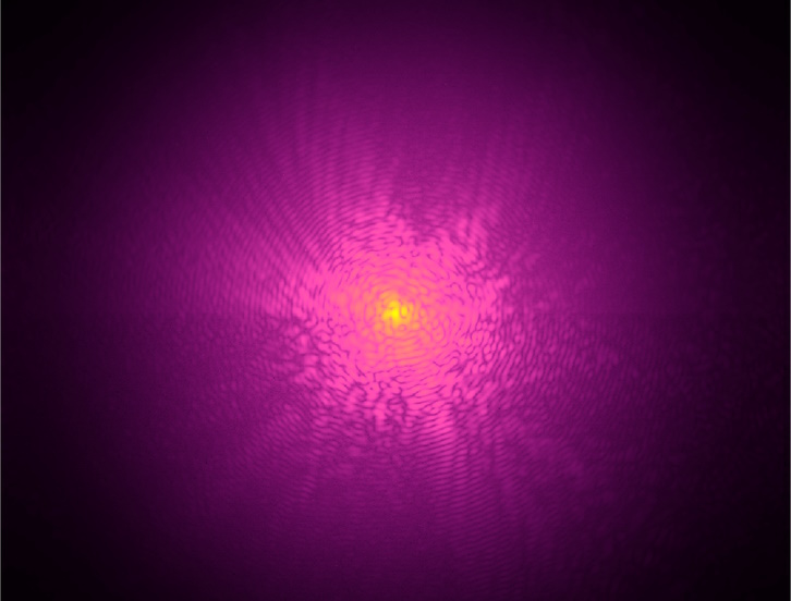
Ptychography over all wavelengths
Theo Murphy meeting organised by Professor John Rodenburg FRS, Professor Ian Robinson and Dr Andrew Maiden.
Ptychography is a form of computational imaging that has distinct advantages over conventional microscopic imaging methods. This meeting brought together researchers from the X-ray community, who have been using ptychography for about 15 years, with more recent adopters of the technique from the visible light, EUV and electron microscopy communities, as well as theoreticians studying novel applications and algorithms relating to the technique.
The schedule of talks and speaker biographies are available below. Speaker abstracts are also available below.
Attending this event
This event has taken place.
To view the programme, please scroll down and select the day on the left-hand side. Click the arrows to view the speakers and talks.
Enquiries: contact the Scientific Programmes team
Organisers
Schedule
Chair
Professor Ian Robinson, University College London, UK
Professor Ian Robinson, University College London, UK
Ian Robinson is a professor in the Condensed Matter and Materials Physics (CMMP) department at UCL and the London Centre for Nanotechnology (LCN). His research is based in the Research Complex at Harwell to develop phase modulation technology for X-ray imaging and he has a Diamond Professorial Fellowship for imaging chromosomes by coherent X-ray diffraction. His research makes extensive use of synchrotron radiation at the Diamond Light Source and elsewhere. He built a beamline at Brookhaven to develop Surface X-ray Diffraction and a second one at Argonne for Coherent X-ray Diffraction. One outcome of the work was the discovery of Crystal Truncation Rods, for which he was awarded the Surface Structure Prize in 2011.
| 09:00-09:05 |
Introduction
|
|---|---|
| 09:05-09:30 |
Beyond the limitations of traditional imaging: advancements in nanoscale 3D X-ray imaging with ptychography and sparsity
Ptychography, and in particular its extension to 3D, is a powerful technique that allows nanoscale material characterization with quantitative electron density contrast. Extensions of the technique to the spectral or time domain offer unique opportunities to study operando materials and to do quantitative chemical mapping. However, being a scanning technique, extensions to higher dimensions can require prohibitive scanning times. Here Professor Guizar-Sicairos will present results that combine ptychographically acquired projections with tomographic sparsity in order to significantly improve the time or spectral resolution, with application to energy conversion materials. Dr Manuel Guizar-Sicairos, Paul Scherrer Institut, Switzerland
Dr Manuel Guizar-Sicairos, Paul Scherrer Institut, Switzerland
"Originally from Mexico, Manuel received a BSc degree in Physics Engineering in 2002 and MSc in Electronic Systems in 2005 from the Tecnológico de Monterrey, Mexico for work in unstable laser resonators, numerical propagation and non-diffracting beams. He later received a MSc in Optics in 2008 and PhD in Optics in 2010 from the Institute of Optics, University of Rochester in NY, with thesis title “Methods for coherent lensless imaging and x-ray wavefront measurement”. Currently he holds a position of beamline scientist at the cSAXS beamline, Paul Scherrer Institut in Switzerland where he develops methods and analysis techniques for 2D and 3D coherent diffraction imaging, and in particular ptychography."
|
| 09:30-09:45 |
Discussion
|
| 09:45-10:15 |
Making every electron count; ptychography at low dose
This lecture will describe recent developments in the use of Electron Ptychography for studies of materials, including biological structures at low electron fluence. Importantly for this application, ptychography is an inherently dose efficient technique, enabling effective phase reconstruction of radiation sensitive samples. Professor Kirkland will particularly highlight recent developments in optimised scanning and data processing using machine learning for reductions in radiation damage and noise reduction in low fluence data sets. In the life sciences Cryo-electron ptychography holds much promise particularly when used with a defocused probe to scan across a specimen with highly overlapped probe positions. This can be applied in a variant of conventional single particle analysis to provide 3D structures taking advantage of the known resolution variation of the effective ptychographic transfer function with convergence angle to provide wide spatial frequency bandwidth transfer. This geometry also allows datasets from wide fields of view to be collected that are suitable for studies of biologically relevant structures in a cellular context. Finally Fourier Ptychography as an alternative data acquisition and processing strategy will be discussed. Angus Kirkland
Angus Kirkland |
| 10:15-10:30 |
Discussion
|
| 10:30-11:00 |
Break
|
| 11:00-11:30 |
Dichroic X-ray ptychography of 3D magnetic textures
Three dimensional magnetic systems promise significant opportunities for both fundamental physics, and technological applications, for example providing higher density devices and new functionalities associated with complex topology and greater degrees of freedom. One of the main challenges comes with characterising complex three dimensional magnetic systems. Here Dr Donnelly will discuss the advances in X-ray magnetic tomographic imaging making use of dichroic X-ray ptychography. Using hard X-ray magnetic tomography, we have been able to map both the static configuration, and dynamical behaviour, of topological magnetic structures. Understanding these complex configurations is challenging: recent advances in analytical techniques have provided new capabilities to locate and identify 3D magnetic solitons, leading to the first observation of nanoscale magnetic vortex rings. While hard X-ray imaging offers high resolution imaging of extended magnetic systems, the weak magnetic dichroism limits the variety of materials that can be imaged. To this end, Dr Donnelly will present recent results of soft X-ray dichroic ptychography where the phase dichroism offers a route to imaging magnetic systems that until now have not been accessible. These new capabilities for the high spatial resolution imaging of three dimensional magnetic configurations opens the door to the exploration of both fundamental and technologically relevant magnetic systems. Dr Claire Donnelly, Max Planck Institute for Chemical Physics of Solids, Germany
Dr Claire Donnelly, Max Planck Institute for Chemical Physics of Solids, GermanyFollowing her MPhys at the University of Oxford, Claire went to Switzerland to carry out her PhD studies at the Paul Scherrer Institute and ETH Zurich. She was awarded her PhD in 2017 for her work on 3D systems, in which she developed X-ray magnetic tomography, work that was recognised by a number of awards. After a postdoc at the ETH Zurich, she moved to the University of Cambridge and the Cavendish Laboratory as a Leverhulme Early Career Research Fellow, where she focused on the behaviour of three dimensional magnetic nanostructures. Since September 2021 she has been a Lise Meitner Group Leader of Spin3D at the Max Planck Institute for Chemical Physics of Solids in Dresden, Germany. Her group focuses on the physics of three dimensional magnetic systems.
|
| 11:30-11:45 |
Discussion
|
| 11:45-12:15 |
Bragg ptychography
Bragg ptychography is a phase retrieval method combining crystalline sensitivity and the possibility to image extended 3D samples. Recent progresses in Bragg ptychography formalism, namely, the introduction of the Bragg projection operator and the advent of 4th generation synchrotron sources have enabled the use of 3D Bragg ptychography with unprecedented accuracy, sensitivity and spatial resolution. Specifically, the probe retrieval and the beam-position refinement are now possible thanks to the amount of information encoded in the diffraction patterns produced with extremely brilliant source. Together with collaborators from ESRF, MAXIV and Diamond, Dr Chamard's group is now pushing forward the integration of Bragg ptychography at synchrotron beamlines to enable the use of this 3D crystalline microscopy approach to a larger x-ray community. In this talk, Dr Chamard will review the history of Bragg ptychography and highlight the pending questions related to the dissemination of the method. 
Dr Virginie Chamard, Institut Fresnel, Aix-Marseille Université, France
Dr Virginie Chamard, Institut Fresnel, Aix-Marseille Université, FranceVirginie Chamard is a CNRS researcher director at Institut Fresnel in Marseille. Together with her group, she develops coherent x-ray and optical ptychography microscopy methods, which she applies to the study of crystalline materials (eg biominerals, implanted metals, phase domains, magnetic systems, etc…). She is now focusing on the integration of 3D Bragg ptychography at several synchrotron beamlines, in parallel to leading research activity on the understanding of biomineralization using physical and chemical microscopy methods. She received an European Research Council grant in 2017. |
| 12:15-12:30 |
Discussion
|
Chair
Dr Laura Clark, University of York, UK
Dr Laura Clark, University of York, UK
Dr Laura Clark obtained her PhD in 2016 from the EMAT group at the University of Antwerp, for research on electron beam shaping, applied to creating electron vortex beams. Her postdoctoral work included time spent in the Findlay group at Monash University developing quantitative methods for DPC-STEM and phase-contrast TEM, and time spent in the Nellist/Kirkland groups at Oxford working on the ESTEEM3 project. She was subsequently a Marie Skłodowska-Curie fellow at the University of Leeds, before taking up her current Royal Society University Research Fellowship at the University of York in 2022. Her research interests are in the field of development of quantitative algorithms for accurate sample characterisation in the (S)TEM, with a particular focus on ptychographic and other phase contrast algorithms.
| 13:30-14:00 |
Evaluating the performance of electron ptychography at low dose
Many materials of interest for energy storage and energy conversion present two important challenges when attempting to characterise at high resolution using electrons. First, they often contain low atomic-number elements such as lithium and oxygen. Second, they are often susceptible to damage under electron irradiation. Electron ptychography is a very promising technique to overcome both these challenges because it allows detection of the small phase shifts induced on a transmitted electron wave and it can make use of a large fraction of the transmission electrons thereby maximising the information per damage event. Comparisons of ptychographic images from a range of samples with those formed using phase contrast TEM (as is used in cryo-TEM for biological samples) suggests that ptychography is very competitive. In this talk Professor Nellist will explore some of the important attributes of electron ptychography for comparison with other techniques. The effects of partial spatial and temporal coherence will be discussed. Suitable metrics to allow comparison between different imaging modes will be explored and initial results presented. The discussion will be illustrated with applications of ptychography to battery materials and polymers. Peter Nellist
Peter Nellist |
|---|---|
| 14:00-14:15 |
Discussion
|
| 14:15-14:45 |
Provably accurate recovery of compactly supported smooth functions from spectrogram measurements
In this talk Professor Iwen will focus on the approximation of smooth functions, up to an unresolvable global phase ambiguity, from a finite set of Short Time Fourier Transform (STFT) magnitude (ie spectrogram) measurements. Algorithms are developed for approximately inverting such measurements, each with theoretical error guarantees establishing their correctness. A detailed numerical study further demonstrates that the algorithms work well in practice and have good numerical convergence behaviour. This is joint work with Mike Perlmutter (UCLA), Nada Sissouno (TUM), and Aditya Viswanathan (UM-Dearborn). 
Professor Mark Iwen, Michigan State University, USA
Professor Mark Iwen, Michigan State University, USAMark Iwen received his PhD in applied and interdisciplinary mathematics from the University of Michigan - Ann Arbor in 2008, after which he completed two postdocs at the University of Minnesota and Duke University. He then joined Michigan State University in 2013 where he is currently an associate professor with a dual appointment in the Department of Mathematics and the Department of Computational Mathematics, Science and Engineering (CMSE). His research interests include computational harmonic analysis, mathematical data science, signal processing, and algorithms for the analysis of large and high dimensional data sets. To date he has graduated 9 PhD students, and authored over 70 journal and conference articles which have been published in interdisciplinary venues ranging from journals such as Applied and Computational Harmonic Analyisis, Foundations of Computational Mathematics and Discrete and Computational Geometry, to computer science and engineering conferences such as the ACM-SIAM Symposium on Discrete Algorithms (SODA) and the IEEE International Conference on Acoustics, Speech, and Signal Processing (ICASSP). |
| 14:45-15:00 |
Discussion
|
| 15:00-15:30 |
Break
|
| 15:30-16:00 |
Framewise network in Ptychographic phase retrieval
Most optimization methods in phase retrieval rely on gradients or proximal operators embedding phase less data onto an image space (and an illumination for blind ptychography), however convergence rate of these methods scales poorly with data size. By analysing the relationship network and minimizing the difference between all pairs of overlapping frames, it is possible to identify the slowly evolving principal modes, and solve large scale position drifts, or slow phase variations. Examples applications will be discussed. Dr Stefano Marchesini, Lawrence Berkeley National Laboratory, USA
Dr Stefano Marchesini, Lawrence Berkeley National Laboratory, USA
"Stefano Marchesini received his Laurea degree in Physics from University of Parma, Italy in 1996, and his PhD at the University J Fourier of Grenoble, France in 2000 on Fluorescence X-ray holography. He then went to work at Lawrence Berkeley National Laboratory (2000-2002, 2007-present) and Lawrence Livermore National Laboratory (2003-2007), on holography and diffractive imaging techniques using synchrotron and FEL sources."
|
| 16:00-16:15 |
Discussion
|
| 16:15-17:00 |
Poster flash talks
|
Chair
Dr Virginie Chamard, Institut Fresnel, Aix-Marseille Université, France
Dr Virginie Chamard, Institut Fresnel, Aix-Marseille Université, France
Virginie Chamard is a CNRS researcher director at Institut Fresnel in Marseille. Together with her group, she develops coherent x-ray and optical ptychography microscopy methods, which she applies to the study of crystalline materials (eg biominerals, implanted metals, phase domains, magnetic systems, etc…). She is now focusing on the integration of 3D Bragg ptychography at several synchrotron beamlines, in parallel to leading research activity on the understanding of biomineralization using physical and chemical microscopy methods. She received an European Research Council grant in 2017.
| 09:00-09:30 |
High-resolution, high-throughput coded ptychography for biomedical imaging
Ptychography has emerged as a transformative coherent imaging technique for both fundamental and applied sciences. Its applications in optical microscopy, however, often fall short for its relatively low imaging throughput and limited resolution. In this talk, Dr Zheng will discuss a coded ptychography technique that achieves an imaging throughput order of magnitude greater than previous demonstrations. In this platform, we translate the samples across the disorder-engineered surfaces for lensless diffraction data acquisition. The engineered surface can be made by smearing a monolayer of blood on top of the image sensor. The entire system can be built using a modified Blu-ray player, where the 405 nm laser from the optical pickup head can be used as a coherent light source for sample illumination. By tracking the phase wraps of the recovered images, the group reports the direct observation of bacterial growth in a 15-s interval at a 120 mm^2 area, with a phase sensitivity like that obtained from interferometric measurements. They also characterize cell growth via longitudinal dry mass measurement and perform rapid bacterial detection at low concentrations. For drug-screening application, they demonstrate antibiotic susceptibility testing and perform single-cell analysis of antibiotic-induced filamentation. The combination of high phase sensitivity, high spatiotemporal resolution, and ultra-large field of view is unique among existing microscopy techniques. Dr Zheng will also discuss several extensions of the coded ptychography technique, including optofluidic ptychography, synthetic aperture ptychography, and depth-multiplexed on-chip imaging, thereby expanding its applicability in different fields. 
Dr Guoan Zheng, University of Connecticut, USA
Dr Guoan Zheng, University of Connecticut, USADr Guoan Zheng received his BS degree in Electrical Engineering from Zhejiang University in 2007, and his MS and PhD degrees in Electrical Engineering from Caltech in 2008 and 2013, respectively. After completing his PhD, he joined the University of Connecticut (UConn) as an assistant professor, and is now the United Technologies Corporation (UTC) Associate Professor in the areas of Biomedical Engineering and Electrical Engineering. Dr Zheng's expertise includes microscopy, ptychography, optical engineering, biophotonics, computational optics, and lab-on-a-chip devices. He has published more than 100 journal papers and has an h-index of 50. His current research efforts focus on the development of novel optical imaging tools to tackle measurement problems in biology and medicine. |
|---|---|
| 09:30-09:45 |
Discussion
|
| 09:45-10:15 |
Ptychography based super-resolution X-ray fluorescence microscopy via deep residual networks
Ptychography is a computational method of microscopic imaging that can generate high spatial resolution complex phase information of an object by processing coherent diffraction patterns scattered from the object. With the development of Ptychography, it has been extensively used to study materials in combination with other imaging techniques, which can give multiple contrast mechanisms of the measured sample – for example, in combination with X-ray fluorescence (XRF) microscopy. While Ptychography can achieve diffraction-limited spatial resolution, the used X-ray probe profile information usually limits the resolution of XRF images. In this talk, Dr Wu will present a machine learning based approach utilizing multimodal measurement (ie the combination of Ptychography and XRF microscopy) to enhance the resolution of the corresponding XRF microscopy by deconvolving the X-ray probe from the XRF signal. The enhanced spatial resolution was observed for both simulated and experimental XRF data, showing superior performance over the state-of-the-art scanning XRF method with different nano-sized X-ray probes. Enhanced spatial resolutions were also observed for the accompanying 3D XRF tomography reconstructions. 
Dr Longlong Wu, Brookhaven National Laboratory, USA
Dr Longlong Wu, Brookhaven National Laboratory, USADr Longlong Wu is an assistant research scientist at the Condensed Matter Physics and Materials Science Department of Brookhaven National Laboratory (BNL). During his PhD, he mainly focused on using surface X-ray scattering techniques to study the structure and dynamic phenomena of soft materials at surfaces. After joining the BNL in July 2019, his research interest focuses on using coherent X-ray diffraction techniques to study the dynamics of correlated materials with X-rays from synchrotron and X-ray Free-Electron Lasers. He also works on the project of the development of Machine Learning phasing algorithms and Bragg ptychography. |
| 10:15-10:30 |
Discussion
|
| 10:30-11:00 |
Break
|
| 11:00-11:30 |
Fourier ptychography and its extensions into 3D polarimetry
Due to the optically transparent nature of many biological specimens, high-resolution microscopic bio-imaging often requires the use of dyes or fluorescent labels to produce sufficient contrast that are not ideal for many in vivo experiments. By measuring the optical phase delay introduced by semi-transparent tissue, quantitative phase imaging methods like ptychography offer an effective label-free imaging platform. Many transparent specimens also exhibit alternative endogenous optical contrast mechanisms, including anisotropic properties such as material birefringence and orientation. Here, Dr Horstmeyer describes several relatively straightforward extensions of ptychography that allow us to measure this phase-sensitive anisotropic information in 3D. The only hardware modifications required to a standard microscope include two polarization-sensitive optical elements and an LED array for specimen illumination. Dr Horstmeyer then extends the standard Fourier ptychography model and inverse solver to account for the vectorial nature of light, the sample, and the microscope itself, which allows us to computationally remove polarization-dependent imaging system aberrations. He demonstrates and validate large-area, high-resolution volumetric reconstructions of specimen refractive index, birefringence, and orientation for muscle fibre sections and diseased heart tissue, the latter of which carries important information for detecting cardiac amyloidosis. 
Dr Roarke Horstmeyer, Duke University, USA
Dr Roarke Horstmeyer, Duke University, USARoarke Horstmeyer is an assistant professor of Biomedical Engineering, Electrical and Computer Engineering, and Physics at Duke University. He develops microscopes, cameras and computer algorithms for a wide range of applications, from forming 3D reconstructions of organisms to detecting blood flow and brain activity deep within tissue. Before joining Duke in 2018, Dr Horstmeyer was a visiting professor at the University of Erlangen in Germany and an Einstein International Postdoctoral Fellow at Charité Medical School in Berlin. Prior to his time in Germany, Dr Horstmeyer earned a PhD from Caltech’s EE department (2016), an MS from the MIT Media Lab (2011), and bachelor’s degrees in Physics and Japanese from Duke in 2006. |
| 11:30-11:45 |
Discussion
|
| 11:45-12:15 |
Experiments in near-field ptychography
Regardless of wavelength, ptychography is conventionally configured as a far-field method, where dark-field signal is processed to realise super-resolution beyond the aperture of the probe-forming optics. Operating in this high-resolution, lens-free imaging mode, it has proven more-or-less unbeatable. However, ptychography has advantages beyond resolution gains, particularly as a high-accuracy, large field-of-view and experimentally undemanding form of quantitative phase imaging (QPI). Applications here range from unstained imaging of live cells at optical wavelengths to imaging electric and magnetic fields in the electron microscope. For these applications, ptychography in a near-field configuration is appealing because the data requirements are radically reduced: the experiment is easier and quicker, and the reconstruction process quicker and less memory-intensive than far-field ptychography. In this talk Dr Maiden will present his group's work on this near-field form of ptychography and multi-slice ptychography, spanning a huge range of wavelengths from the optical, through X-ray to the electron regimes. 
Dr Andrew Maiden, University of Sheffield, UK
Dr Andrew Maiden, University of Sheffield, UKDr Maiden studied Electronic and Electrical Engineering at the Universities of Birmingham and Melbourne, before moving to Durham University to complete his PhD on the use of computer-generated holograms for lithography. He is now a Senior Lecturer in Computational Imaging at the University of Sheffield and a Visiting Researcher at the Diamond Light Source synchrotron. For the past decade, Dr Maiden has worked together with Professor John Rodenburg on novel experimental implementations and algorithms for ptychography. His current projects are exploring new varieties of 3D and multislice ptychography for X-ray imaging of large volumes, near-field electron ptychography for imaging electric and magnetic fields, and distributed ptychographic algorithms for efficient processing of big ptychographic data sets. |
| 12:15-12:30 |
Discussion
|
Chair
Dr Andrew Maiden, University of Sheffield, UK
Dr Andrew Maiden, University of Sheffield, UK
Dr Maiden studied Electronic and Electrical Engineering at the Universities of Birmingham and Melbourne, before moving to Durham University to complete his PhD on the use of computer-generated holograms for lithography. He is now a Senior Lecturer in Computational Imaging at the University of Sheffield and a Visiting Researcher at the Diamond Light Source synchrotron. For the past decade, Dr Maiden has worked together with Professor John Rodenburg on novel experimental implementations and algorithms for ptychography. His current projects are exploring new varieties of 3D and multislice ptychography for X-ray imaging of large volumes, near-field electron ptychography for imaging electric and magnetic fields, and distributed ptychographic algorithms for efficient processing of big ptychographic data sets.
| 13:30-14:00 |
Exploring ptychography with high-energy X-rays
To date, scanning coherent X-ray microscopy (X-ray ptychography) has been largely limited to X-ray energies below 20 keV, mainly due to the available coherent photon flux at current third generation synchrotron-radiation sources and the capabilities of existing detectors. The first problem of low coherent flux will be solved by future diffraction-limited synchrotron radiation sources that will allow coherent imaging experiments at these higher X-ray energies, and the second problem could be solved by a new generation of high-energy pixel detectors. In this talk, an introduction to the Hard X-ray Micro/Nano-Probe Beamline P06 at PETRA III (DESY) will be given, and various ptychographic imaging results obtained in the hard X-ray regime will be summarized. In a recent test experiment, an Eiger2 CdTe photon counting detector was integrated into the instrument to extend the energy range accessible for X-ray ptychography beyond 20 keV. Since the penetration depth is large at these high X-ray energies, initial measurements were performed on thicker industrial catalyst samples that produce a significant phase shift of multiple π in X-ray transmission. To avoid strong phase wrapping effects, a so-called refractive-index update is used, which directly refines the real and imaginary part of the refractive index instead of amplitude and phase of the object transmission function. The performance of the CdTe detector is discussed and some preliminary ptychographic imaging results of an 80μm-thick Pt/Rh-wire measured at an X-ray photon energy of E = 35 keV are summarized. 
Dr Andreas Schropp, Deutsches Elektronen-Synchrotron DESY, Germany
Dr Andreas Schropp, Deutsches Elektronen-Synchrotron DESY, GermanyDr Schropp studied physics at RWTH Aachen before obtaining his PhD on coherent X-ray diffraction imaging at the Universität Hamburg. By combining this technique with scanning microscopy using a nanofocused X-ray beam, the first ptychographic images were obtained during a postdoctoral stay at TU Dresden. Ptychography enabled a comprehensive characterization of nanofocusing X-ray optics at synchrotron radiation sources and XFELs, which contributed significantly to the further improvement of modern diffraction-limited X-ray optics. After an extended stay of three years at the SLAC National Accelerator Laboratory (Stanford University) as a visiting scientist to investigate X-ray imaging capabilities at XFELs with high temporal and spatial resolution, he is now working enthusiastically in the field of high-resolution X-ray microscopy as a research associate in the 'X-ray Nanoscience and X-ray Optics' group at DESY. |
|---|---|
| 14:00-14:15 |
Discussion
|
| 14:15-14:45 |
Multi-wavelength ptychography with high-harmonic generation sources
High-harmonic generation (HHG) driven by ultrafast lasers is an effective way to produce broadband coherent extreme ultraviolet (EUV) radiation with a table-top setup. Such sources open up the prospect of spectroscopic nanoscale imaging of integrated circuits and biological structures. Given the limitations of high-resolution optical components for EUV radiation, lensless imaging concepts such as ptychography are essential for efficient HHG-based imaging. To use the full bandwidth of HHG sources effectively, ptychography is ideally performed with multiple wavelengths in parallel. As perhaps the main requirement for robust image reconstruction is diversity, an important question is how to control the HHG source properties to optimize diversity, among scan positions as well as between wavelengths. A unique property of ptychography is its ability to retrieve the probe field and object response separately, making ptychography a powerful wavefront sensing technique. This feature enables a detailed investigation of the properties of HHG beams, which can be used to study the physics of HHG. Dr Witte will present the group's recent work on multi-wavelength HHG-based ptychography, and its applications in EUV imaging and wavefront sensing. Specifically, he will discuss strategies towards efficient spectroscopic imaging, and how the group used ptychographic wavefront sensing to characterize complex HHG beams, and study how properties such as wavefronts and orbital angular momentum transfer from the fundamental beam to the harmonics. 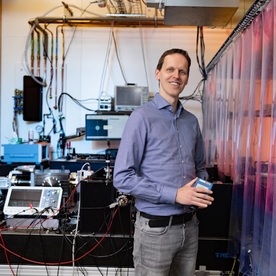
Dr Stefan Witte, Advanced Research Center for Nanolithography, The Netherlands
Dr Stefan Witte, Advanced Research Center for Nanolithography, The NetherlandsStefan Witte received his PhD in 2007 from the Vrije Universiteit Amsterdam, for research on intense ultrafast laser development and precision spectroscopy. He did postdoctoral work at on nonlinear microscopy and biomedical imaging (Vrije Universiteit) and on ultrafast electron dynamics and lensless imaging with high-harmonic sources (JILA, University of Colorado). Since 2014 he has worked at the Advanced Research Center for Nanolithography (ARCNL), where he leads the EUV Generation and Imaging group at ARCNL and is the head of the Metrology Department. He is also an associate professor at the Vrije Universiteit Amsterdam. He was awarded an ERC Starting Grant in 2014, and an ERC Consolidator Grant in 2019. His present research interests include coherent diffractive imaging with visible and EUV radiation, high-harmonic generation and its applications, photo-acoustic imaging, and laser development. |
| 14:45-15:00 |
Discussion
|
| 15:00-15:30 |
Break
|
| 15:30-16:00 |
What are the ultimate resolution limits for ptychography?
Electron microscopes use electrons with wavelengths of a few picometers, and are potentially capable of imaging individual atoms in solids at a resolution ultimately set by the intrinsic size of an atom. Even with the rapid advances in aberration-corrector technology, both residual aberrations in the electron lenses and multiple scattering of the incident beam inside the sample, the best resolution possible was an order of magnitude worse than this limit. However, with recent advances in detector technology and ptychographic algorithms to unscramble multiple scattering, the resolution of the electron microscope is now limited only by the dose to the sample, and thermal vibrations of the atoms themselves. At high doses, these approaches have allowed us to image the detailed vibrational envelopes of individual atom columns as well as locating interstitial dopant atoms that would be hidden by channelling of the probe with conventional imaging modes. Even the location of all atoms in thin amorphous films now seems within reach. However, as the dose is reduced, so is our ability to robustly reconstruct the object. Challenges in characterizing and predicting performance will be discussed. 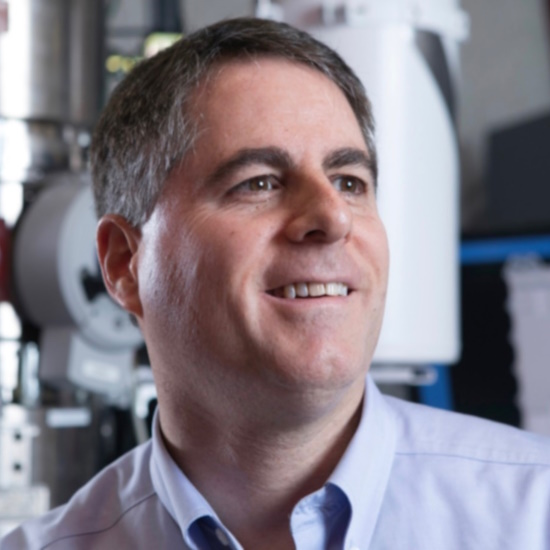
Professor David Muller, Cornell University, USA
Professor David Muller, Cornell University, USABiography not available |
| 16:00-16:15 |
Discussion
|
| 16:15-17:00 |
Panel discussion
|