Ribosome heterogeneity and specialisation
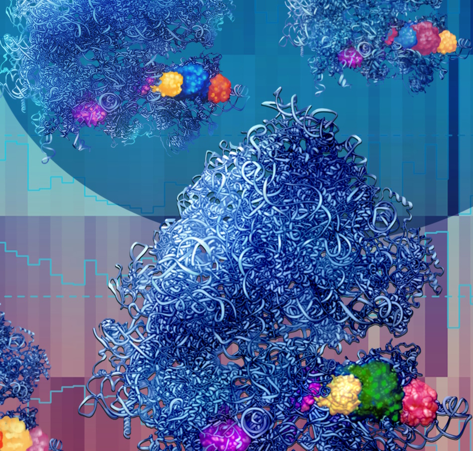
Scientific discussion meeting organised by Dr Julie Aspden, Dr Maria Barna, Dr William Faller, and Professor Anders Lund.
Ribosomes were thought to be homogeneous, passively translating mRNA into proteins. However, recent work has discovered that the composition of ribosomes is highly heterogeneous and that different ribosome populations can regulate the translation of specific mRNAs. This meeting discussed the latest advances, covering all types of ribosome heterogeneity in a variety of organisms and systems, including impact on human disease.
The schedule of talks, speaker biographies and abstracts are available below.
The meeting papers have been published in Philosophical Transactions of the Royal Society B.
Attending this event
This meeting has now taken place. A recording of the meeting will be available in due course.
Enquiries: contact the Scientific Programmes team.
Organisers
Schedule
Chair

Dr William Faller, Netherlands Cancer Institute, The Netherlands

Dr William Faller, Netherlands Cancer Institute, The Netherlands
William studied for his BSc at the National University of Ireland, Galway, and graduated in 2003. He followed this with a PhD under the supervision of Professor William Gallagher at the UCD Conway Institute in Dublin, where his project involved the study of DNA methylation in melanoma cells. He continued his focus on cancer in his Post Doctoral studies with Professor Owen Sansom at the CRUK Beatson Institute in Glasgow. During this time, he began to work on mTOR signalling, particularly the regulation of mRNA translation. In 2017 he became a Group Leader at the NKI, where his lab focuses on non-canonical forms and functions of the ribosome.
| 09:05-09:30 |
Biochemical and genetic characterisation of ribosome biogenesis and functional diversity
Baker’s yeast ribosomes are composed of four structural RNAs and 79 proteins that are selected from a panoply of ~150 ribosomal RNA (rRNA) genes and 137 ribosomal protein genes (RPGs). Most RPGs are duplicated (dRPGs) and the selective pressure maintaining these gene-duplications and their functional significance remain largely unexplored. It was initially believed that RPGs have remained duplicated over the aeons to satisfy the high demand for ribosome production by providing a sufficient dose of duplicated protein paralogs with identical functions. However, our studies of ribosome production revealed that most ribosomal protein paralogs are independently regulated and that they respond to different growth conditions. We proved this by separately deleting the duplicated paralogs and observing distinct phenotypic effects and functions that could not be complemented by their evolutionary partner genes. As a clue to the evolved functional divergence, we found that the ratio of expressed dRPGs depends on growth conditions, leading to ribosomes with altered make-ups and consequently to changes in the program of mRNAs that are selected for translation. Together these observations argue against an equal and redundant role for dRPGs and propose a new model where each paralog is uniquely regulated to serve a particular function during ribosome biogenesis and protein synthesis. 
Professor Sherif Abou Elela, University of Sherbrooke, Canada

Professor Sherif Abou Elela, University of Sherbrooke, CanadaSherif Abou Elela is a Professor of Microbiology at the University of Sherbrooke in Quebec, Canada and a member of the oncology group at the university's Hospital Clinical Research Centre. He is also director of the school's Functional Genomics’ Laboratory, the Scientific Rirector of the U of Sherbrooke RNomics Platform, director of the protein purification platform, and the coordinator of the RiboClub and the Canadian Consortium for RNA Research (C2R2). He holds Canada Chair in RNA Biology and Cancer Genomics. Born in Egypt, Abou Elela earned a bachelor's degree in Zoology from the University of Qatar in 1988. Relocating to Canada, he completed a PhD in Molecular Biology and Genetics at the University of Guelph in Ontario in 1994 where he studied ribosome function and created the first system to mutate and study rRNA repeats in vivo using plasmid system. During this time, he uncovered a new role of 5.8S rRNA modification in the regulation of translation. From 1994 to 1997, he was a postdoctoral fellow at the RNA Biology Center at the University of California Santa Cruz (UCSC) where he discovered Rnt1p the first eukaryotic member of the Dicer/Drosha family of ribonucleases in yeast and studied its role in rRNA processing. In 1997, Abou Elela joined the Sherbrooke faculty, and in 2013, he was elevated to the school's Canada Research Chair in RNA Biology and Cancer Genomics. His independent laboratory focuses on study different aspect of RNA and cancer biology including the mechanism and contribution of splicing and introns to cancer development and the effect of non-coding RNA and rRNA modification on translation and cancer cell aggressiveness. Abou Elela's research projects have received more than $20 million in grants from Genome Canada, Genome Quebec, CIHR, NSERC and CFI. He holds three patents and has published more than 100 articles, reviews, book chapters and other published work. Abou Elela's research is at the 'forefront of RNA biology and its diagnostic and therapeutic applications.' His research group 'aims to understand the basic mechanisms that control the synthesis and stability of RNA in eukaryotic cells in order to understand how genes are regulated.' Their current applied research focus is on identifying novel markers for cancer. |
|---|---|
| 09:30-09:55 |
Building a case for 'functional identity fraud' in eRpL22 paralogue-specific ribosomes in Drosophila germline development
Co-expression of eukaryotic-specific ribosomal protein paralogues, eRpL22 and eRpL22-like, within the male germline generates heterogeneous ribosomes are shown by paralogue-specific polysome immunoprecipitation and mRNA characterisation by RNAseq to translate distinct mRNAs. Dr Ware developed a model Drosophila S2 cell system that recapitulates the enrichment pattern for model mRNAs detected on eRpL22-like ribosomes in the testis to explore the mechanism of targeted translation. The mechanism for targeting specific mRNAs to- or excluding mRNAs from- paralogue-specific ribosomes is unknown. For targeted mRNAs, the structural characteristics governing association remain unclear. No clear consensus structural features are detected among enriched mRNAs shown by GO analysis to function in the same biological process. Additional computational analyses may uncover conserved features for further study. On the paralogue side, eRpL22 and eRpL22-like are structurally distinct, most notably at the N terminus that projects from the subunit surface. 60S subunit models position eRpL22/eRpL22-like on the solvent side at the subunit base, removed from the subunit interface. eRpL22/eRpL22-like N- and C- terminal extensions are theoretically available for interactions that could create an interface between the base and the preinitiation complex. Dr Ware hypothesises that the N terminal extension is required for specific mRNA enrichment on eRpL22-like ribosomes. She is using the S2 system transfected with constructs lacking the eRpL22-like N-terminal domain to quantify and compare model mRNA association with engineered eRpL22-like ribosomes to mRNAs associated with wild type eRpL22-like ribosomes. These experiments will provide insights into the potential role of the N terminus of eRpL22 paralogues in specifying specific translation. 
Professor Vassie Ware, Lehigh University, USA

Professor Vassie Ware, Lehigh University, USAVassie C Ware is a Professor of Molecular Biology in the Department of Biological Sciences at Lehigh University where her laboratory investigates functional diversification of ribosomal protein (Rp) paralogues of the eRpL22 family and the impact of ribosome heterogeneity in Drosophila melanogaster germline development. Her work in the ribosome field started as a postdoctoral associate in Susan Gerbi’s laboratory at Brown University where she determined the first primary and secondary structures for a multicellular organismal 28S rRNA (Xenopus laevis), defined eukaryotic-specific expansion segments, and mapped species-specific processing sites within Drosophila and Sciara 28S rRNA expansion segments. Her current work seeks to understand mechanisms governing translation specificities of paralogue-specific ribosomes in spermatogenesis to define specialised ribosomes with unique roles and/or to define new roles for eRpL22 paralogues in extra-ribosomal pathways. She received a BA degree in Human Biology from Brown University and MPhil and PhD degrees in Biology from Yale University. |
| 09:55-10:20 |
Diversity of ribosomes at the level of rRNA variation in human health and disease
Ribosomal DNA and RNA (rDNA and rRNA) sequences are usually discarded from sequencing analyses, but with hundreds of copies of rDNA genes it is unknown whether they possess sequence variations that form different types of ribosomes that affect human physiology and disease. Dr Barna and her team have developed a new accurate variant-calling algorithm (termed RGA) and compared rDNA variations found in short- and long-read sequencing data from the 1,000 Genomes Project (1KGP) and Genome In A Bottle (GIAB). They additionally developed a novel protocol for long-read sequencing full-length rRNA (RIBO-RT) from actively translating ribosomes. Our analyses identified hundreds of rDNA variants, most of which, surprisingly, are short insertion-deletions (indels) and dozens of highly abundant rRNA variants that are incorporated into translationally active ribosomes. To visualise variant ribosomes at the single cell level, they developed an in-situ rRNA sequencing method (SWITCH-seq) which revealed that variants are co-expressed within individual cells. Strikingly, by analysing rDNA, Dr Barna and her team found that variants assemble into distinct ribosome subtypes encoded on different chromosomes. They discovered that these subtypes acquire different rRNA structures by successfully employing dimethyl sulphate (DMS) probing of full length rRNA. With this atlas they investigated the impact of rRNA variation on health and disease. Across human tissues, Dr Barna and her team observed tissue-specific rRNA subtype expression in endoderm/ectoderm-derived tissues. In cancer, low abundant rRNA variants can become highly expressed, which suggests the presence of cancer-specific ribosomes. Together, this study identifies and comprehensively characterises the diversity of ribosomes at the level of rRNA variants, their chromosomal location and unique structure as well as functionally linking ribosome variation to tissue-specific biology and cancer. 
Dr Maria Barna, Stanford University, USA

Dr Maria Barna, Stanford University, USADr Barna obtained her BA in Anthropology from New York University and her PhD from Cornell University, Weill Graduate School of Medicine. Dr Barna was subsequently appointed as a UCSF Fellow through the Sandler Fellows program, which enables exceptionally promising young scientists to establish independent research programs immediately following graduate school. She is presently an Associate Professor in the Genetics Department at Stanford University. Dr Barna has received a number of distinctions including being named a Pew Scholar, Alfred P Sloan Research Fellow, and top ’40 under 40’ by the Cell Journal. She has received the Basil O’ Connor Scholar Research Award and the NIH Directors New Innovator Award. She is the recipient of the Elizabeth Hay Award, H W Mossman Award, Tsuneko and Reiji 'Okazaki Award', American Society for Cell Biology Emerging Leader Prize, the Rosalind Franklin Young Investigator Award, and the RNA Society Early Career Award. She is presently a NYSCF Robertson Stem Cell Investigator. |
| 10:20-10:30 |
Discussion
|
| 10:30-11:00 |
Break
|
| 11:00-11:25 |
Neuronal ribosome localisation and heterogeneity
Owing to their morphological complexity and dense network connections, neurons modify their proteomes locally, using mRNAs and ribosomes present in dendrites and axons. Professor Schuman will discuss the group's efforts to understand the neuronal ribosome population present in dendrites and axons, including work on the abundance and dynamics of ribosomes. Dr Erin Schuman, Max-Planck Institute for Brain Research, Germany
Dr Erin Schuman, Max-Planck Institute for Brain Research, Germany
"Erin Schuman was born in 1963 in California. After completing her BA in Psychology at the University of Southern California in 1985, Erin Schuman received her PhD. in Neuroscience from Princeton University in 1990. She conducted postdoctoral studies in the Department of Molecular and Cellular Physiology at Stanford University. She was appointed to the Biology Faculty at the California Institute of Technology (Caltech) in 1993 and stayed there until 2009. In 2009, she moved to Frankfurt, Germany to found the Department of Synaptic Plasticity in Max Planck Institute for Brain Research.
In 1997 Erin Schuman was appointed Investigator at the Howard Hughes Medical Institute (HHMI). She received several awards and grants, including the Pew Scholars Award, the Beckman Young Investigator Award, and an Alfred P. Sloan Fellowship. In 1995, she was named as the American Association of University Women’s Emerging Scholar. In 2013, she gave the Cruikshank Lecture at the GRC on Dendrites and received the Hodgkin Huxley Katz Prize Lecture by the Physiological Society (UK)." |
| 11:25-11:50 |
Exploring the function and structure of heterogeneous ribosomes in the gonads of Drosophila melanogaster
Dr Aspden has previously profiled the composition of ribosomes from different Drosophila tissues. She discovered the enrichment of specific RP paralogs within the ribosomes of Drosophila gonads, with 4 paralogs enriched in ovary and 6 in testis. One of these testis-specific paralogs, RpL22-like, is incorporated into ~50% of ribosomes in the testis and shares 45% aa sequence identity with the canonical paralog, RpL22. To assess the impact of RpL22/RpL22-like paralog switching RNAi was performed. RpL22 knockdown in the ovary affected germ cell maintenance, while knockdown in the testis had no observable effect. This ovary phenotype was not fully rescued by ectopic expression of RpL22-like. RpL22-like RNAi did not impact spermatogenesis and had no effect on the ovary. Immunostaining has revealed that RpL22 and RpL22-like localise to different cellular subtypes and different sub-cellular compartments within the testis. Together these experiments suggest that RpL22 and RpL22-like possess different functional roles during gametogenesis. To dissect the contribution of RpL22-like to mRNA translation RNAi followed by quantitative mass spectrometry was performed. The levels of 18 proteins change upon knockdown, suggesting that the incorporation of RpL22-like may alter the ribosome’s translational preference. Levels of DGP-1 protein, an ortholog of mammalian ribosome rescue factor, GTPBP1, were reduced ~50% upon RpL22-like RNAi, while mRNA levels were unaffected. This suggests that RpL22-like may translationally regulate DGP-1 mRNA. Work is underway to understand the mechanism by which RpL22 and RpL22-like containing ribosomes regulate the translation of specific mRNAs, and how specialised ribosomes contribute to gametogenesis. 
Dr Julie Aspden, University of Leeds, UK

Dr Julie Aspden, University of Leeds, UKJulie read Biochemistry at Oxford before undertaking a PhD in Biochemistry at the University of Cambridge on the initiation of mRNA translation. During her first postdoc at Berkeley, her work focused on alternative mRNA splicing in Drosophila. Her second postdoc was at the University of Sussex, where she became interested in identifying novel sites of translation. In 2015 Julie established her independent research group at Leeds. Her group addresses questions on the regulation of mRNA translation, combining biochemistry, genomics, molecular biology and genetics. Julie has 21 years of experience and expertise in RNA biology, and an active member of the RNA Society. She has become a UK leader in ribosome profiling. Her research has been funded by both the Medical Research Council and the Biotechnology and Biological Sciences Council. In 2022 as PI, Julie was awarded, a £5.6 million BBSRC sLoLa grant to 'unlock the secrets of specialised ribosomes across eukaryotes'. |
| 11:50-12:05 |
Molecular visualisation of ribosome populations in mammalian cells
Structural studies have addressed eukaryotic ribosomes at atomic resolution but the techniques available so far required their isolation from the native cellular context, which abolishes spatial relationships in the cell and is known to affect the composition of ribosomal intermediates. Recent method development in the field of cryo electron tomography now opens the door to the molecular visualisation of ribosome heterogeneity within their native cellular context. In their recent work, Dr Fedry and her team visualised the mRNA translation process in situ, in intact mammalian cells and analysed its reorganisation under persistent collision stress, indicating how perturbations in initiation, elongation and quality control processes contribute to an overall reduced protein synthesis (publication under review). Their results notably highlight the presence of a Z-site bound tRNA on 80S complexes, increased under stress and stabilised on collided disomes, as well as the accumulation of an off-pathway 80S complex resulting from collision splitting events. To further visualise the translocation of secretory protein nascent chains into the Endoplasmic Reticulum (ER) on native membranes, they used cryoET on vesicles derived from the rough ER of mammalian cell lines (Gemmer et al, 2023). Dr Fedry and her team characterised the distinct populations of ribosome translocon complexes assembled in the ER membrane and revealed their clustering according to polysomes translating different types of nascent chains. Future work elaborating on these studies will provide molecular visualisation of specialised ribosomes in different cell types and tissues. 
Dr Juliette Fedry, MRC Laboratory of Molecular Biology, UK

Dr Juliette Fedry, MRC Laboratory of Molecular Biology, UKJuliette Fedry used cryo electron tomography and subtomogam averaging to provide subnanometer visualisation of protein translocation at the mammalian Endoplasmic Reticulum membrane. This work allowed the identification of the different specialised ribosome translocon complexes, as well as their quantification and spatial analysis on native membranes. Dr Fedry further implemented Focused Ion Beam milling approaches to study mRNA translation and its reorganisation upon ribosome collision stress in intact mammalian cells. These studies revealed a higher diversity of ribosome complexes than previously anticipated from purified samples opening the way for mechanistic investigations. |
| 12:05-12:20 |
A subcellular map of translational machinery composition and regulation at the single-molecule level
Ribosomes are ubiquitous translation machineries that were detected as dense particles under the electron microscope over 60 years ago. Our perception of all ribosomes as the same kind of dense particles has not changed much since then, even though growing evidence strongly suggests that different ribosomal populations exist to play distinct roles in translational control. However, quantitative spatial distributions of these ribosomes characterised at the single-molecule level has not yet been described. Here, Zijian Zhang will present an expansion microscopy approach to visualise individual ribosomes and an optogenetic proximity-labelling technique to characterise their composition. Alongside his team, he generated a super-resolution ribosomal map that revealed distinct spatial 'hot spots'' containing free subunits and translating polysomes. Notably, the large subunits showed enrichment near polysomes, especially at the endoplasmic reticulum (ER). By characterising ribosome composition at the ER, they identified Lsg1 as being enriched on large subunits and crucial for the selective translation of transmembrane proteins. Additionally, they discovered ribosome heterogeneity on the mitochondria outer membrane, that is linked to the translation of metabolism-related transcripts. Finally, they visualised ribosomes in neuronal processes, which revealed the predominant role of monosomes in neuronal translation and further confirmed the existence of ribosome heterogeneity in distal neurites. Considering the great diversity of ribosomal populations that have been largely understudied, Zijian Zhang and his team have provided a powerful approach to investigate global ribosomal localisation and composition at unprecedented resolution. 
Mr Zijian Zhang, Stanford University, USA

Mr Zijian Zhang, Stanford University, USAZijian is a graduate student in the Department of Chemical and Systems Biology at Stanford University. During his undergraduate research at Peking University and Caltech, he discovered his passion for quantitative microscopy. He then joined Dr Maria Barna’s laboratory to develop novel imaging techniques to illuminate cellular communication and translational control. Currently, he is dedicated to developing expansion microscopy techniques to visualise the diverse ribosomal populations in mammalian cells at the single-molecule level. |
| 12:20-12:30 |
Discussion
|
Chair

Professor Anders Lund, University of Copenhagen, Denmark

Professor Anders Lund, University of Copenhagen, Denmark
Anders H Lund is Professor and Director at the Biotech Research and Innovation Centre at University of Copenhagen. The group is addressing the hypothesis that modifications of the ribosomal RNA alter ribosome function and impose selective translation. Hence, specifically modified ribosomes may be functionally specialised to carry out translational programs of importance for establishing cellular identity and maintain cell homeostasis. He is exploring this in cell culture model systems, mouse models and cancer organoids using a broad spectrum of genetic, computational and biochemical techniques.
| 13:30-14:30 |
Keynote: From origin of life to next generation RNA-based therapeutics
The site for peptide bond formation in the ribosomes, the PTC, is located within a highly conserved internal pocket made exclusively of rRNA. The high conservation implies its existence irrespective of environmental conditions and indicates that it may represent a prebiotic RNA machine, which could be the kernel around which life originated. Lab constructs imitating this pocket possess capabilities for peptide bond formations, thus indicating that a molecular prebiotic bonding entity still exists and functions within ribosomes of all living cells. In contrast, other ribosomal components undergo genetic variability that led to the creation of specific structural features. Among them, those related to ribosomal genetic diseases, or specific to antibiotics resistant pathogens, are being used as bases for the design of RNA based next generation therapeutics. Professor Ada Yonath, Weizmann Institute of Science, Israel
Professor Ada Yonath, Weizmann Institute of Science, IsraelAda Yonath studied chemistry at the Hebrew University, earned Ph.D. degree from Weizmann Institute of Science (WIS) and carried postdoctoral education at Carnegie-Melon university and MIT, USA. Currently she is a professor of structural biology at WIS, holds the Kimmel Professorial Chair, and directing the Kimmelman Center for Biomolecular Structure and Assembly. In 1986-2004 she also headed a Max-Planck-Research Unit in Hamburg, Germany. Among others, she is a member the US National Academy of Sciences; the Israel Academy of Sciences and Humanities; the American Academy for Art and Scjence, the European Molecular Biology Organization. She also holds honorary doctorates from Tel Aviv, Ben Gurion, Bar Ilan and Oxford Universities. Her awards include the 1st European Crystallography Prize; the Israel Prize; The Paul Karrer Gold Medal; the Louisa Gross Horwitz Prize; the Paul Ehrlich Ludwig Darmstaedter Medal; the Wolf Prize; the UNESCO Award for Women in Science; the Albert Einstein World Award of Science; the Erice Prize for Peace and the Nobel Prize for Chemistry. |
|---|---|
| 14:30-14:45 |
NAC-mediated ribosome localisation regulates metabolism in intestinal stem cells
Intestinal stem cells (ISCs) face the challenge of integrating metabolic demands with unique regenerative functions. Studies have shown an intricate interplay between metabolism and stem cell capacity, however it is still not understood how this process is regulated. Combining ribosome profiling and CRISPR screening in intestinal organoids, Sofia Ramalho's research shows that RNA translation is at the root of this interplay. Her team identify the nascent polypeptide-associated complex (NAC) as a key mediator of this process, and show that it regulates ISC metabolism by re-localising ribosomes to the mitochondria. Upon NAC inhibition, intestinal cells show decreased import of mitochondrial proteins, which are needed for oxidative phosphorylation, and consequently, maintaining a stem cell identity. Furthermore, they show that overexpression of NACα (one of the two subunits of NAC) is sufficient to increase mitochondrial respiration and promote ISC identity. Their results unveil the pivotal role of ribosome specialisation through localisation in regulating metabolism and ISCs function. 
Ms Sofia Ramalho, Netherlands Cancer Institute, The Netherlands

Ms Sofia Ramalho, Netherlands Cancer Institute, The NetherlandsSofia Ramalho is a final year PhD student at the lab of Ribosome Dynamics run by Dr William Faller at the Netherlands Cancer Institute (NKI). Sofia did her undergraduate studies in Biochemistry in Lisbon, Portugal. Then, she decided to specialise in oncobiology and got her MSc degree from the Faculty of Medicine of the University of Lisbon. She worked for two years at the Institute of Molecular Medicine (first as a Masters student then as a research technician) in the group of Professor Joao T Barata where she studied cell signalling pathways and intracellular localization of IL-7 receptor in leukaemia. Currently, Sofia focuses her work on ribosome specialisation and heterogeneity in colorectal cancer and she is interested in understanding how ribosome specialisation through intracellular localisation impacts intestinal stem cell biology and metabolism. She also works on how ribosome protein-mediated heterogeneity impacts the development of colorectal cancer. |
| 14:45-15:00 |
Structural and functional insights into ribosomes lacking a single RNA modification
Trypanosomatids, including Leishmania, are unicellular protozoan parasites that cycle between insect and mammalian hosts and are the causative agent of sleeping sickness, cutaneous and visceral diseases affecting millions worldwide. Here, Dr Rajan will describe the changes of pseudouridine (Ψ) modification on rRNA in the two life stages of these parasites and functional analysis of a single snoRNA guiding Ψ on helix 69 (H69) of the large ribosomal subunit. Loss of a single Ψ had species-specific changes in the ribosome that affected the translation of only a subset of proteins. To decipher the molecular mechanism of these phenotypes, Dr Rajan and his team determined the structure of ribosomes lacking the single Ψ and its parental strain at ~2.4 Å resolution in both T. brucei and L. major using cryo-EM techniques. Our findings demonstrate the significance of a single Ψ on H69 structure, and its interactions with helix 44 and specific tRNAs. Based on the high-resolution structures, they propose a novel mechanism explaining how ribosomes lacking a single RNA modification can filter specific tRNAs and thus affect the translation of only a subset of mRNA. 
Dr K Shanmugha Rajan, Weizmann Institute of Science, Israel

Dr K Shanmugha Rajan, Weizmann Institute of Science, IsraelDr K Shanmugha Rajan is an Indian post-doctoral fellow residing in Israel. He completed his master’s and doctoral studies with Professor Shulamit Michaeli, Bar-Ilan University, researching the role of non-coding RNAs and RNA modification in mammalian parasites such as Trypanosomes and Leishmania. Recently Rajan started his post-doctoral training with Professor Ada Yonath, Weizmann Institute of Science, studying ribosome structure using cryogenic electron microscopy. In Yonath’s lab, Rajan investigates the mechanistic insights of single rRNA modification towards the function of ribosomes that can be harnessed to combat infectious diseases. |
| 15:00-15:30 |
Break
|
| 15:30-15:55 |
Endogenously encoded ribosomal RNA Sequence variation can regulate gene expression and phenotype
Research in the Blanchard lab has historically focused on the development of single-molecule fluorescence imaging methods that enable quantification of transient interactions and conformations that underpin the function and regulation of diverse biological systems. The protein synthesis mechanism, a process defined by rapid transactions and directional movements, has served as an exemplar in this regard. Using single-molecule imaging methods, the Blanchard lab has gained fundamentally new insights into transient interactions that define the genetic code, the directional movements of the ribosome along messenger RNA templates and the mechanisms of small-molecule action on these processes. In this context, published and anecdotal observations over the past several decades have led the Blanchard lab to ask whether the translation mechanism may be influenced by endogenously encoded sequence variations that exist within the ribosomal RNA component of the assembled ribosome. Professor Blanchard's team’s findings have revealed that the functioning ribosome pool in most species is intrinsically heterogeneous at the level of its ribosomal RNA composition (Kurylo et al, 2018; Parks et al, 2018). They also show that the relative expression levels of variant ribosomal RNA alleles within the assembled ribosome can vary with physiological context and that specific variant alleles have the capacity to modulate transcriptional control of gene expression and phenotype (Kurylo et al, 2018). Their findings also suggest that opportunities for selective drug targeting of variant alleles may be possible. Conserved sequence variations within the assembled ribosome – confirmed both at the level of ribosomal DNA and ribosomal RNA sequencing – have also been evidenced in mouse and human, including tissue-specific expression of specific variant ribosomal RNA alleles in the brain, lungs, liver and ovaries of mouse littermates. These observations reveal the expression of distinct ribosome sub-types in these organs, leading us to speculate that endogenously encoded sequence variations within the hundreds of ribosomal DNA alleles present in the mammalian genome have the capacity – at least in principle – to provide a mechanism for templating and regulating heterogeneity into the expressed ribosome pool. As Professor Blanchard's team's findings further indicate that ribosome-biogenesis upregulation is a hallmark of metastatic disease (Prakash et al, 2019), they seek to develop and implement strategies to identify when and where cancer-specific ribosomes are expressed and to target them specifically for therapeutic purposes. 
Professor Scott Blanchard, St Jude Children's Research Hospital, USA

Professor Scott Blanchard, St Jude Children's Research Hospital, USAScott C Blanchard received his PhD in the Program in Biophysics from Stanford University School of Medicine in 2002. He completed his post-doctoral research in the Department of Applied Physics at Stanford University in the laboratory of Dr Steven Chu. From 2004-2019, Scott was a faculty member in the Department of Physiology and Biophysics at Weill Cornell Medicine (WCM) in New York City, where he served as Associate Director of The Tri-Institutional PhD Program in Chemical Biology within the Tri-Institutional Network that includes Memorial Sloan-Kettering Cancer Center and The Rockefeller University. In June of 2019, Scott was appointed Full Member and Endowed Chair in Molecular Imaging in the Department of Structural Biology at St Jude Children's Research Hospital, where he leads a Center for Single-Molecule Imaging seeking to develop and enable the methods and tools required to delineate the physical basis of molecular recognition and biological regulation. |
| 15:55-16:20 |
Transcript- and species-specific translation by ribosome expansion segments
Ribosomes have recently emerged to directly regulate gene expression. Roles for ribosomal RNA (rRNA) in gene regulation remain largely unexplored. Expansion Segments (ESs) are rRNA regions exposed on the outer ribosomal shell that consist of many highly variable, tentacle-like rRNA structures that extend from the conserved rRNA core in eukaryotes; with largely unknown roles in translation. Dr Leppek found a role for ESs in mRNA-specific binding and translation. She identified a single 18S rRNA ES, ES9S, as the interaction site for an Internal Ribosome Entry Site (IRES)-like RNA structure of the Homeobox a9 (Hoxa9) 5’ UTR that directs spatiotemporal, tissue-specific translation control of HOXA9 expression in the mouse embryo. As ESs are highly variable across evolution, Dr Leppek engineered chimeric, 'humanised' yeast ribosomes for ES9S in the yeast 18S rRNA, that endow reconstituted, species-specific rRNA ES binding of 5’ UTR motifs. She next developed VELCRO-IP (variable expansion segment-ligand chimeric ribosome-IP) RNA-seq to site-specifically map the interactome of mRNA regions in the embryo transcriptome that preferentially associate with hES9S. This work identified multiple hES9S-selective mRNAs that undergo cap-independent translation control. Dr Leppek also confirmed IRES-like activity of Hox IRES-like elements with circular RNA reporters. Further, the Hox stem-loop RNA top-ranked in her unbiased screen of mRNA features for optimised RNA therapeutics. This work unravels species-specific, selective mRNA-rRNA interactions on the ribosome for translation. Dr Leppek's own lab focuses on how rRNA-directed specialised translation is employed in the innate immune response. 
Dr Kathrin Leppek, University Clinic Bonn, Germany

Dr Kathrin Leppek, University Clinic Bonn, GermanyKathrin Leppek is an Assistant Professor at the University Clinic Bonn, Germany, since 2022. She did her PhD in Georg Stoecklin’s lab at DKFZ and Heidelberg University, Germany, where she discovered a class of functional mRNA structures that interact with Roquin proteins for mRNA decay in innate immune regulator mRNAs. In her postdoc with Maria Barna at Stanford University, USA, she discovered a new mode of gene regulation by which ribosomal RNA (rRNA) regions exposed on the outer ribosome shell bind to selective transcripts to control mRNA- and species-specific translation. Her lab works on how gene regulation is directly executed by the ribosome, particularly in the innate immune system. They combine innovative RNA biochemistry and RNA-based technology development with model systems ranging from yeast to macrophages. Ultimately, they aim to decipher how rRNA-directed specialised translation shapes gene expression to understand the role of the ribosome in innate immune responses. |
| 16:20-16:30 |
Discussion
|
| 16:30-17:00 |
Poster flash talks
|
| 17:00-18:00 |
Poster session
|
Chair

Dr Julie Aspden, University of Leeds, UK

Dr Julie Aspden, University of Leeds, UK
Julie read Biochemistry at Oxford before undertaking a PhD in Biochemistry at the University of Cambridge on the initiation of mRNA translation. During her first postdoc at Berkeley, her work focused on alternative mRNA splicing in Drosophila. Her second postdoc was at the University of Sussex, where she became interested in identifying novel sites of translation. In 2015 Julie established her independent research group at Leeds. Her group addresses questions on the regulation of mRNA translation, combining biochemistry, genomics, molecular biology and genetics. Julie has 21 years of experience and expertise in RNA biology, and an active member of the RNA Society. She has become a UK leader in ribosome profiling. Her research has been funded by both the Medical Research Council and the Biotechnology and Biological Sciences Council. In 2022 as PI, Julie was awarded, a £5.6 million BBSRC sLoLa grant to 'unlock the secrets of specialised ribosomes across eukaryotes'.
| 09:00-09:25 |
rRna modifications in health and disease
Abstract available soon. 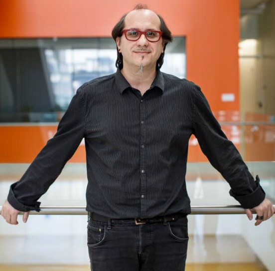
Dr Davide Ruggero, University of California, USA

Dr Davide Ruggero, University of California, USADr Davide Ruggero, PhD, is a Professor in the Department of Urology and Cellular & Molecular Pharmacology, at the University of California, San Francisco (UCSF). Dr Ruggero’s research is centred on understanding post-transcriptional control of gene expression in both normal health and disease in a cell- and tissue-specific manner, with a particular focus on cancer biology. He has made numerous breakthrough discoveries in the area of mammalian translational control as well as how deregulation in translation control leads to cancer. His research has been instrumental in the design of a new generation of cancer therapeutic agents that modulate the cellular proteome at a post-genomic level. He has garnered noteworthy distinctions including the prestigious V-Scholar Foundation's Award for Cancer Research for his work on deregulations in protein synthesis during lymphomagenesis. Most recently he was a recipient of the Outstanding Investigator Award (R35) from the National Institutes of Health. Dr Ruggero also recently received the prestigious American Cancer Society Professor Award. |
|---|---|
| 09:25-09:50 |
Specialised ribosomes induced by cancer associated ribosomal protein mutations
Somatic ribosomal protein defects such as missense mutations in RPL10 (uL16) and RPS15 (uS19) as well as somatic copy number losses of RPL5 (uL18), RPL11 (uL5) and RPL22 (eL22) have been described in a variety of hematologic and solid malignancies. We previously characterised the T-cell leukemia associated RPL10-R98S mutation using a genomewide translatome analysis (proteome, polysomal RNA-seq, Ribo-seq and total mRNA-seq). This revealed that RPL10-R98S generates a specialised ribosome that hypertranslates oncoproteins as JAK-STAT signaling components, anti-apoptotic protein BCL2 and serine/glycine synthesis enzyme PSPH. To verify whether other cancer-associated somatic ribosome defects rewire translation in a similar manner, we generated an onco-ribosome cell line library by CRISPR-Cas9 containing isogenic lymphoid cell clones (wild type, Rpl5+/-, Rpl11+/-, Rpl22+/-, Rpl22-/-, Rpl10-R98S, Rps15-P131S and Rps15-H137Y). Genomewide translatome analysis of this library reveals little translational changes in the RP knock-out cell lines. By contrast, the analysed RP point mutations are associated with profound translational rewiring, which is most pronounced for the Rps15 mutants. A significant impact of these Rps15 mutations on the ribosomal structure and translation dynamics is further supported by cryo-EM analyses. The genes presenting significant alteration of translation efficiency in Rpl10 and Rps15 point mutants show an enrichment for transcriptional regulators, revealing extensive cross-talk between translational and transcriptional regulation in RP point mutant cells. Furthermore, Dr De Keersmaecker's results support that loss of function mutations in Rpl5, Rpl11 and Rpl22 impose little translational rewiring, suggesting that these genotypes rather result in extra-ribosomal pro-oncogenic effects. 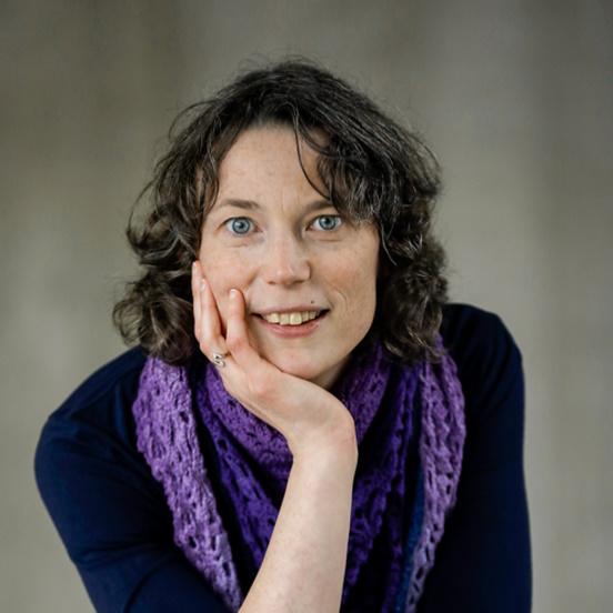
Dr Kim De Keersmaecker, KU Leuven, Belgium

Dr Kim De Keersmaecker, KU Leuven, BelgiumDuring her PhD at KU Leuven, Belgium (2004-2008), Kim De Keersmaecker worked on the role of oncogenic kinases in T-cell leukaemia. From 2008 until 2010, she performed postdoctoral research at Columbia University (New York, USA), studying the role of transcription factor TLX1 in T-cell leukaemia. During a second postdoc (KU Leuven, 2010-2013), she discovered somatic mutations in ribosomal proteins in cancer. In 2014, Kim received an ERC Starting Grant and established an independent research lab at KU Leuven studying the role of somatic ribosome defects in cancer. This successful project was followed by an ERC Consolidator Grant in 2019, which is aimed at exploring the role of synonymous mutations in cancer, with special attention for novel modes of translational dysregulation. The impact of her research is illustrated by publications in leading journals such as Nature Genetics, Nature Medicine, Nature Communications and Molecular Cell. |
| 09:50-10:15 |
Tuning the ribosome in health and disease?
Translational regulation impacts both pluripotency maintenance and cell differentiation. To what degree the ribosome exerts control over this process remains unanswered. Accumulating evidence has demonstrated heterogeneity in ribosome composition in various organisms. 2'-O-methylation (2'-O-me) of rRNA represents an important source of heterogeneity, where site-specific alteration of methylation levels can modulate translation. Here, Professor Lund examines changes in rRNA 2'-O-me during mouse brain development and tri-lineage differentiation of human embryonic stem cells (hESCs). He found distinct alterations between brain regions, as well as clear dynamics during cortex development and germ layer differentiation. Professor Lund identifies a methylation site impacting neuronal differentiation. Modulation of its methylation levels affects ribosome association of the fragile X mental retardation protein (FMRP) and is accompanied by an altered translation of WNT pathway-related mRNAs. Together, these data identify ribosome heterogeneity through rRNA 2'-O-me during early development and differentiation and suggest a direct role for ribosomes in regulating translation during cell fate acquisition. 
Professor Anders Lund, University of Copenhagen, Denmark

Professor Anders Lund, University of Copenhagen, DenmarkAnders H Lund is Professor and Director at the Biotech Research and Innovation Centre at University of Copenhagen. The group is addressing the hypothesis that modifications of the ribosomal RNA alter ribosome function and impose selective translation. Hence, specifically modified ribosomes may be functionally specialised to carry out translational programs of importance for establishing cellular identity and maintain cell homeostasis. He is exploring this in cell culture model systems, mouse models and cancer organoids using a broad spectrum of genetic, computational and biochemical techniques. |
| 10:15-10:30 |
Increased fidelity of protein synthesis extends lifespan
Loss of proteostasis is a fundamental process driving aging. Proteostasis is affected by the accuracy of translation, yet the physiological consequence of having fewer protein synthesis errors during multi-cellular organismal aging is poorly understood. Dr Bjedov and he team's phylogenetic analysis of RPS23, a key protein in the ribosomal decoding center, uncovered a lysine residue almost universally conserved across all domains of life, which is replaced by an arginine in a small number of hyperthermophilic archaea. When introduced into eukaryotic RPS23 homologs, this mutation leads to accurate translation, as well as heat shock resistance and longer life, in yeast, worms, and flies. Furthermore, they show that anti-aging drugs such as rapamycin, Torin1, and trametinib reduce translation errors, and that rapamycin extends further organismal longevity in RPS23 hyperaccuracy mutants. This implies a unified mode of action for diverse pharmacological anti-aging therapies. These findings pave the way for identifying novel translation accuracy interventions to improve aging. 
Dr Ivana Bjedov, UCL Cancer Institute, UK

Dr Ivana Bjedov, UCL Cancer Institute, UKIvana earned a BSc degree in Molecular Biology from University of Zagreb. She did her PhD at the University Pierre and Marie Curie in Paris, in the laboratory of Professor Miroslav Radman, where she studied DNA repair in E. coli. Her interest in ageing developed at University College London, where she was a postdoctoral EMBO fellow in the laboratory of Professor Linda Partridge, and where she manipulated the mTOR pathway using rapamycin to improve healthspan and lifespan in Drosophila. She joined UCL Cancer Institute upon obtaining ERC Starting Grant and her laboratory is interested in DNA repair and protein synthesis. |
| 10:30-11:00 |
Break
|
| 11:00-11:25 |
The nonessential ribosomal protein eS25 is required for the cell cycle
Dr Thompson's group has previously shown that eS25 is required for efficient translation by non-canonical mechanisms of initiation. In order to understand the role of eS25 in cellular homeostasis they performed a proteomics analysis in primary human renal proximal epithelial cells. These studies revealed that cell cycle proteins were decreased upon eS25 knockdown. Indeed, a cell cycle analysis showed that eS25 knockdown decreased the percent of cycling cells. Specifically, there was a decrease in S phase cells and an increase in the G2/M population. The S phase decrease could be overcome by BK polyomavirus infection, which is known to push cells into the cell cycle, suggesting that exit from the cell cycle can be overcome. Furthermore, knockdown of p16, a repressor of cell cycle entry also rescued the delay in cell cycle entry as well as viral titres in BKPyV infected primary RPTE cells. Finally, although the co-knockdown of p16 in eS25 knockdown cells rescued S phase and viral titres, a pulse chase analysis revealed that this was the result of an increase in the number of cycling cells from the non-cycling population instead of reversing the eS25 cell cycle block. Therefore, these data suggest that eS25 knockdown results in a robust cell cycle arrest independent of senescence-associated inhibitors, such as p16. The cell cycle arrest resulted in an accumulation of cells in G2/M in the eS25 knockdown cells. These studies suggest that eS25 impacts the cell cycle state. 
Dr Sunnie Thompson, University of Alabama, USA

Dr Sunnie Thompson, University of Alabama, USADr Sunnie Thompson received her PhD in Marvin Wickens’ laboratory studying translational control during early development. As a post-doctoral fellow in Peter Sarnow's laboratory she developed expertise in internal ribosome entry site (IRES) mechanisms of initiation. Her research focuses on understanding how IRESs recruit ribosomes and initiate translation. She discovered that a small ribosomal protein, eS25 (RPS25), was essential for IRES-mediated translation, but had no effect on cap-dependent translation initiation. She showed that a cellular protein was essential for diverse mechanisms of non-canonical initiation by demonstrating that eS25 is also required for ribosomal shunting. Dr Thompson identified novel host factors for poliovirus and dengue virus using their thio-uridine cross-linking-mass spectrometry method (TUX-MS). Her laboratory developed new tools to perform single-cell virology and cell cycle studies using immunofluorescence and scripts for image analysis. |
| 11:25-11:50 |
The ribosomal P-stalk regulates HLA Class I antigen presentation
HLA Class I antigen processing and presentation (APP) is a highly regulated process that enables CD8+ T cell immunosurveillance. APP begins with the ribosomal synthesis of a source antigen, yet the role of ribosomes, particularly specialised ribosomes, in antigen presentation is poorly understood. Here, Dr Faller will show that the presence of the 'P-stalk' on the ribosome enhances antigen presentation. The addition of the P-stalk to the ribosome is stimulated by cytokines that upregulate APP components, and knockdown of one of the P-stalk proteins (P1) reduces T cell recognition of tumour cells. Mechanistically, he shows that P1-containing ribosomes exhibit enhanced translation of HLA Class I molecules and accessory APP components. Finally, analysis of patient data reveals that the mRNA expression of the P-stalk proteins positively correlates with CD8+ T cell infiltration, a trend not seen for other ribosomal proteins. In all, Dr Faller demonstrates that the presence of the P-stalk defines a specialised ribosome population that enhances antigen presentation, something that may be exploited by cancer cells to escape immunosurveillance. 
Dr William Faller, Netherlands Cancer Institute, The Netherlands

Dr William Faller, Netherlands Cancer Institute, The NetherlandsWilliam studied for his BSc at the National University of Ireland, Galway, and graduated in 2003. He followed this with a PhD under the supervision of Professor William Gallagher at the UCD Conway Institute in Dublin, where his project involved the study of DNA methylation in melanoma cells. He continued his focus on cancer in his Post Doctoral studies with Professor Owen Sansom at the CRUK Beatson Institute in Glasgow. During this time, he began to work on mTOR signalling, particularly the regulation of mRNA translation. In 2017 he became a Group Leader at the NKI, where his lab focuses on non-canonical forms and functions of the ribosome. |
| 11:50-12:15 |
Decoding the ribosomal epitranscriptome and its dynamics at single molecule resolution
The dynamic deposition of chemical modifications into RNA is a crucial regulator of temporal and spatial accurate gene expression programs. A major difficulty in studying these modifications, however, is the need of tailored protocols to map each RNA modification individually. In this context, direct RNA nanopore sequencing (DRS) has emerged as a promising technology that can overcome these limitations, as it is in principle capable of mapping all RNA modifications simultaneously, in a quantitative manner, and in full-length native RNA reads. Here, Dr Novoa will present the latest work on how we can use DRS to identify RNA modifications with single nucleotide and single molecule resolution, to then study the biological functions and dynamics of the epitranscriptome, their interplay with other regulatory layers, as well as to decipher how and why epitranscriptomic dysregulation is often associated to human disease. 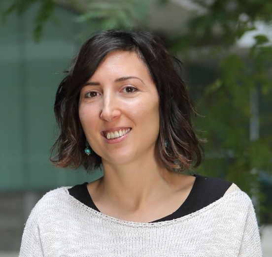
Dr Eva Novoa, Centre for Genomic Research, Spain

Dr Eva Novoa, Centre for Genomic Research, SpainDr Eva Maria Novoa leads the ‘Epitranscriptomics and RNA Dynamics laboratory’ at the Centre for Genomic Regulation (CRG) in Barcelona, Spain since 2018. Eva Maria’s laboratory is focused on deciphering the language of RNA modifications, and how its orchestration can regulate our cells in a space-, time- and signal-dependent manner. Her laboratory has pioneered the use of nanopore sequencing to study RNA modifications and post-transcriptional regulation. Specifically, her laboratory has developed novel algorithms to map and quantify RNA modifications, optimised the library preparations to make the technology applicable to low-input patient-derived samples, and expanded its applicability beyond coding mRNAs. She has published 40 peer-reviewed publications and has filed 2 patents. Eva Maria’s work has been awarded with grants from ERC (2022), AECC (2021), MERCK (2021), MICINN (2022; 2018) and ARC (2018). |
| 12:15-12:30 |
Discussion
|
Chair

Dr Maria Barna, Stanford University, USA

Dr Maria Barna, Stanford University, USA
Dr Barna obtained her BA in Anthropology from New York University and her PhD from Cornell University, Weill Graduate School of Medicine. Dr Barna was subsequently appointed as a UCSF Fellow through the Sandler Fellows program, which enables exceptionally promising young scientists to establish independent research programs immediately following graduate school. She is presently an Associate Professor in the Genetics Department at Stanford University. Dr Barna has received a number of distinctions including being named a Pew Scholar, Alfred P Sloan Research Fellow, and top ’40 under 40’ by the Cell Journal. She has received the Basil O’ Connor Scholar Research Award and the NIH Directors New Innovator Award. She is the recipient of the Elizabeth Hay Award, H W Mossman Award, Tsuneko and Reiji 'Okazaki Award', American Society for Cell Biology Emerging Leader Prize, the Rosalind Franklin Young Investigator Award, and the RNA Society Early Career Award. She is presently a NYSCF Robertson Stem Cell Investigator.
| 13:30-13:55 |
Making Rps26-deficient ribosomes
Rps26-deficient ribosomes, a physiologically relevant ribosome population which arises during high Na+ stress to specifically support the translation of mRNAs involved in the response to high salt stress, are generated via dissociation of Rps26 from fully assembled ribosomes. The chaperone Tsr2 binds ribosomes to directly release Rps26 when intracellular Na+ concentrations rise. Tsr2-mediated Rps26 release is reversible, enabling a rapid response that also conserves ribosomes. However, because the concentration of Tsr2 relative to ribosomes is low, how the released-Rps26 Tsr2 complex is managed, and how Rps26-deficient ribosomes accumulate over time to nearly 50% of all ribosomes, remains unclear. Here, Professor Karbstein shows the existence of a feed-forward loop that enables the accumulation of Rps26-deficient ribosomes in response to prolonged exposure to high salt stress. Her data shows that under high salt stress released Rps26 is degraded via the Pro/N-degron dependent pathway and she has identified the E3-ligases Gid4 and Gid10 as mediators of Rps26 degradation. Moreover, Gid10, which is induced transcriptionally in response to high salt stress also is enriched on Rps26-deficient ribosomes. Finally, substitution of the proline in position 2 of Rps26 to serine increases the stability of free Rps26 and limits the accumulation of Rps26-deficient ribosomes under high salt. Yeast with this mutation, or yeast lacking Gid4 and Gid10 are more sensitivity to high salt stress. Together Professor Karbstein's results demonstrate that degradation of Rps26 released from ribosomes via the Pro/N-degron pathway is important for the generation of Rps26-deficient ribosomes under prolonged salt stress. 
Professor Katrin Karbstein, Scripps Institute, University of Florida, USA

Professor Katrin Karbstein, Scripps Institute, University of Florida, USABorn and raised in Germany, Professor Karbstein received a Vordiplom from the Ruhr-Universität Bochum, and a Diplom from the Universität Witten/Herdecke. For her PhD, she joined the laboratory of Dan Herschlag at Stanford University, where she studied catalysis and conformational changes in RNA enzymes. During her postdoc in Jennifer Doudna’s lab in Berkeley, she developed a biochemical system to study ribosome assembly. In 2006, she joined the faculty at the University of Michigan in Ann Arbor and moved from there to Scripps Florida in 2010. She is now a Full Professor at The Scripps Research Institute and at The Herbert Wertheim UF Scripps Institute for Biomedical Innovation & Technology in Jupiter, Florida, where she is also the DEI liaison. In addition to her science, Dr Karbstein is passionate about science education and promoting access to science opportunities for everyone. Dr Karbstein is a single mother of two teenage daughters, one in college, and one finishing up high school. |
|---|---|
| 13:55-14:20 |
Towards analysis of remodelled ribosomes in proliferating plant tissue during cold stress
Plant acclimation to low temperature occurs through system-wide mechanisms that include proteome changes and altered transcript-to-protein translation. Rather than monolithic executing machines that operate on altered mRNA abundances, ribosomes can contribute to selective translation. Root apices from germinating seedlings of the monocot plant barley were chosen as a model to study changes in protein abundance and synthesis rates during cold acclimation. Knowledge of metabolic and physiological parameters allowed to compare protein synthesis rates in different physiological states, specifically the cold acclimated compared to the optimal temperature state. Professor Kopka shows that during acclimation, specific ribosomal proteins are synthesised and assembled into ribosomal complexes from root proliferative tissue and associate ribo-proteome shifts with changes in other translation-related and non-translation-related macromolecular protein complexes. He works towards understanding how a reconfigured ribosome population may confer selectivity to translation under altered temperature conditions. 
Professor Joachim Kopka, Max Planck Institute, Germany

Professor Joachim Kopka, Max Planck Institute, GermanyJoachim Kopka (JK) is Research Group Leader at the Max-Planck-Institute of Molecular Plant Physiology (MPIMP). In 1998 JK co-initiated GC-MS based profiling technology for applications in plant metabolomics at the MPIMP and the Metanomics GmbH company. Since 2001 the Applied Metabolome Analysis group of JK at the MPIMP has provided metabolomics infrastructure to the MPIMP and external partner groups with initial focus on the Golm-Metabolome-Database. JK develops metabolome analysis tools and stable isotope labelling applications with a specific interest in stress physiology and biotechnology of plants and photosynthetic microorganisms. Cytosolic plant ribosome biogenesis is a recently added research interest that was sparked by the discovery of cold sensitive Arabidopsis reil mutants that are deficient for the plant homologs of the yeast REI1 ribosome biogenesis factor. Investigation of reil mutants and plant ribosomes links to JK´s longstanding experience in systems analyses of plant stress responses including plant cold tolerance. |
| 14:20-14:45 |
Regulation of ribosome biogenesis and translation by RNA modifications
Ribosome biogenesis in eukaryotes is a complex and highly regulated process involving the action of over 200 assembly factors bringing together a total of 79 proteins and 4 ribosomal RNAs in yeast. The maturation of rRNAs from precursor transcripts is a critical aspect of ribosome biogenesis involving the precise processing steps facilitated by essential nucleases, among which the RNA exosome complex plays a pivotal role. Additionally, over hundred RNA chemical modifications, the majority of which are guided by small nucleolar RNAs, are deposited on the ribosomal RNAs co- and post-transcriptionally. Despite tight regulatory steps, rRNA processing and modifications go awry in several human diseases. Missense mutations in genes encoding structural subunits of the RNA exosome cause a growing family of diseases with diverse pathologies, collectively termed RNA exosomopathies. Similarly, pathogenic mutations that impact rRNA modifications are associated with neurodevelopmental disorders and implicated in the onset and progression of cancer. Data presented here will highlight recent discoveries made by my research team and our collaborators that shed light on the molecular consequences of dysregulated rRNA processing and rRNA modifications. These molecular defects have a profound impact on both the quantity and quality of ribosomes within the translating pool, thereby disrupting the delicate balance of cellular protein homeostasis. The implications of Dr Ghalei research underscore the intricate interplay between ribosome biogenesis, RNA processing, modifications, and disease pathogenesis, providing crucial insights and a deeper understanding of these complex cellular processes. 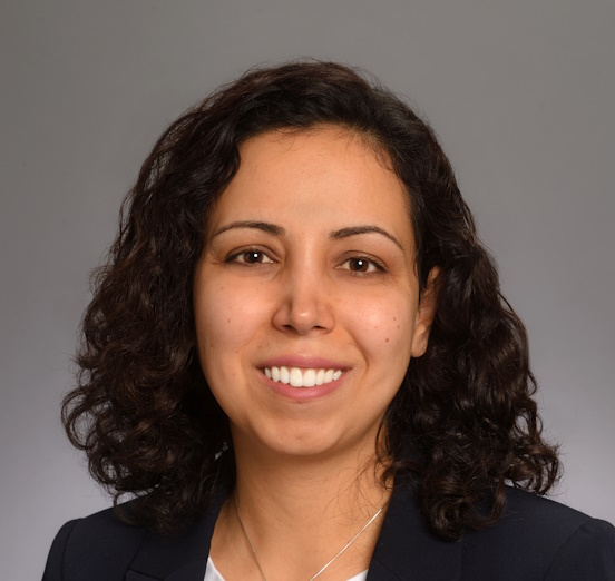
Dr Homa Ghalei, Emory University School of Medicine, USA

Dr Homa Ghalei, Emory University School of Medicine, USADr Homa Ghalei received her undergraduate degree in Cell and Molecular Biology from Tehran University in Iran, graduating as the Valedictorian. As an International Max Planck Research Fellow, she conducted her PhD thesis work in the Department of Cellular Biochemistry at the Max Planck Institute for Biophysical Chemistry in Göttingen, Germany, in laboratories of Professors Reinhard Lührmann and Markus Wahl. Her doctoral research, focusing on investigation of structurally similar RNA-protein complexes, was distinguished with Suma Cum Laude Honours. For her postdoctoral research, Dr Ghalei joined the laboratory of Professor Katrin Karbstein at the Scripps Research Institute in Florida, where she studied ribosome biogenesis and eukaryotic translation regulation. Presently, Dr Ghalei serves as an Associate Professor in the Department of Biochemistry at Emory School of Medicine. Her laboratory investigates the regulation of gene expression, with a specific focus on the impact of non-coding RNAs and RNA modifications. |
| 14:45-15:00 |
Discussion
|
| 15:00-15:30 |
Break
|
| 15:30-15:45 |
Closing in on ribosomal RNA modifications
The ability of ribosomes to translate the genetic code into protein requires a finely tuned chemical ecosystem with ions, water molecules, post-transcriptional and post-translational modifications all playing a role. Ribosomal RNA modifications, strategically positioned at essential functional sites on the ribosome, such as the decoding centre, mRNA channel and tRNA binding sites, are linked to a range of human diseases including Bowen Conradi syndrome, cancer, and obesity. However, the lack of high-resolution structures has precluded accurate positioning of all the functional elements of the ribosome and limited our understanding of the specific role of ribosomal RNA chemical modifications in modulating ribosome function in health and disease. Using a new sample preparation methodology based on functionalised graphene-coated grids, Dr Faille and his team solved the cryo-EM structure of the human large ribosomal subunit to a global resolution of 1.58 Å. The accurate assignment of water molecules, magnesium and potassium ions, and chemical modifications makes for an exhaustive structural model. In the core of the large subunit, where most rRNA modifications are, hydrogen atoms can be directly visualised in the cryo-EM map. Analysis of hydrogen bonds involving modified RNA residues highlight the fundamental biological role of ribosomal RNA methylation in harnessing unconventional carbon-oxygen interactions to maintain structural stability and fine-tune the functional interplay with tRNA. 
Dr Alexandre Faille, Cambridge Institute for Medical Research, UK

Dr Alexandre Faille, Cambridge Institute for Medical Research, UKAlex has completed his PhD at the Université de Toulouse Paul Sabatier in Lionel Mourey's group. His work focused on establishing the tridimensional structure of polyketide synthases from Mycobacterium tuberculosis using X-ray crystallography. He has then moved to the University of Cambridge in Alan Warren's group in 2015 where he began working on ribosomes. He started to learn cryo-electron microscopy and used it to solve high-resolution structures of human ribosomes. He is currently studying the role of ribosomal RNA modifications in the structure and function of ribosomes in the context of cancer and ribosomopathies. |
| 15:45-16:00 |
Ribosomal DNA heterogeneity is essential for female differentiation in zebrafish
Recent publications have highlighted a ribosomal RNA-encoding locus in zebrafish (Danio rerio) that undergoes massive extrachromosomal amplification during oocyte growth and ovary differentiation (termed 45S-M, or ’fem-rDNA’ (Ortega-Recalde et al, 2019)) and is distinct from the regular rDNA locus encoding rRNA found in somatic tissues (45S-S, (Locati et al, 2017)). Although the 45S-M locus falls within the most prominent sex-linked region in zebrafish (Wilson et al, 2014), its role in female sexual differentiation is unclear. Dr Hore and his team used CRISPR-Cas9 gene editing to alter 45S-M sequence in zygotes, and found there was no effect on subsequent embryo growth or development. At 29 days post fertilisation, all juvenile individuals displaying modification of fem-rDNA lacked fem-rDNA amplification. Morphological analysis after 100 days revealed treated individuals to be almost exclusively male and phenotypically indistinguishable from untreated males belonging to the same clutch. These fem-rDNA edited males produced phenotypically normal sperm and were able to fertilise eggs from wildtype females, with resulting embryos once more displaying normal development. Together, this shows 45S-M is essential for female differentiation, but is of limited importance to male development and fertility. Dr Hore concludes that use of specialised rDNA loci is a fundamental driver of ribosome function in many vertebrate species.
Dr Tim Hore, University of Otago, New Zealand

Dr Tim Hore, University of Otago, New ZealandDr Tim Hore is an Associate Professor and research group leader at the University of Otago in New Zealand. His research team is interested in long-held developmental puzzles – such as how do cells erase identity (Hore et al, 2016; Ravichandran et al, 2022); how does sex impact on DNA and longevity (Sugrue et al, 2021); and how does the germline change in preparation for the next generation (Ortega et al, 2019; Bond et al, 2023). Dr Hore’s interests in ribosome specialisation are stimulated by the observation of rDNA amplification in the female germline of zebrafish and other non-mammalian vertebrates. |
| 16:00-16:15 |
Discussion
|
| 16:15-17:00 |
Panel discussion
|
