Cell lineages across scales, space and time

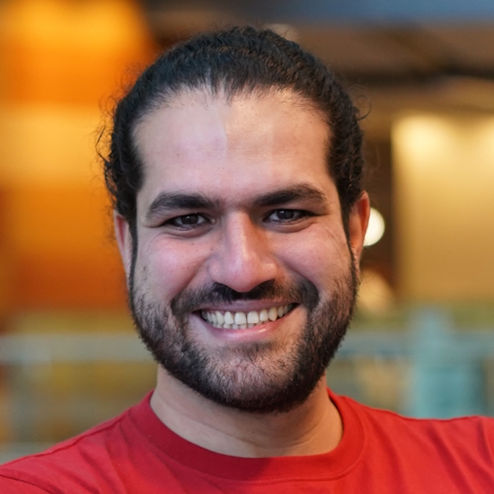
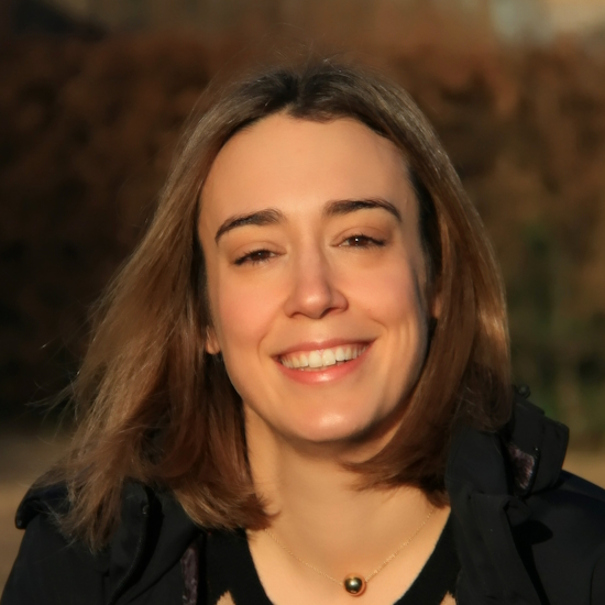
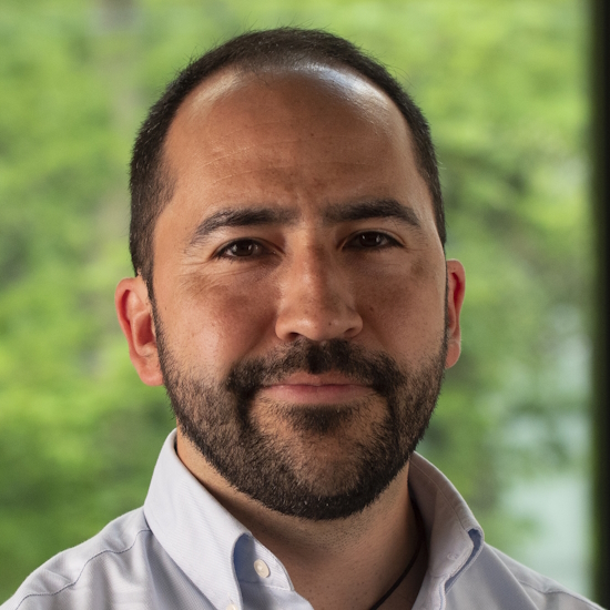
Theo Murphy meeting organised by Dr Giulia LM Boezio, Dr Afnan Azizi, Dr Claudio Cortés and Dr John Russell.
Lineage trajectories and cellular decisions coordinate embryonic development and growth. Similarly, cellular origin and clonal trajectories are crucial elements to understanding and modulating the response to injury, disease, and therapies. This meeting will showcase cutting-edge research spanning developmental biology, organ regeneration, and cancer, in light of recently developed techniques to study lineage trajectories and cellular decisions.
The schedule of talks and speaker biographies will be available soon. Speaker abstracts will be available closer to the meeting date.
Poster session and short talks
There will be a poster session on Tuesday 7 May. If you would like to apply to present a poster, please submit your proposed title, abstract (not exceeding 200 words and in the third person), author list, name of the proposed presenter, and institution to the Scientific Programmes team no later than Monday 15 April.
We also welcome applications for short talks with the same abstract. Please indicate with your abstract submission if you wish to present your findings in a short talk.
Include the text 'Abstract submission - Cell lineage' in the email subject line. Please note that spaces are limited, and the selection of posters and presentations will be at the discretion of the scientific organisers.
Attending this event
This event is intended for researchers in relevant fields, and is a residential meeting taking place at the Edgbaston Park Hotel and Conference Centre, 53 Edgbaston Park Road, Birmingham, B15 2RS.
- Free to attend
- Advance registration essential (please request an invitation)
- This is an in person meeting
- Catering options are available to purchase during registration. Participants are responsible for their own accommodation booking.
Enquiries: contact the Scientific Programmes team
Organisers
Schedule
Chair

Dr Afnan Azizi, King’s College London and The Francis Crick Institute, UK

Dr Afnan Azizi, King’s College London and The Francis Crick Institute, UK
Afnan obtained a BSc in Biochemistry with a minor in Mathematics and an MSc in Cellular and Molecular Medicine from University of Ottawa, Canada. He then moved to the University of Cambridge to pursue his PhD in the laboratory of Professor Bill Harris, looking at the role of interkinetic nuclear migration in morphogenesis of the vertebrate retina. He is now a postdoctoral fellow at King’s College London and The Francis Crick Institute working between the Houart and Guillemot laboratories to tackle the problem of how the human brain is formed at early embryonic stages and how it is different from that of other vertebrates, using 3D microscopy of intact tissue, single cell transcriptomics, and brain organoids.
| 09:00-09:05 |
Introduction

Dr Giulia LM Boezio, The Francis Crick Institute, UK

Dr Giulia LM Boezio, The Francis Crick Institute, UKGiulia Boezio is a postdoctoral researcher in the Developmental Dynamics lab at the Francis Crick Institute, London. She obtained a MSc in Molecular Biology from the University of Milan (Italy) with a thesis focusing on Sox transcription factor control of zebrafish angiogenesis. For her PhD, she joined the lab of Didier Stainier at the Max Planck Institute for Heart and Lung Research, in Germany, to study intercellular and intertissue crosstalk in the context of heart development in zebrafish. For her postdoctoral research, Giulia joined James Briscoe’s lab to continue pursuing her interest in developmental biology and organ formation. In particular, she now focuses on understanding the lineage trajectories and the principles of cell fate commitment in the spinal cord, using a combination of imaging and genomics lineage tracing tools in chicken embryos and human embryonic tissues. |
|---|---|
| 09:10-09:40 |
Principles of Neural Stem Cell Lineage Progression
The concerted production of the correct number and diversity of neurons and glia by neural stem cells is essential for intricate neural circuit assembly. In the developing cerebral cortex, radial glia progenitors (RGPs) are responsible for producing all neocortical neurons and certain glia lineages. Clonal analysis by exploiting the single cell resolution of the genetic MADM (Mosaic Analysis with Double Markers) technology revealed an inaugural quantitative framework of RGP behaviour in the developing neocortex. However, the cellular and molecular mechanisms controlling RGP lineage progression through proliferation, neurogenesis and gliogenesis remain largely unknown. To this end we use quantitative MADM-based experimental paradigms at single RGP resolution to define the cell-autonomous functions of candidate genes and signalling pathways controlling RGP-mediated neuron and glia genesis. Ultimately, our results shall translate into a deeper understanding of brain function and why human brain development is so sensitive to the disruption of particular signalling pathways in pathological neurodevelopmental and psychiatric disorders. 
Professor Simon Hippenmeyer, Institute of Science and Technology, Austria

Professor Simon Hippenmeyer, Institute of Science and Technology, AustriaSimon Hippenmeyer studied Molecular Biology and obtained a PhD in Neurobiology at Biozentrum, University of Basel, Switzerland. He was a postdoctoral fellow at Stanford University, USA. In 2012 he joined IST Austria (ISTA) as Assistant Professor and became Professor in 2019. Since 2020 he is also the Life Sciences Research Area Chair. His research focuses on fundamental neurobiological questions related to neural stem cell biology, neural development and circuit assembly. |
| 09:40-10:10 |
Cell lineages in development and evolution
During embryogenesis, numerous cell lineages arise from a common progenitor and, along the ontogeny, diversify, interact with each other, integrate, and jointly build high-dimensional phenotypes. Our group employs the development of the face as a model to study cell lineages, their dynamics and underlying molecular drivers. We focus on the neural crest cells (NCCs) that give rise to an array of cell types and are essential contributors to facial morphogenesis. How the neural crest cells maintain the multipotency and commit to different fates was recently reassessed using new technologies. We employed single-cell transcriptomics to explore how multipotent NCCs make decisions and how the progression of NCC-derived lineages is coordinated along facial development. With this knowledge, we aim to reconstruct and compare the developmental history of the face in different species and identify evolutionary mechanisms altering development (such as heterochrony and heterotopy) to understand how morphological variation arises. I will also briefly mention how detailed knowledge of cell lineages enables better insight into the evolution of cell types. Here, we employ single-cell transcriptomes of skeletogenic lineage (cells forming cartilage and bone) with genomic phylostratigraphy (gene birth-dating) to reveal the assembly of cell type-specific gene expression programs. We provide evidence for the evolution of ancestral skeletogenic cell type at the onset of Bilateria and demonstrate the subsequent transcriptome elaboration and individuation. In particular, we show that taxon-restricted genes enabled the cells to control ancient functions and gave rise to the osteoblasts and hypertrophic chondrocytes. Interestingly, our analyses suggest that chondrocyte hypertrophy (thus endochondral ossification) evolved earlier than previously believed, which is supported by the recently discovered fossil evidence. 
Dr Markéta Kaucká, Max Planck Institute for Evolutionary Biology, Germany

Dr Markéta Kaucká, Max Planck Institute for Evolutionary Biology, GermanyWith a PhD in Animal Physiology and a master's degree in Molecular Biology and Genetics, Markéta moved to Sweden to conduct developmental biology research at Karolinska Institute. She revealed fundamental processes behind early head development and skull growth. Markéta did a second postdoc at the Centre for Brain Research at the Medical University Vienna to investigate the tight developmental link between the brain and skull formation. In 2019, Markéta was appointed an independent Group Leader at the Max Planck Institute for Evolutionary Biology in Germany. She leads the group Evolutionary Developmental Dynamics, and her research is at the intersection of Evo-Devo and molecular and cellular biology. Markéta employs an interdisciplinary skillset including single-cell omics, high-resolution and 3D imaging, bioinformatics and in vivo models to investigate the complex process of face formation and shaping, with a particular focus on how evolution altered these processes along vertebrate phylogeny. |
| 10:10-10:40 |
Regulation of temporal patterning in the developing retina
The generation of cell diversity and neural circuit assembly in the central nervous system requires precise spatiotemporal coordination of mechanisms regulating cell proliferation, specification, and differentiation. While much progress has been made in the last decades to elucidate how neural progenitors choose between alternative fates at any given time during development, much less is known about the mechanisms regulating how progenitors change over time to generate the right cell types at the right time. To study this question, Professor Cayouette and team use the mouse retina as a model system, where multipotent retinal progenitors give rise to seven major classes of retinal cell types in a precise chronological order. In this seminar, Professor Cayouette will present their latest work identifying a conserved cascade of transcription factors that encode temporal identity in mouse retinal progenitors. Their data indicate that these factors are necessary and sufficient to generate retinal cell types associated with their temporal window of expression. Professor Cayouette will also present recent results showing that temporal identity factors can reprogram terminally differentiated glia into neuron-like cells, bridging the concept of temporal patterning with somatic cell state maintenance. 
Professor Michel Cayouette, Montreal Clinical Research Institute (IRCM) and Université de Montréal, Canada

Professor Michel Cayouette, Montreal Clinical Research Institute (IRCM) and Université de Montréal, CanadaMichel Cayouette (PhD) is Director of the Cellular Neurobiology Research Unit and Vice-President, Research and Academic Affairs at the Montreal Clinical Research Institute (IRCM). He is also a Full Research Professor in the Department of Medicine at Université de Montréal, and Adjunct Professor in the Department of Anatomy and Cell Biology at McGill University. He is Director of the FRQS Vision Health Research Network, an initiative dedicated to promoting research capacity and international visibility for more than 140 vision scientists in Quebec. He is also Chief Scientific Advisor for Fighting Blindness Canada and member of the International Scientific Advisory Board of Institut de la Vision (Paris, France). His research focuses on the cellular and molecular mechanisms regulating nervous system development and regeneration, with a particular emphasis on the retina. |
| 10:40-11:00 |
Break
|
| 11:00-11:30 |
Horizontal gene transfer in transmissible cancer
Although somatic cell genomes are usually entirely clonally inherited, sporadic exchange of nuclear DNA between cells of an organism by a process of cell fusion, phagocytosis, or through other mechanisms, can occur. This phenomenon has long been noted in the context of cancer, where it could be envisaged that horizontal gene transfer plays a functional role in disease evolution. However, an understanding of the frequency and significance of this process in naturally occurring tumours is lacking. I will describe the results of a screen that searched for horizontal gene transfer in transmissible cancers occurring in dogs and Tasmanian devils. 
Professor Elizabeth Murchison, University of Cambridge, UK

Professor Elizabeth Murchison, University of Cambridge, UKElizabeth Murchison is Professor of Comparative Oncology and Genetics at the University of Cambridge, Department of Veterinary Medicine. Her laboratory, the Transmissible Cancer Group, studies the genetics, evolution and host interactions of clonally transmissible cancers in dogs and Tasmanian devils. Elizabeth grew up in Tasmania, where she loved to catch glimpses of the island’s unique wildlife during hikes in the rugged wilderness. She obtained her undergraduate degree from the University of Melbourne, and performed doctoral research at Cold Spring Harbor Laboratory, New York. After a postdoctoral fellowship at the Wellcome Sanger Institute, where she sequenced the genome of the Tasmanian devil and its transmissible cancer, she joined the University of Cambridge in 2013. She is a keen science communicator and in 2011 she delivered a TED talk entitled Fighting a Contagious Cancer which has been translated into 29 languages and viewed by a global audience more than 500,000 times. |
| 11:30-12:30 |
3x selected from abstracts
|
| 12:30-13:30 |
Lunch
|
Chair

Dr Giulia LM Boezio, The Francis Crick Institute, UK

Dr Giulia LM Boezio, The Francis Crick Institute, UK
Giulia Boezio is a postdoctoral researcher in the Developmental Dynamics lab at the Francis Crick Institute, London. She obtained a MSc in Molecular Biology from the University of Milan (Italy) with a thesis focusing on Sox transcription factor control of zebrafish angiogenesis. For her PhD, she joined the lab of Didier Stainier at the Max Planck Institute for Heart and Lung Research, in Germany, to study intercellular and intertissue crosstalk in the context of heart development in zebrafish. For her postdoctoral research, Giulia joined James Briscoe’s lab to continue pursuing her interest in developmental biology and organ formation. In particular, she now focuses on understanding the lineage trajectories and the principles of cell fate commitment in the spinal cord, using a combination of imaging and genomics lineage tracing tools in chicken embryos and human embryonic tissues.
| 13:30-14:00 |
Single-cell multi-omics map of human foetal blood in Down's Syndrome
Down’s Syndrome (DS) predisposes individuals to haematological abnormalities, such as increased number of erythrocytes and leukaemia in a process that is initiated before birth and is not entirely understood. To understand dysregulated haematopoiesis in DS, we integrated single-cell transcriptomics of over 1.1 million cells with chromatin accessibility and spatial transcriptomics datasets using human foetal liver and bone marrow samples from three disomic and 15 trisomic foetuses. We found that differences in gene expression in DS were both cell type- and environment-dependent. Furthermore, we found multiple lines of evidence that DS haematopoietic stem cells (HSCs) are “primed” to differentiate. We subsequently established a DS-specific map linking non-coding elements to genes in disomic and trisomic HSCs using 10X Multiome data. By integrating this map with genetic variants associated with blood cell counts, we discovered that trisomy restructured regulatory interactions to dysregulate enhancer activity and gene expression critical to erythroid lineage differentiation. Further, as DS mutations display a signature of oxidative stress, we validated both increased mitochondrial mass and oxidative stress in DS and observed that these mutations preferentially fell into regulatory regions of expressed genes in HSCs. Altogether, our single-cell, multi-omic resource provides a high-resolution molecular map of foetal haematopoiesis in Down’s Syndrome and indicates significant regulatory restructuring giving rise to co-occurring haematological conditions. |
|---|---|
| 14:00-14:30 |
High-resolution mapping of cell lineages during mammalian embryogenesis
Understanding the routes through which a single cell populates the adult organism is one of the most fundamental yet elusive areas of biology. In recent years, immense progress has been made in cataloguing cell identity during mouse development through single-cell RNA sequencing, though these data alone do not shed light on the ancestry and fate choices taken by cells. To address this, we have recently developed and published new mouse models, named CARLIN and DARLIN, that use CRISPR-mediated cellular barcoding to trace thousands of cells in vivo with unique, transcribed tags in an inducible manner. Using these systems, we have mapped the clonal and anatomical origins of the hematopoietic system. Our data demonstrate the existence of diverse embryonic origins for both foetal and long-lived blood cells and shed light on the drivers of hematopoietic heterogeneity in adult tissues. Furthermore, we have generated single-cell RNA sequencing libraries of whole barcoded early mouse embryos, allowing us to create a blueprint of lineage decisions taken by cells during gastrulation. Our work sheds light on long-standing questions in developmental biology and can be used to understand the cell-of-origin of paediatric diseases, and to bring insight into very basic biological questions concerning cell fate commitment. 
Dr Sarah Bowling, Stanford University School of Medicine, USA

Dr Sarah Bowling, Stanford University School of Medicine, USADr Bowling completed her PhD at Imperial College London in the labs of Tristan Rodriguez and Jesus Gil, where her project focused on understanding the mechanisms and roles of cell competition in early mouse development. For her postdoctoral research, she co-developed new mouse models that enable the simultaneous tracing of thousands of cells in vivo with unique, transcribed cellular barcodes. Using these models, she has mapped the origins of the hematopoietic system and created a blueprint of lineage decisions taken by cells during gastrulation. She will be starting her lab in the Developmental Biology Department at Stanford University School of Medicine in early 2024. Her laboratory will study mechanisms of plasticity and resilience in the mammalian embryo, and use this knowledge to extend our understanding of regeneration and disease. |
| 14:30-15:00 |
Next-generation lineage and circuit tracing to uncover principles of neuronal network assembly
Mammalian brain development involves the generation of many neuronal and non-neuronal cell types from a presumably small pool of progenitor cells. Single-cell RNA-seq has been widely used for building cell type atlases of developing and adult brains, but the lineage relationships between mature cell types and progenitor cells are not well understood. I will present our efforts for massively parallel in vivo barcoding of early progenitors to profile gene expression and clonal relations with single-cell and spatial transcriptomics. I will demonstrate how this technology can be used to reveal clonal divergence and convergence across different cell classes in the mouse forebrain and discuss our recent findings on clonally inherited gene expression patterns in the mouse cortex. Finally, I will discuss how cellular barcoding approaches could be utilized for high density tracing of synaptic networks. 
Dr Michael Ratz, Karolinska Institutet, Sweden

Dr Michael Ratz, Karolinska Institutet, SwedenMichael Ratz is a group leader at Karolinska Institute in Stockholm, Sweden. He started his lab in 2023 to study the molecular mechanisms of mammalian brain development in health and disease using cutting edge tools in molecular and synthetic biology, single-cell and spatial transcriptomics as well as computational biology. Michael conducted postdoctoral research with Jonas Frisén (Karolinska Institute, Sweden) and Joakim Lundeberg (KTH, Sweden), where he pioneered the development of next-generation clonal tracing to reveal the developmental origins of the mammalian brain at the level of single cells. During his postdoc, Michael has also been a visiting researcher in the lab of Karl Deisseroth (Stanford University, USA) where he got acquainted with 3D intact-tissue RNA-sequencing. Michael received a PhD in molecular biology from the University of Göttingen, where he worked in the labs of Stefan Jakobs and Stefan W. Hell at the Max Planck Institute for Biophysical Chemistry. |
| 15:00-15:30 |
Break
|
| 15:30-15:45 |
1x selected from abstracts
|
| 15:45-16:15 |
Recording Notch signaling activity during brain development
CRISPR/Cas genome editing tools and scRNA-seq have been combined for clonal tracing and lineage tree reconstruction with cell type resolution. We reasoned that CRISPR-Cas molecular recorders can be adapted to investigate a new biological paradigm – signal transduction during development. Briefly, the system works as follows: when a cell receives sufficient input from a signalling source of interest, Cas protein expression is activated and induces irreversible mutations to a genomic CRISPR barcode array. The edits permanently mark cells and their progenies, maintaining a record of the signalling events over time. The edited barcodes are expressed as mRNA and sequenced at single-cell resolution together with the cell’s transcriptome. Thus, the transcriptional identity of the cell, its lineage and a record of signalling history are simultaneously determined. This technology, SABER-seq (signal-activated barcode editing recorder), is a novel platform for rapid, scalable and high-resolution mapping of brain-wide signalling activity during development. We applied SABER-seq to record Notch signalling in developing zebrafish brains. SABER-seq has two components: a signalling sensor and a barcode recorder. The sensor activates Cas9 in a Notch-dependent manner with inducible control while the recorder accumulates mutations that represent Notch activity in founder cells. The Notch pathway regulates neural cell fates. However, our understanding of its roles in various neural sublineages and developmental windows is not complete. We are investigating which neuron subtypes and progenitor classes are derived from ancestors stimulated by the Notch pathway. We anticipate our method will be broadly applicable to other signalling pathways and disease states. 
Dr Bushra Raj, University of Pennsylvania, USA

Dr Bushra Raj, University of Pennsylvania, USABushra Raj, PhD, is an Assistant Professor in the department of Cell and Developmental Biology at the University of Pennsylvania Perelman School of Medicine. Dr Raj obtained her PhD at the University of Toronto and completed her postdoctoral training at Harvard University. During her postdoc, Dr Raj developed a new technology that enables simultaneous readouts of cell identity and cell lineage relationships during zebrafish brain development at high throughput and with single-cell resolution. The Raj lab studies cell specification and gene regulatory programs underlying brain development using zebrafish as a model. Specifically, the Raj lab is developing novel genomics and CRISPR/Cas tools to investigate how Notch signaling, progenitor cell transcriptional identities and lineage histories coordinate cell fate decisions in the brain. Dr Raj is the recipient of a K99/R00 grant from NICHD and the DP2 New Innovator Award from NINDS. |
| 16:20-18:40 |
1min posters flash talks
|
| 16:40-20:15 |
Poster session
|
Chair
Dr John Russell, Cambridge University, UK
Dr John Russell, Cambridge University, UK
| 09:00-09:05 |
Welcome to day 2
|
|---|---|
| 09:05-09:40 |
Digital ascidian embryos: natural variation and the logical rules of animal embryogenesis
Ascidians are marine invertebrates which belong to the vertebrate sister group. While adult ascidians show remarkable regenerative capacities, their embryos seem to be living on a different planet: they develop without growth or apoptosis, with a quasi-invariant cell lineage, conserved since the emergence of the group around 400 MY ago (Lemaire, 2011). Ascidian genomes, however, evolve particularly rapidly. To understand how distinct ascidian species can form very similar embryos despite the divergence of their genomes, we are combining experimental, mathematical, and physical approaches (e.g., Guignard, Fiuza et al., 2020). During the talk, I will present our ongoing efforts to quantify natural variation within and between species during ascidian embryonic development, and our expectation of what natural variation in ascidian embryonic development could tell us more generally about the logic of animal embryonic development. Our analyses include methods to measure distances between cell lineages within and across embryos. 
Dr Patrick Lemaire, Centre de Recherche en Biologie cellulaire de Montpellier, France

Dr Patrick Lemaire, Centre de Recherche en Biologie cellulaire de Montpellier, FrancePatrick Lemaire initially trained as an engineer before embarking on a PhD at EMBL with Patrick Charnay, followed by a Post-doc in Cambridge with Sir John B Gurdon. He established a developmental biology team lab in Marseille 30 years ago, first working on the formation of the Xenopus organiser, then switching to ascidian embryology. Patrick is now based at CRBM, Montpellier, France. His team uses a systems biology-inspired approach to study the transparent and stereotyped embryos of the ascidian Phallusia mammillata. Their overall goal is to bridge the molecular and cellular scales of analysis in order to provide an integrated view of a developmental program in time and space and to study its natural variations and evolution. |
| 09:40-10:10 |
Tracking the origin of cancer in space and time
Oncogenic mutations are abundant in tissues of healthy individuals, but rarely form tumours. Yet, how healthy tissue organization and dynamics may impact on the fate of these mutant cells remains unclear. Using the mammary gland as a model, we try to resolve these mechanisms. Making use of lineage tracing and (intravital) imaging approaches, we trace the fate of epithelial cells that acquire oncogenic mutations in their intact environment. We find that tissue hierarchy and dynamics, such as oestrous cycle driven remodelling, pregnancy and lactation, impact on mutant cell fate and behaviour. Interestingly, the impact of tissue remodelling is oncogene dependent, and may either be protective against or promoting mutant cell survival and spread. For example, in the context of Brca1-/-Trp53-/- cells, we find that rounds of local tissue remodelling, driven by the oestrous cycle leads to the stochastic and collective loss of mutant cells throughout the epithelium, and the elimination of the majority of mutant clones. However, it simultaneously enables a minority of mutant clones, that by chance survive, to geometrically expand. This expansion leads to cohesive fields of mutant cells spanning large parts of the mammary ducts. Eventually, this process of clone expansion becomes restrained by the one-dimensional geometry of the ducts, limiting uncontrolled colonization. Together, we reveal layers of protection that serve to eliminate mutant cells in healthy tissues, at the expense of the expansion of a minority of cells, which may spread, thereby predisposing the tissue to transformation. 
Dr Colinda Scheele, VIB-KU Leuven Centre for Cancer Biology, Belgium

Dr Colinda Scheele, VIB-KU Leuven Centre for Cancer Biology, BelgiumColinda Scheele is a group leader at VIB-KU Leuven Center for Cancer Biology as of June 2020, Belgium. Colinda received a Master in Biomedical Sciences from Utrecht University. She performed her PhD research in the lab of Professor Jacco van Rheenen (at the Hubrecht Institute (Utrecht) and the Netherlands Cancer Institute (Amsterdam)) which she completed in 2020 with highest honours. She pioneered unbiased lineage tracing approaches, 3D whole organ imaging techniques and intravital microscopy strategies to study mammary stem cell dynamics and branching morphogenesis and received several awards for her work, including the Boehringer Ingelheim Fonds fellowship and the Antoni van Leeuwenhoek award. Directly after her PhD she established her independent lab at VIB Centre for Cancer Biology and KU Leuven Department of Oncology (Belgium). Her lab studies how healthy tissue architecture prevents or promotes the different steps of tumorigenesis and uses advanced (intravital) imaging approaches and organoid technology. These tools are complemented with omics and quantitative modelling to further elucidate the mechanisms of tissue transformation. |
| 10:10-10:40 |
PhOTO-Bow: An optical lineage-tracing system combining single-cell tracking with clonal analysis
Tracking the fate of individual cells and their progeny through lineage tracing has been widely used to investigate various biological processes including embryonic development, homeostasis, regeneration, and disease. Fluorescent reporter-based lineage tracing approaches based on the original 'Brainbow' method, which mark each cell with various colour combinations, have been fundamental to our understanding of developmental biology and stem cell research. Although they have provided impressive insights into cell dynamics, they have disadvantages: the Cre–loxP recombination event does not lead to instantaneous cell labelling, which makes precise spatiotemporal tracking of a cell population from the clonal founder challenging. Here, the scientists designed an optical lineage-tracing system, termed PhOTO-Bow, which combines the beneficial features of primed conversion(1-8) of photoconvertible fluorescent proteins (precise and instantaneous cell labelling for single-cell tracking) with the Brainbow system (indelible labelling for identification of all progenies of a single cell). The PhOTO-Bow system fulfils the requirements to establish comprehensive lineage trees and can infer much needed spatiotemporal axes for reconstructing cell state transitions during development and disease. 
Dr Periklis (Laki) Pantazis, Imperial College London, UK

Dr Periklis (Laki) Pantazis, Imperial College London, UKDr Periklis (Laki) Pantazis is a Reader in Advanced Optical Precision Imaging (equiv. Associate Professor) at the Department of Bioengineering at Imperial College London and the Director of the Imperial College London and LEICA Microsystems Imaging Hub, a strategic collaboration in the field of optical imaging and its uses in research and innovation between Imperial College London and Leica Microsystems. He studied Biochemistry at the Leibniz University of Hannover, Hannover/Germany followed by a PhD in Biology and Bioengineering at the Max Planck Institute of Molecular Cell Biology and Genetics in Dresden/Germany. He pursued then postdoctoral studies at the California Institute of Technology/Pasadena/CA/USA before joining as an Assistant Professor the ETH Zurich Department of Biosystems Science and Engineering in Basel/Switzerland. In 2018/2019, he was appointed a Royal Society Wolfson Research Merit Award holder and established his Laboratory of Advanced Optical Precision Imaging at Imperial College London. The aim of his research activity is to develop advanced imaging technologies (nanoprobes (Sonay et al., ACS Nano 2021) (press release), imaging modality (Dempsey and Georgieva et al., Nat Meth 2015) (press release), and activity sensors (Yaganoglu and Kalyviotis et al., Nat Comm 2023) (press release) to establish an effective acquisition and interpretation workflow i) for the mechanistic analysis of biological systems in animal models such as mouse and zebrafish and ii) for the use in novel diagnostic and therapeutic strategies. His team fosters interdisciplinary projects in the fields of Developmental and Stem Cell Biology, Engineering, Chemistry and Optics and has secured 14 active/11 pending patents. |
| 10:40-11:00 |
Break
|
| 11:00-11:30 |
Deconstructing an organ architecture - multicolour cell labelling in the liver
Organs depend on highly specialized architectures to perform their distinctive functions. Yet, for most tissues it is poorly understood how progenitors self-organize in vivo to establish the functional 3D tissue organization during development. We investigate this fundamental question focusing on the vertebrate liver, which in addition has the capacity to rebuild its architecture following injury. In the liver, hepatocytes are the main cell type and are arranged between the vascular and biliary ductal networks. In addition, hepatocytes connect apically with small canaliculi to the terminal branches of the biliary network. Despite the importance for hepatic function, little is known about cell type ratios and how they subsequently arrange in 3D. We employ the transparency of zebrafish embryos combined with novel lineage tracing tools and live-imaging to capture the cell behaviours and tissue interactions directing the progenitor-to-functional 3D-architecture transition at single cell resolution. Findings of how the correct cell type proportions for a functional tissue organization are set up, and how hepatocytes connect to the biliary system, including novel modes of cell-cell communication between different hepatic cell types will be presented. Such insights will aid our understanding of congenital bile duct abnormalities and further generating 3D functional hepatic architecture by tissue engineering approaches. 
Dr Elke Ober, FAU Erlangen-Nürnberg, Germany

Dr Elke Ober, FAU Erlangen-Nürnberg, Germany |
| 11:30-12:15 |
3x selected from abstracts
|
| 12:20-13:30 |
Lunch
|
Chair

Dr Claudio Cortés, University of Oxford, UK

Dr Claudio Cortés, University of Oxford, UK
Dr Cortes is fascinated by how cells move and how tissues/organs acquire their shape. He is particularly interested in mechanobiology and how biological systems encode their exquisite complexity, using the embryonic heart as a model. He focuses on live imaging and modeling cell behaviour, using human, mouse and in-vitro models. He’s currently based in the University of Oxford.
| 13:30-14:00 |
Regeneration at your fingertips: the role of the extracellular matrix
While some vertebrates are able to regenerate their appendages, this regenerative capacity is limited in mammals. Remarkably, the distal portion of the mammalian digit tip can regenerate following amputation. In contrast, amputations further along the finger or toe, removing critical structures such as the nail unit, results in fibrosis. The cues that direct one wound recovery process instead of the other is largely unknown. Our work examines the role of the extracellular matrix (ECM) element hyaluronic acid (HA) in promoting regeneration in mice. Through single-cell transcriptomics and immunohistochemistry, we discovered that regenerating stromal cells construct an ECM niche that harbours markedly more cross-linked, aggrecan-HA complexes. This is critical for forming pericellular coats of ECM called the glycocalyx, which can drastically alter the mechanical microenvironment and ligand binding to cell-surface receptors. Knockdown of hyaluronic acid using 4-methylumbelliferone leads to a wound healing response that resembles a non-regenerative amputation. Through in situ and in vitro studies we explore the mechanisms which inhibit regeneration from a tissue mechanics level and propose that a hyaluronic acid-rich environment can promote responsiveness to regenerative cues such as BMP signalling. Altogether, our work identifies a major difference in HA response in the regenerative ECM niche, which if therapeutically augmented, may be able to improve wound healing and mitigate fibrosis. 
Dr Mekayla Storer, Cambridge University, UK

Dr Mekayla Storer, Cambridge University, UKMekayla Storer is a developmental biologist focusing on how stem cells regenerate mammalian digits. She obtained her PhD at the Centre for Genomic Regulation in Barcelona, Spain, under the guidance of Dr Bill Keyes where she discovered that senescence evolved as a way to build mammalian limbs and is required for normal development. In 2015, she joined the laboratory of Drs Freda Miller and David Kaplan at the Hospital for Sick Children in Canada to pursue her post-doctoral training. Here, she spent five years studying stem cell behaviour during neural development and digit tip regeneration using genetic lineage tracing techniques and single cell transcriptomic approaches. Mekayla is currently a Group Leader at the Wellcome-MRC Cambridge Stem Cell Institute and affiliate at the University of Cambridge. |
|---|---|
| 14:00-14:30 |
Barcoding the mouse germline clones from development to the next generation
Germline is the only cell lineage transmitted to the next generation. De novo mutations occurring in germline will be conveyed by sperm and eggs to the next generation. However, albeit being critical for transmitting the mutations, germline cells’ clonal dynamics remains underexplored in mammals. Using a DNA barcoding methodology, Professor Yoshida and team are analysing the clonal lineage behaviour of the entire germ cell population in male mice – from their initial population in early embryos through migrating primordial germ cells, sex determination, spermatogonial stem cell establishment and maintenance, leading to spermatogenesis. They further analysed the transmission of barcoded genomes to the next generation. Professor Yoshida and team found that a considerable fraction of clones are extinct during early development, while those surviving this stage maintain their clonal diversity until adulthood, proportionally transmitted to the next generations. Professor Yoshida will discuss the potential significance and cellular mechanisms underlying these observed clonal dynamics. 
Professor Shosei Yoshida, National Institute for Basic Biology, Japan

Professor Shosei Yoshida, National Institute for Basic Biology, JapanShosei Yoshida’s interest is in germ cells, transmitting genome and other heritable information to the next and subsequent generations through eggs and sperm. Using mice, Shosei has been investigating cellular identity and fate dynamics of sperm stem cells (SSCs), by developing intravital live-imaging and pulse labelling-based cell fate tracing assays combined with mathematical modeling. Shosei’s discoveries include the reversible potential of differentiation-primed SSCs returning back to the self-renewing pool both in homeostasis and regeneration, active SSC migration in between differentiating progeny in a vasculature-associated open niche, and the mechanism of SSC density homeostasis by competing mitogens. Shosei is extending his research targets to broader germline events from a perspective of clonal cellular lineage evolution for a deeper understanding of genetic and epigenetic inheritance across generations. |
| 14:30-15:00 |
Fluctuating methylation as a high temporal-resolution molecular clock to track lineages in vivo.
In vitro cell barcoding methods are a powerful tool to track cell lineage relationships, but they are inappropriate for use in human clinical samples. To understand cancer evolution in vivo, we must instead rely on naturally occurring heritable lineage tracing markers that encode the evolutionary history of a population of cells. Here, Dr Gabbutt shall discuss his team’s recent work on identifying selectively neutral methylation sites and employing them as a molecular clock to characterise the evolutionary history of almost 2000 lymphoid cancers (Gabbutt et al, 2023). 
Dr Calum Gabbutt, The Institute of Cancer Research, UK

Dr Calum Gabbutt, The Institute of Cancer Research, UKDr Gabbutt is a computational biologist interested in leveraging mathematical techniques to resolve the clonal relationships between cells, particularly in the context of cancer. His work focuses on building mathematical models to describe how the patterns observed in patient data vary according to the underlying evolutionary dynamics of cells. He is also interested in the development of Bayesian inference methods to learn the parameters of these models from data. Dr Gabbutt received an integrated Master of Physics degree from the University of Oxford. Following this, he completed a PhD at Barts Cancer Institute, where he developed methods to quantify clonal evolution in healthy human tissues. Thereafter, he began a Postdoctoral Fellowship at the Institute of Cancer Research, where he extended the lineage tracing methods he developed during his PhD to the study of cancer. |
| 15:00-15:30 |
Break
|
| 15:30-15:45 |
1x selected from abstracts
|
| 15:45-16:15 |
Dynamics of spermatogenic stem cell regulation
To replenish cells lost through exhaustion or damage, tissue stem cells must achieve a perfect balance between renewal and differentiation. To study the factors that control such fate asymmetry, emphasis has been placed on mechanisms in which stem cell competence relies on signals from a discrete anatomical niche. However, in many tissues, stem cell maintenance takes place in an open or facultative niche, where stem cells disperse among their differentiating progenies. Using mouse spermatogenesis as a model, we present evidence that stem cell density regulation relies on a feedback mechanism, reminiscent of “quorum sensing” in bacterial populations, in which cells transition reversibly between states biased for renewal and primed for differentiation. Using a modelling-based approach, we show that this mechanism provides predictive insights into stem cell dynamics during steady-state, as well as under perturbed and transplantation conditions. We discuss the potential implications of these findings for the regulation of stem cell density in other epithelial contexts, as well as their ramifications for the elucidation of dynamic information from single-cell gene expression profiling data. 
Professor Ben Simons, University of Cambridge, UK

Professor Ben Simons, University of Cambridge, UKBenjamin D Simons is a Group Leader and Director of the Gurdon Institute, the Herchel Smith Chair in Physics and holds a Royal Society EP Abraham Professorship in the Department of Applied Mathematics and Theoretical Physics at the University of Cambridge. His research combines mathematical and computational approaches with genetic manipulation, fate mapping and single-cell approaches to study mechanisms of cell fate in epithelial tissues using mouse models and stem cell-derived human organ cultures. His research spans a wide range of applications, from the development, maintenance, and regeneration of epithelial tissues to how these programmes become dysregulated during the transition to diseased and cancerous states. |
| 16:15-17:00 |
Panel discussion and future perspectives
|
