The pulsing brain
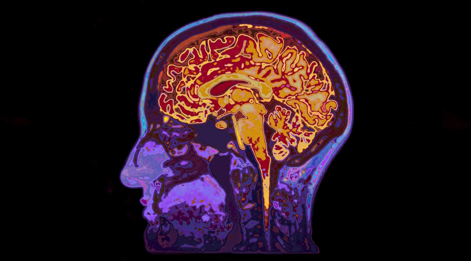
Theo Murphy meeting organised by Dr Andrea Lecchini-Visintini, Dr Emma Chung, Dr Jatinder Minhas and Professor Stephen Payne
Understanding the brain’s pulsing dynamics is an emerging research theme. Medical imaging and ultrasound researchers have developed methods to generate relevant data efficiently. Mathematical models require merging biomaterials and biofluids and transport frameworks. The main objectives of this meeting are to showcase existing data, to initiate the development of models, and to highlight key questions relevant to clinical translation.
Venue
This event is intended for researchers in relevant fields, and is a residential meeting taking place at the Leonardo Royal Brighton Waterfront hotel, King's Road, Brighton, BN1 2GS
Attending this event
- Free to attend and in-person only
- Please request an invitation using the above link. When requesting an invitation, please briefly state your expertise and reasons for attending. Requests are reviewed by the meeting organisers on a rolling basis. You will receive a link to register if your request is successful
- Catering options will be available to purchase upon registering. Participants are responsible for booking their own accommodation. Please do not book accommodation until you have been invited to attend the meeting by the meeting organisers
Enquires: please contact the Scientific Programmes team.
Organisers
Schedule
Chair
Dr Samantha Holdsworth, Mātai Medical Research Institute & University of Auckland, New Zealand

Dr Samantha Holdsworth, Mātai Medical Research Institute & University of Auckland, New Zealand
Dr Samantha Holdsworth, an MRI physicist, currently holds the position of CE/Research Director at Mātai Medical Research Institute, and serves as an Associate Professor at the University of Auckland and Principal Investigator at the Centre for Brain Research in New Zealand.
With 23 years of experience in MRI acquisition, post-processing, and analysis, she has developed fast, high-resolution MRI methods and pioneered amplified MRI, a novel technique for visualising brain motion.
During her 11-year tenure as a scientist at Stanford, Dr Holdsworth streamlined and translated various MR imaging and imaging reconstruction methods into clinical practice, enhancing the detection of brain disorders and diseases. Now based at Mātai in her hometown of Gisborne, New Zealand, Dr Holdsworth has assembled a team of local community, national, and international collaborators. Together, they are actively involved in numerous medical imaging research initiatives aimed at clinical translation for the benefit of the local community and to improve outcomes globally.
| 09:05-09:30 |
Probing the pulsing brain with MRI
Heartbeat-induced brain pulsations are thought to be important for maintaining brain homeostasis in the healthy brain. Changes in brain pulsations due to aging and/or vascular disease go hand in hand with disease processes that lead to dementia. Improving our understanding of the role of brain pulsations in health and disease requires methods that can non-invasively map these pulsations in humans. MRI has unique potential to measure multiple facets of brain pulsations. This lecture will start by sketching the cranial cavity and its contents, which set the stage for brain pulsations. Next, it will provide an overview of MRI methods that can probe the various aspects of pulsations in the intracranial cavity, including blood flow velocity pulsations, blood volume pulsations, brain tissue motion and deformation, and pulsations in the cerebrospinal fluid. By going over all these aspects, the lecture aims to provide an overview of the current understanding, together with the MRI methods that are available to further this understanding. Open questions and unmet needs regarding measuring brain and intracranial pulsations in its multiple facets will be highlighted. 
Dr Jaco J M Zwanenburg, Translational Neuroimaging Group, Center for Image Sciences, UMC Utrecht, The Netherlands

Dr Jaco J M Zwanenburg, Translational Neuroimaging Group, Center for Image Sciences, UMC Utrecht, The NetherlandsJaco Zwanenburg is a magnetic resonance imaging physicist with the drive to create dedicated MRI techniques that break new grounds for assessing anatomy and function in humans, with a focus on the cerebrovasculature. His research group is uniquely positioned in the Center for Image Sciences of the UMC Utrecht, with access to state-of-the-art equipment (including 7T MRI), cerebrovascular patient cohorts, and animal models of cerebrovascular disease. His ambition is to shift the MRI assessment of cerebrovascular diseases away from markers that reflect irreversible brain damage (e.g. infarcts, haemorrhages and atrophy), towards markers that identify vascular and parenchymal changes before irreversible damage occurs. As such, he aspires to contribute to future improvement of diagnosis, prognosis and treatment of cerebrovascular diseases. |
|---|---|
| 09:45-10:15 |
Intrinsic brain motion as an imaging marker for diseases
The human brain is the continuous subject of extensive investigation aimed at understanding its behavior and function. Despite an overwhelming interest and major research initiatives on how our brain operates, comparatively little is known about how it functions at the mechanical level. Recent findings have directly linked major brain development, mechanisms, and diseases to the mechanical response of the brain both at the cellular and tissue levels. Despite clear evidence that mechanical factors play an important role in regulating brain activity, current research efforts focus mainly on the biochemical or electrophysiological activity of the brain, mostly due to the difficulty of probing the brain physically. 
Associate Professor Mehmet Kurt, University of Washington, USA

Associate Professor Mehmet Kurt, University of Washington, USAProfessor Mehmet Kurt is the director of Kurtlab (www.kurtlab.com) and an Associate Professor at the Department of Mechanical Engineering (and by courtesy, Radiology and Neuroscience) at the University of Washington. His primary research area of interest is brain biomechanics and neuromechanics imaging. He received his PhD in Mechanical Science and Engineering from the University of Illinois at Urbana-Champaign in 2014 on developing novel nonlinear system identification methods. He was a postdoctoral scholar in the Departments of Bioengineering and Radiology at Stanford University from 2014-2017. His awards include NSF CAREER Award (2022), Fortune Magazine’s 40 under 40 list in Turkey (2021), Provost’s Early Career Award for Research Excellence (2020), NSF Vizzies Best Scientific Visualization Award, People's Choice (2018), Annals of Biomedical Engineering "Editor's Choice Award" (2017), Thrasher Research Foundation Early Career Award (2015), and the Thomas Bernard Hall Prize for the Best Paper of the Year (2011). His research has been highlighted in various media and his research group is currently sponsored by multiple grants from NSF, NIH and DoD. Dr Kurt also organizes various activities for increasing LGBTQ+ visibility in STEM and currently manages the Peer Review for Inclusion, Diversity, and Equity (PRIDE) program. |
| 10:30-11:00 |
Break
|
| 11:00-11:30 |
Dynamic diffusion-weighted imaging of pulsatile CSF dynamics in the paravascular space
Paravascular CSF dynamics have gained renewed research interest in neuroscience due to its recently discovered waste clearance function. Pulsation is considered a driving force behind its flow dynamics, yet challenges remain due to the limited non-invasive imaging techniques for studying the human brain. 
Assistant Professor Quiting Wen, Indiana University School of Medicine, USA

Assistant Professor Quiting Wen, Indiana University School of Medicine, USADr. Qiuting Wen, pronounced "chiu-ting," is a MR physicist who earned her PhD in Bioengineering from UC San Francisco. Her research focuses on developing advanced MRI techniques to understand brain fluid dynamics, its physiological drivers, and associated waste clearance functions. She is recognized for her work in dynamic diffusion-weighted imaging, which she applies to study fluid dynamic changes in both normal and diseased brains. |
| 11:45-12:15 |
Unveiling tissue secret from natural brain vibrations
Elastography, sometimes referred as seismology of the human body, is an imaging modality now implemented on medical ultrasound systems, on MRI and recently in optical coherence tomography devices. It allows to measure shear wave speeds within soft tissues and gives a tomography reconstruction of the shear elasticity. The shear elasticity being the elasticity felt by fingers during palpation, elastography is thus a palpation tomography. In the first part of this presentation, a passive elastography method is described. Inspired by noise correlation seismology and time reversal, it allows to extract from natural shear waves produced in the human body by heart beatings, muscles activities, arterial pulsations, a shear wave speed estimation. Therefore, an elasticity palpation mapping with no shear wave source is conducted. 
Professor Stefan Catheline, University of Lyon, France

Professor Stefan Catheline, University of Lyon, FranceStefan Catheline received his PhD degree in physics (1998) from University of Paris VII (Denis Diderot) for his work on transient elastography. His research conducted him from University of Paris to University of Lyon, France, via University of California, San Diego, USA, and University of Montevideo, Uruguay. Travelling was the occasion to learn about medical ultrasounds, underwater acoustics, wave physics, and seismology among other things. He holds 12 patents in the field of ultrasounds and seismology and wrote more than 100 research articles. He has been co-founder of two companies: Sensitive Object in the field of acoustic interactivity and SEISME in the field of elastography. |
Chair
Professor Stephen Payne, National Taiwan University, Taiwan
Professor Stephen Payne, National Taiwan University, Taiwan
Stephen Payne is Professor and Yushan Fellow at National Taiwan University, Taipei, Taiwan. Prior to this he was Professor at the University of Oxford. He is the current Editor-in-Chief of the journal Medical Engineering and Physics. He has published around 140 papers in international journals, written 3 books, supervised over 30 PhD students to successful completion, and raised over £3.1M in research funding.
| 13:30-14:00 |
Human brain tissue mechanics: experiments, modelling, and simulation
The brain is not only one of the most important but also the arguably most complex and compliant organ in the human body. While long underestimated, increasing evidence confirms that mechanics plays a critical role in modulating brain function and dysfunction. Computational models based on nonlinear continuum mechanics can help understand the basic processes in the brain, e.g., during development, injury, and disease, and facilitate early diagnosis and treatment of neurological disorders. 
Professor Silvia Budday, Institute of Continuum Mechanics and Biomechanics Friedrich-Alexander-Universität Erlangen-Nürnberg, Germany

Professor Silvia Budday, Institute of Continuum Mechanics and Biomechanics Friedrich-Alexander-Universität Erlangen-Nürnberg, GermanySilvia Budday studied Mechanical Engineering at the Karlsruhe Institute of Technology (KIT), and spent one year abroad at Purdue University, Indiana, USA. For her PhD on “The Role of Mechanics during Brain Development” at Friedrich-Alexander-Universität Erlangen-Nürnberg (FAU), she was awarded the GACM Best PhD Award, the ECCOMAS Best PhD Award, and the Bertha Benz-Prize from the Daimler und Benz Stiftung as a woman visionary pioneer in engineering. For her research focusing on experimental and computational soft tissue biomechanics with special emphasis on brain mechanics, she received several awards, including the Heinz Maier-Leibnitz-Prize by the DFG and BMBF in 2021. Since 2023, she is a Full Professor heading the Institute of Continuum Mechanics and Biomechanics (LKM) at FAU and recently received an ERC Starting Grant for her project “Mechanics-augmented brain surgery (MAGERY)”. |
|---|---|
| 14:15-14:45 |
Computational models of the glymphatic system
The glymphatic theory propose a clearance system for the brain that is particularly active during sleep. The system is important as many neurodegenerative diseases are related to accumulation of metabolic waste. This talk will present an overview of computational models, current challenges and open questions. Professor Kent-Andre Mardal, University of Oslo, Norway
Professor Kent-Andre Mardal, University of Oslo, NorwayProfessor Kent-Andre Mardal graduated from the University of Oslo in 1999 in applied and industrial mathematics. He obtained his PhD in 2003 at Simula Research Laboratory / University of Oslo, developing numerical methods with applications to fluid mechanics. His research interests concern mathematical modelling of the fluid flows of the brain in health, disease and during ageing. Particular focus the last years has been the interaction between the cerebrospinal fluid and both the macro- and micro-circulation. |
| 15:00-15:30 |
Break
|
| 15:30-16:00 |
Neuron multiphysics, a new paradigm for the building block of the brain
Neurons are traditionally considered solely through their electrophysiological function. They are however multiphysics systems whereby their physical properties – electrophysiological, mechanical, biochemical, etc. – are in fact working collaboratively and concurrently. In this presentation, we will demonstrate how this is indeed the case, and what the implications are for the general field of neuromodulation. 
Professor Antoine Jerusalem, University of Oxford, UK

Professor Antoine Jerusalem, University of Oxford, UKProfessor Antoine Jérusalem graduated in 2004 with a double degree from the Ecole Nationale Supérieure de l’Aéronautique et de l’Espace with a Diplôme d’Ingénieur, and from the Massachusetts Institute of Technology with a Master of Science in Aeronautics and Astronautics. In 2007, he obtained his Ph.D. in Computational Mechanics of Materials from MIT, where he stayed as a Postdoctoral Associate for a year. Antoine was the group leader of the Computational Mechanics of Materials Group in Madrid’s Advanced Studies Institute of Materials (IMDEAMaterials) from 2008 to 2012, and is currently a Professor of Mechanical Engineering at the University of Oxford. He is also an Affiliate Researcher in the Mathematical Institute at Oxford and the codirector of the International Brain Mechanics and Trauma Lab. Antoine’s research activities focus on computational modelling of many types of materials and structures, ranging from metals to composite materials with a major focus on the multiphysics of neurons and brain and applications in Ultrasound Neuromodulation and TBI. His modelling activities involve the development and use of state-of-the-art advanced numerical techniques. Professor Jérusalem has active collaborations with different institutes and universities around the world. |
| 16:15-16:45 |
CSF dynamics in perivascular spaces (online talk)
Flow of cerebrospinal fluid (CSF) in the in the perivascular spaces that surround cerebral blood vessels plays an important role in clearance of waste from the brain and is linked to diseases such as Alzheimer’s and small vessel disease. Recently, artificial intelligence velocimetry (AIV), which integrates sparse two-dimensional (2D) in vivo velocity measurements with physics-informed neural networks, has been used to infer high-resolution pressure, volumetric flow rates, and shear stresses of CSF in pial PVSs, providing a wealth of information about the dynamics of these flows. However, in the initial work, the PVS and vessel wall boundaries were assumed to be stationary, contrary to what has been observed in experiments. In fact, the motion of the vessel wall directly adjacent to the PVS is closely coupled to the CSF dynamics and is thought to drive the CSF flow. This talk will describe new results from an updated AIV model that incorporates moving blood vessel walls and residual-based attention to the physics-informed neural networks, quantifying the importance of including vessel wall motion in AIV inferences. The types and extent of vessel wall motion and the impact on CSF flow in the adjacent PVSs will also be described and quantified. The impact of these results on brain-wide models of CSF flow will also be discussed. 
Research Assistant Professor Kimberly A S Boster, University of Rochester, USA

Research Assistant Professor Kimberly A S Boster, University of Rochester, USAKimberly Boster is a Research Assistant Professor at the University of Rochester in the Mechanical Engineering Department. Her background is in fluid mechanics, heat transfer, and image processing. After experiencing a minor traumatic brain injury, she became fascinated with brain fluid mechanics and after completing her PhD decided to begin studying fluids in the brain. She first focusing on cerebrovascular flows before turning her attention to the glymphatic system in 2020. Her work focuses on building numerical models of CSF flow, analysis of in vivo experiments detailing CSF flow, and combining experimental measurements and knowledge of the underlying physics to answer fundamental questions related to CSF flow in the brain. |
Chair
Dr Emma Chung, King's College London, UK

Dr Emma Chung, King's College London, UK
Emma is a Senior Lecturer with King’s College London and Research Lead for the University Hospitals of Leicester NHS Trust Medical Physics Department. Emma is a registered Clinical Scientist, and Fellow of the Institute of Physics with a special interest in Ultrasound Education and clinical translational ultrasound research. In 2020, Emma was highly commended by England's Chief Scientific Officer for her research using Doppler ultrasound for the detection of brain injury. Emma is a former Editor-in-Chief of the journal 'Ultrasound' and is a former Honorary Secretary of the British Medical Ultrasound Society (BMUS).
| 09:00-09:30 |
Ultrasound brain tissue pulsation (BTP) in brain disorders
The recent development of ultrasound, both in probe technology and signal processing, has allowed the precise measurement of Brain Tissue Pulsations (BTP). Our team has been particularly involved in the characterization of BTP in clinical conditions. In several studies, we observed that BTP is involved in the pathophysiology of cerebrovascular diseases, cognitive and psychiatric disorders, orthostatic hypotension, normal aging, brain volume reduction, etc. Our most prolific work with BTP has focused on depression. We indeed found excessive BTP amplitudes in major depressive episode that tends to reduce with successful antidepressant treatment. In addition, we found that BTP could be used as a prognostic marker for treatment response with new generation antidepressant agent. More recent works include the technological coupling of BTP with EEG for a multimodal assessment of brain physiology in both healthy participants and patients with brain disorders. Ultrasound BTP has a high potential to being implemented in clinical routine because it is portable, non-invasive, easily accessible and low cost. However, BTP has also limitations and further clarifications in both the technique itself and in the experimental studies are required. 
Professor Thomas Desmidt, University Hospital of Tours, France

Professor Thomas Desmidt, University Hospital of Tours, FranceMD, PhD in Psychiatry, specialized in Old Age Psychiatry. Head of the Department of Old Age Psychiatry, University Hospital of Tours, France. Member of the INSERM Unit "IBrain". Focuses on the development of new treatments for Depression and Alzheimer's disease, as well as characterizing the neuropathology of Depression, Alzheimer's disease and related disorders, especially using neuroimaging, including ultrasound. |
|---|---|
| 09:45-10:15 |
Measuring BTP with Trans-cranial Tissue Doppler
Healthy brain tissue pulsations (BTPs) are known to occur with every cardiac cycle. In this talk, the development of Brain Tissue Velocimetry (Brain TV), a novel transcranial tissue Doppler (TCTD) ultrasound prototype, will be presented. Brain TV was created in collaboration with Nihon Kohden, Japan, and monitors brain tissue motion over the cardiac cycle. Brain TV is a portable system that can record BTPs in real-time, from multiple positions on the head, using 2 MHz transcranial Doppler (TCD) probes, together with other synchronised physiological measurement data, such as blood pressure, heart rate, and end-tidal carbon dioxide. Using Brain TV, it was possible to record BTPs from up to 30 sample depths within the brain, ranging from 22 to 80 mm below the probe’s surface, in ultrasound phantom brain models, healthy volunteer studies, and in stroke patients. This presentation will illustrate the use of the device, summarise results and insights obtained in past studies, and present the ideas behind current and future studies. 
Miss Jennifer Nicholls, University of Leicester, UK

Miss Jennifer Nicholls, University of Leicester, UKJennifer Nicholls is a post-doctoral research assistant in the department of Cardiovascular Sciences at the University of Leicester. Jennifer has recently submitted her PhD thesis, which aimed to understand the impact of large vessel occlusion on brain tissue pulsatility, measured using a novel ultrasound technique known as transcranial tissue Doppler ultrasound. Jennifer’s work involved determining the parameters which affect brain tissue pulsations (BTPs) in an ultrasound phantom brain model, determining the effect of raised intracranial pressure on BTPs in a phantom model and healthy subjects, and determining the effect of stroke on BTPs in a phantom and in patients. This year, Jennifer won the University of Leicester's Cardiovascular Sciences departmental 3 Minute Thesis Competition, highlighting her enthusiasm for her work. |
| 10:30-11:00 |
Break
|
| 11:00-11:30 |
Brain ultrasonology in acute brain injured patients (online talk)
Brain ultrasonology stands out as a valuable diagnostic tool due to its non-invasive nature and real-time imaging capabilities, making it an efficient method for evaluating cerebral blood flow dynamics and detecting various pathologies promptly. By offering quick and bedside assessments, this technology enables healthcare professionals to make rapid decisions and interventions that can significantly impact patient outcomes. In light of these advantages, this talk aims to further delve into the potential applications and benefits of brain ultrasonology in clinical practice. Discussing this topic can enhance our understanding of how this tool can empower healthcare providers to optimize patient care and improve outcomes, especially in situations where time is of the essence. This presentation will delve deeper into the importance of brain ultrasonology as a rapid and bedside tool for gaining insights into cerebral dynamics. Such a discussion could not only raise awareness about the capabilities of this technology but also foster dialogue on how we can leverage its advantages to enhance patient care. Associate Professor Chiara Robba, Policlinico San Martino, University of Genoa, Italy
Associate Professor Chiara Robba, Policlinico San Martino, University of Genoa, ItalyChiara Robba is Associate Professor and Senior Consultant at the University of Genoa, Italy. Her main interests are neurocritical care, cross talk between brain and peripheral organs and mechanical ventilation. She is currently the Chair of the Neurointensive Care section of the European Society of Intensive Care. She is author of more then 400 peer reviewed papers. |
| 11:45-12:15 |
Ultrasound elastography during neurosurgery
Neurosurgery is a complex and intricate procedure that benefits from intraoperative guidance. During tumour resection for example, a surgeon will subjectively evaluate tumour stiffness and adherence to surrounding normal brain, using the information to optimise the resection procedure and achieve maximal resection. The outcomes for the patient, for both brain tumour and focal epilepsy surgery, depend on the extent of resection. Magnetic resonance imaging (MRI) and x-ray computed tomography provide valuable information but have not solved this problem, lacking the real-time feedback needed for intraoperative decision making. Ultrasound elastography is a promising tool that may improve a surgeon’s appreciation of tissue properties during surgery. Various forms of ultrasound elastography have been explored over the past 20-25 years. These include passive methods which use the brain’s natural pulsations, strain imaging which employs hand-induced palpation, and shear wave elastography which watches the progression of shear waves initiated by acoustic radiation force. Findings from research with these methods suggest that ultrasound elastography can effectively differentiate tumours and other lesions from healthy brain (in some cases when MRI is negative), determine the degree of adherence between tumour and brain, locate cleavage plains, and enhance the detection of residual tumour for increasing the completeness of resection. The method has considerable potential for further development and holds promise in neurosurgery, providing real-time information on tissue properties and enhancing surgical decision-making to improve safety and outcomes for patients. 
Professor Jeff Bamber, Institute of Cancer Research, UK

Professor Jeff Bamber, Institute of Cancer Research, UKJeffrey Bamber is Professor of Physics Applied to Medicine, Deputy Dean (Biomedical Sciences) and has been Head of Ultrasound and Optical Imaging Physics since 1986 at The Institute of Cancer Research and The Royal Marsden Hospital, London. He has a BSc in Physics, an MSc in Biophysics and Bioengineering, and a PhD in Biophysics. He has been a Gledden Short Stay Fellow at the University of Western Australia, Guest Professor at the Tokyo Institute of Technology and Visiting Scientist at the Medical Products Group, Hewlett-Packard, Andover, MA, USA. Several of his inventions are available on commercial ultrasound systems, and he has served as scientific advisor to various companies. He is vice-president of the IBUS Breast Imaging School, past president of the International Association for Breast Ultrasound, past vice-president of the International Society of Skin Imaging and past chair of the British Medical Ultrasound Society Science and Education Committee. |
Chair
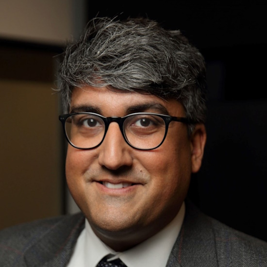
Dr Jatinder S Minhas, University of Leicester, UK

Dr Jatinder S Minhas, University of Leicester, UK
Dr Minhas’s research endeavours to bridge the gap between technical studies in cerebrovascular physiology (particularly acute intracerebral haemorrhage), and delivery of clinical stroke care and research. He strongly believes this niche is vital for delivering technically excellent and innovative translational programmes of research with the potential to deliver significant improvements in stroke care, in reasonable time frames.
| 13:30-14:00 |
Digital twins of the brain and in-silico clinical trials
Digital twins, a term often used in engineering, aim to develop computer simulations of a real-world object such that the digital twin of the object can predict what will happen to the object in the future. Increasingly, this concept is being applied with humans (or aspects of their physiology) as the object to be simulated. This allows us to better understand how to treat an individual, leading to personalised healthcare, and improved patient outcomes. Furthermore, it is hoped such an approach to healthcare can lead to a greater focus on preventative medicine to ensure people live healthier lives. From this concept, there also arises the concept of in silico clinical trials, or clinical trials that are run entirely on a computer using digital twins of humans. The development of this technology will allow for quicker, cheaper clinical trials reducing drug and medical device costs, lowering the economic burden of healthcare. 
Dr Wahbi K El-Bouri, University of Liverpool, UK

Dr Wahbi K El-Bouri, University of Liverpool, UKDr Wahbi El-Bouri works at the cutting-edge of Digital Twin development as a Lecturer (Assistant Professor) at University of Liverpool. He heads up his research team, the Virtual Vascular Human research group, where he develops Digital Twin technology of the vascular system for the purpose of in silico clinical trials. An engineer by training, he received both his MEng and DPhil in biomedical engineering from University of Oxford before transitioning to Department of Cardiovascular Medicine in Liverpool to begin the process of translating these Digital Twins to clinical environments. Combining our understanding of the physics and mathematics that describe the human body with patient data, Wahbi seeks to develop advanced Digital Twins that can be used for risk prediction as well as product testing to speed-up and de-risk drug and medical device development. |
|---|---|
| 14:15-14:45 |
Hemodynamics and solute exchange in personalised brain models
Intracranial dynamics concerns physical, chemical and biological interactions between cerebral vasculature, cerebrospinal fluid (CSF) and the brain cells. Despite progress in medical imaging, organ wide patterns of fluid and solute exchange between blood, CSF and neural tissue in normal and especially pathological states remain inadequately quantified; key principles of transport and control functions of brain metabolism are still poorly understood. We propose to harness predictive mathematical models informed by dynamic imaging data to close existing knowledge gaps in cerebral blood flow and CSF dynamics. We outline mathematical models of the cerebral circulation using graph theoretical approaches. We predict blood flow patterns across multiple length scales from large blood vessels down to individual capillaries in digital analogues of mouse and human brains. Digital twins of the mouse and human brains are constructed by fusing subject-specific medical image data with hemodynamically inspired network synthesis methods. Our mechanistic modelling approach provides a rigorous platform for translating in vivo imaging observations from animals to humans and between individual subjects. The impact of these experimental and computational results on neurodegenerative diseases including dementia, Alzheimer’s disease and hydrocephalus will be highlighted. We will also show clinically relevant applications concerning the quantification of solute dispersion after intrathecal drug administration. 
Professor Andreas A Linninger, University of Illinois, USA

Professor Andreas A Linninger, University of Illinois, USAAndreas Linninger is a Full Professor of Biomedical Engineering and Adjunct Faculty of Neurosurgery at the University of Illinois at Chicago. He also held appointments at the University of California at Berkeley, Massachusetts Institute of Technology and Rijksuniversiteit Gent. His work includes pioneering contributions to mathematical modelling of the intracranial dynamics of cerebrospinal fluid, blood flow, hydrocephalus and drug delivery systems for the Central Nervous System. He has developed algorithms for in silico synthesis of anatomically accurate digital replicas of the whole mouse and human brain. His Fourier-based dual mesh techniques for reactive diffusion-convection problems enable metabolic simulations of unprecedented resolution in multi-scale, multi-physics, mixed domain problems that arise in solute exchange across the blood brain barrier. He has authored more than 170 peer reviewed publications and a dozen chapters in mathematical and biomedical textbooks. |
| 15:00-15:30 |
Break
|
| 15:30-16:00 |
Closing remarks
|
| 16:00-17:00 |
Open discussion: Future directions
|

