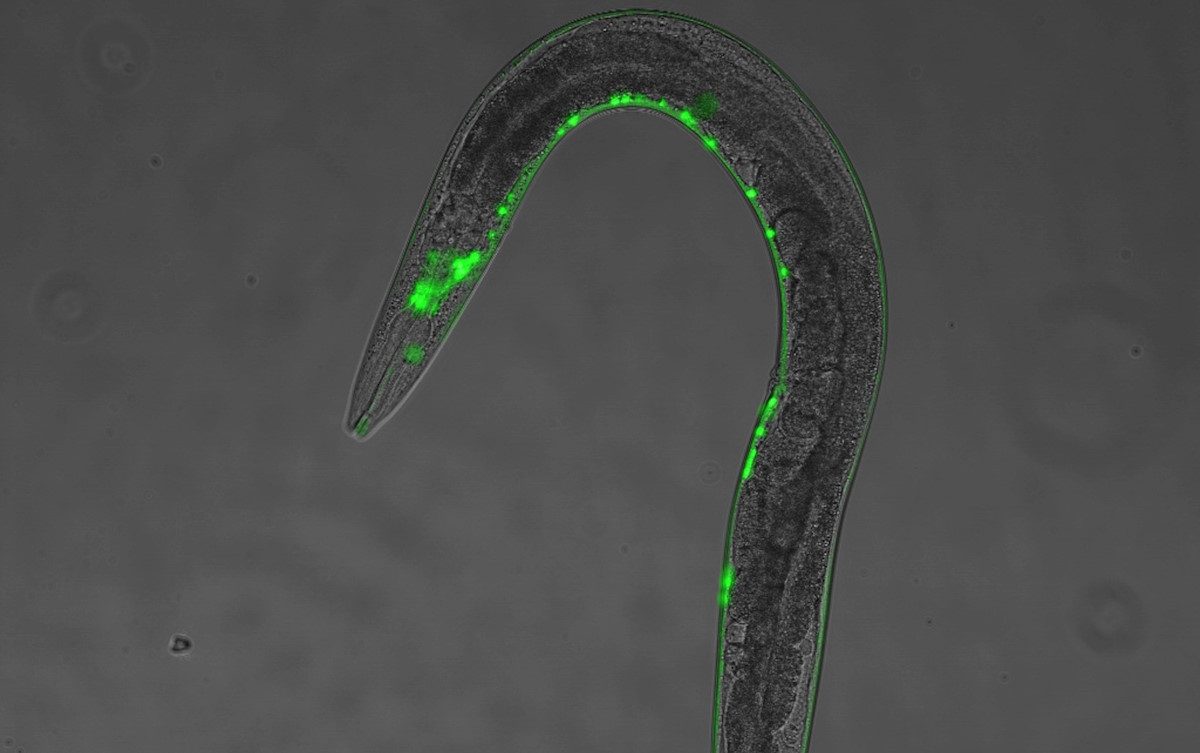
ProteostaSys: a systems view of proteostasis
.png)


Theo Murphy meeting organised by Dr Ritwick Sawarkar, Professor Rick Morimoto, Professor Lea Sistonen, and Dr Anat Ben-Zvi.
Proteostasis underpins the homeostasis of the functional proteome and is implicated in most age-related disease conditions. This meeting capitalised on the recent definition of the human proteostasis network to bring together experts from molecular cell biology and quantitative systems biology. The meeting facilitated interdisciplinary discussions about proteostasis to speed up fundamental discoveries with biomedical implications.
Organisers


Schedule
Chair
Dr Laura Itzhaki
University of Cambridge, UK
Dr Laura Itzhaki
University of Cambridge, UK
Dr Laura Itzhaki’s background is in protein folding and protein engineering. Her group’s research has helped to elucidate the folding-related phenomenon of three-dimensional domain swapping. They resolved the mechanism by which domain swapping occurs, delineated how this capability is encoded in the amino-acid sequence, and revealed the potential for domain swapping to regulate protein function and to drive disease-associated protein misfolding. Subsequent studies on several proteins have shown their findings to constitute universal principles of domain swapping. More recently their research has focused on understanding the rules governing the biophysics of a special protein class, so-called tandem-repeat proteins. These proteins have a simple and distinctive modular architecture, one consequence of which is that they can be subjected to gross perturbations (multi-site mutations, repeat deletions/insertions) yet remain correctly folded. This extraordinary malleability has enabled Dr Itzhaki’s group to resolve in unprecedented detail the multitude of inter-converting structures that comprise their energy landscapes and to apply methods originally designed for small proteins to investigate giant repeat arrays leading to unique insights. The simple, modular architecture of repeat proteins allows their properties to be engineered in a strikingly predictable way, and Dr Itzhaki’s group are currently exploiting this design-ability to dissect cellular mechanisms of folding and unfolding, and to engineer repeat proteins as a new therapeutics platform.
| 08:45-09:00 |
Welcome by the lead organisers
|
|---|---|
| 09:00-09:20 |
AI-based discovery of pharmacological chaperones
Chemical chaperones play a central role in the protein homeostasis system by stabilising native protein conformations. Over the past two decades, small molecules with a similar mechanism of action, known as pharmacological chaperones, have emerged as effective therapeutics. In this lecture, we will present an AI-based method to screens ultra-large chemical libraries to identify compounds that bind target proteins with high affinity and specificity. We illustrate this approach in the case of Alzheimer’s disease and steatotic liver disease, demonstrating how it accelerates the path from virtual hits to viable lead compounds. 
Professor Michele VendruscoloUniversity of Cambridge, UK
Professor Michele VendruscoloUniversity of Cambridge, UK Michele Vendruscolo is Professor of Biophysics, Director of the Chemistry of Health Laboratory and Co-Director of the Centre for Misfolding Diseases at the Department of Chemistry of the University of Cambridge, where he moved over 20 years ago. His work is aimed at establishing the fundamental principles of protein homeostasis and protein aggregation, and at exploiting these principles to develop methods for drug discovery in neurodegenerative diseases. He has published over 500 scientific papers and 20 patents, and given over 500 invited lectures at international meetings. He has been founder and chief scientific officer of Wren Therapeutics (now WaveBreak Therapeutics). |
| 09:20-09:40 |
Regulation of the Huntingdon folding landscape by Hsc70, DNAJB1 and Apg2
Molecular chaperones play crucial roles in protein folding, targeting, sequestration, disaggregation, and degradation of misfolded proteins. Their intervention is critical in preventing the conversion of proteins from their native states into amyloid fibrils. Key components in this chaperone network are the HSPA complexes, particularly involving J-domain proteins (JDPs), which drive the HSPA folding machinery by delivering substrates and activating the HSPA ATPase. A focus of Janine Kirstein’s research is the analysis of the versatility of the incredibly diverse JDP family with 50 different JDPs identified in humans. A well-studied JDP-HSPA chaperone complex, comprising DNAJB1, Hsc70, and the NEF Apg2, can suppress Huntingtin Exon1Q48 (HTT) aggregation in an ATP-dependent manner. DNAJB1 binds with a specific motif located between CTDI and CTDII to the polyQ-adjacent proline rich domain of HTT. Mutation of the highly conserved H244 of the motif in DNAJB1 completely abrogates the suppression of HTT aggregation and disaggregation of HTT fibrils by the trimeric chaperone complex. Notably, this mutation does not affect the binding and remodelling of any other tested protein substrate including amyloid proteins, suggesting that the generalist DNAJB1 uses unique site for the interaction with distinct protein substrates such as HTT. Surprisingly, while an overexpression of DNAJB1 is protective in HTT expressing cultured cells, it is detrimental on an organismal level. Overexpression of the DNAJB1 homolog, DNJ-13 leads to an increase in HTT aggregation, spreading and toxicity in C. elegans. Increased levels of DNJ-13 might boost the disaggregation activity of the trimeric chaperone complex in vivo and thereby generate seeding-competent HTT species that favour aggregation and spreading of HTT. 
Professor Janine KirsteinLeibniz Institute on Ageing, Germany
Professor Janine KirsteinLeibniz Institute on Ageing, Germany Janine Kirstein studied Biology at the University of Greifswald (Germany), obtained her PhD at the Freie Universität Berlin (Germany) and then joined the lab of Rick Morimoto at Northwestern University (USA) as Postdoc. She started her own lab in 2013 in Berlin at the Leibniz Institute for Molecular Pharmacology (Germany). In 2019 she was appointed as Professor at the University of Bremen and in 2023 at the Friedrich-Schiller-University in Jena (Germany) with a joint affiliation with the Leibniz Institute on Aging. Janine Kirstein is an Allen Distinguished Investigator (2023), is and has been a PI of many structured research consortia (SPP2453, FOR 5228, SPP1623, SFB740 and EXC257) and co-organised twice the European C. elegans meeting. |
| 10:10-10:30 |
Discussion
|
| 10:30-11:15 |
Break
|
| 11:15-11:35 |
CBFA2T2 regulates lipid metabolism in complex with the transcription factor HSF1 and histone demethylase KDM3C
The heat shock response (HSR) is an evolutionarily conserved mechanism to maintain proteostasis and is characterised by induction of heat shock proteins (HSPs) in proteotoxic stress conditions. This induction is regulated mainly at transcriptional level by heat shock transcription factors (HSFs). HSF1 is a master regulator of the HSR in human cells and regulate the expression of not only HSP genes but also a variety of genes involved in cell proliferation and metabolism under conditions without environmental stresses. HSF1 could access to and activate these target genes probably by recruiting histone-modifying enzymes and chromatin-remodelling complexes. However, HSF1-mediated regulatory mechanisms of metabolism-related genes are not well known. Here, we identified a transcriptional co-regulator CBFA2T2 as an HSF1-interacting protein irrespective of heat shock. CBFA2T2 and HSF1 up-regulate a common set of genes involved in lipid metabolism, including SREBP1, SREBP2 and SOAT1, in melanoma MeWo cells. Remarkably, HSF1 binding to the promoters of these genes required CBFA2T2, whereas CBFA2T2 occupied them even in the absence of HSF1. CBFA2T2 accesses to the target gens through the recruitment of PBAF chromatin remodelling complex and transcription factors such as PRDM1. CBFA2T2 and HSF1 co-operate to recruit the histone demethylase KDM3C, which regulates lipid metabolism. Blockage of the interaction between CBFA2T2 and HSF1 inhibited lipid droplet formation and proliferation of melanoma cells. Our observations provide regulatory mechanisms of HSF1-mediated expression of lipid metabolism-related genes, which support melanoma tumorigenesis. 
Professor Akira NakaiYamaguchi University, Japan
Professor Akira NakaiYamaguchi University, Japan Professor Akira Nakai was born in Hyogo Prefecture, Japan. 1981-1987, Undergraduate at Tottori University School of Medicine, Tottori Prefecture, Japan; 1987-1991, Graduate student at Second Department of Internal Medicine, Tottori University; 1991-1993, Postdoctoral fellow, Department of Biochemistry, Molecular Biology and Cell Biology, Northwestern University, IL, USA; 1993-2000, Assistant Professor, Department of Cell Biology, Chest Disease Research Institute, Kyoto University and Department of Molecular and Cell Biology, Institute for Frontier Medical Sciences, Kyoto University, Kyoto Prefecture, Japan; 2000-present, Professor, Department of Biochemistry and Molecular Biology, Yamaguchi University School of Medicine, Yamaguchi Prefecture, Japan. |
| 11:35-11:55 |
Regulation of protein homeostasis in oocyte aging
Female reproductive aging starts in humans at the average age of 35, when a significant number of oocytes are still present while oocyte quality rapidly declines. During postnatal oogenesis, the primary oocytes arrested at meiotic prophase I undergo robust protein synthesis in the growth phase to prepare the machinery for meiotic maturation and maternal factors for subsequent embryogenesis, making oogenesis particularly reliant on proteostasis. Data from Dr Li and colleagues suggest proteostasis decline is a driving force of oocyte aging. In both C. elegans and mice, loss of HSF1 activities is an early event of oocyte aging, leading to reduced expression of chaperone genes and insufficient protein folding capacity. Depletion of HSF-1 from C. elegans oocytes phenocopies the maladaptive stress responses observed in maternal aging, which ultimately undermines oocyte proteostasis and quality. Conversely, over-expression of HSF-1 in the germline preserves oocyte quality and reduces chromosomal errors in maternal aging. Remarkably, Dr Li and colleagues found that three well-established pro-reproductive longevity signaling pathways in C. elegans (insulin/IGF-1, TGF-b-Sma/Mab, and mTORC2-SGK-1) converge on the regulation of protein synthesis in germ cells to suppress the reproductive defects associated with HSF-1 depletion and preserve oocyte proteostasis in maternal aging. 
Dr Jian LiNew York Medical College, USA
Dr Jian LiNew York Medical College, USA Jian Li, PhD, completed his graduate study in Dr David Gilmour's lab at Penn State University. His thesis work, using biochemical and next-generation sequencing approaches, uncovered mechanisms underlying promoter-proximal pausing of RNA polymerase II, an essential regulatory step of transcription in metazoans. Dr Li then moved to Dr. Rick Morimoto’s lab at Northwestern University for his postdoctoral training, where he started using the nematode C. elegans as a model. His work discovered the transcriptional program of HSF1, the key regulator of cellular heat shock response, in animal development independent of temperature stress. Dr Li started his lab at the Oklahoma Medical Research Foundation in 2017 and joined New York Medical College in 2022. His lab combines genetics and cellular biology with multi-omics (functional genomics, transcriptomics, proteomics, and metabolomics) to study proteostasis pathways at cellular and organismal levels. Recent publications from his lab revealed how insulin/IGF-1 signaling and HSF1 interact to control germline proteostasis via cell-autonomous and non-cell-autonomous mechanisms. Ongoing research projects in Dr. Li’s lab include exploring how to improve oocyte proteostasis and quality in maternal aging using C. elegans and mouse models, and understanding how aberrant activation of HSF1 supports malignancy through remodeling mitochondria and energy metabolism. |
| 11:55-12:05 |
Short talk
Dr Luca CirilloThe Institute of Cancer Research, UK Dr Luca CirilloThe Institute of Cancer Research, UK |
| 12:05-12:30 |
Discussion
|
Chair

Professor Stefan Marciniak
University of Cambridge, UK
Professor Stefan Marciniak
University of Cambridge, UK
Stefan studied medicine at the University of Cambridge as part of its MB/PhD program. After training in respiratory medicine in Cambridge, London and Edinburgh, he undertook post-doctoral research in New York University for three years and then again in Cambridge. In 2012, he established his lab in the Cambridge Institute for Medical Research (CIMR) to study diseases caused by abnormal protein folding, including alpha1-antitryspin deficiency. In 2016, he became Professor of Respiratory Science at the University of Cambridge. He is also an active respiratory physician at Cambridge University Hospitals and Royal Papworth Hospital. In Cambridge, he directs the MB/PhD program, while nationally he directs the NHS England Rare Disease Collaborative Network (RDCN) in Familial Pneumothorax and co-leads the LifeArc Rare Respiratory Disease Centre.
| 13:30-13:50 |
Unveiling non-cell autonomous mechanisms in alpha-synuclein pathobiology: insights from C. elegans
Pathological alpha-synuclein condensation and toxicity are associated with a range of age-related neurodegenerative diseases, including Parkinson’s disease and multiple system atrophy (MSA). However, the biological triggers and consequences of these pathological changes are not completely understood. Previous screens in a C. elegans model, capturing both condensation- and toxicity features of alpha-synuclein, have revealed several biological interventions that counter alpha-synuclein pathology. These interventions include metabolic rewiring, such as the inhibition of organismal tryptophan degradation, and microbiome replacement. However, the protective mechanisms underlying these interventions remain unclear. This presentation highlights ongoing research aimed at identifying the mechanisms of such non-cell-autonomous protection. Using diverse biological screens in both C. elegans and protective microbiota, previously unknown regulators and microbial metabolites were identified that modify alpha-synuclein pathobiology. These results provide insight into the impact of systemic factors and host-microbiome interactions in the development of synucleinopathies. 
Professor Ellen NollenUniversity Medical Center Groningen, The Netherlands
Professor Ellen NollenUniversity Medical Center Groningen, The Netherlands Ellen Nollen is a professor of Molecular Biology of Aging at ERIBA and University Medical Center Groningen. She specialises in the mechanisms underlying aging and neurodegenerative diseases. Nollen leads the Laboratory of Molecular Neurobiology of Ageing at ERIBA. Her team’s research focuses on understanding protein condensation, aggregation, and toxicity in aging and age-related diseases using the model organism C. elegans. She ultimately aims to translate new insights into clinical applications in collaboration with clinicians and industrial partners. Nollen previously conducted research at the UMCG's Genetics Department, Hubrecht Laboratory, and Northwestern University. After completing her PhD in 2000 at the University of Groningen, she received several awards, including the Rosalind Franklin Fellowship, an Alfred Tissières Young Investigator Award, and an ERC Starting Independent Researcher Grant. |
|---|---|
| 13:50-14:10 |
Alpha-synuclein and tau aggregates polymorphism and the structural molecular basis of diverse synucleinopathies and tauopathies
The aggregation of the proteins alpha-synuclein (a-Syn) and tau within the central nervous system is deleterious and associated to diverse neurodegenerative disorders. How a-Syn and tau aggregates target defined cells, how those aggregates traffic between them, multiply by recruiting their endogenous counterparts and cause distinct synucleinopathies and tauopathies is still poorly understood. Proteostasis decline favors a-Syn and tau aggregation. Evidences for a-Syn and tau aggregates-mediated sequestration of important proteins, among which molecular chaperones and members of the cellular clearance machinery that mimick their loss of function, will be presented and discussed. A-Syn and tau aggregates are also released from affected neuronal cells after multiplication, they bind to naïve neurons cell membranes and redistribute protein receptors with functional consequences that will be detailed. The reason why the aggregation of a-Syn causes distinct synucleinopathies was unknown until we showed this protein to form structurally different fibrillar aggregates in test tubes and proposed that the different structures may cause different pathologies. How the diversity of a-Syn and tau fibrillar aggregates, including those derived from patient brains, cause distinct diseases will be discussed. The importance of targeting the surfaces of aggregated a-Syn and tau from the diagnostic and therapeutic standpoints will be highlighted and discussed. 
Dr Ronald MelkiCentre national de la recherche Scientifique, France
Dr Ronald MelkiCentre national de la recherche Scientifique, France Ronald Melki is Director of Research at CNRS. He established a team involved in molecular chaperone-mediated folding in 1994 and has been engaged in documenting prion and prion-like aggregates formation, disaggregation and propagation since 1999. He chaired several Institutes in France and is recipient of several awards, among which the Coups d'Elan pour la Recherche Française by the Bettencourt Schueller Fondation for excellence in research on PD in 2011, the Grand Prix Scientifique of the Fondation Simone et Cino Del Duca of the French Academy for excellence in research on PD and MSA. His team characterised the aggregation of the baker’s yeast prions Ure2p and Sup35p in the absence and the presence of molecular chaperones revealing an unsuspected diversity in prion particles. Since 2008, he contributed to the demonstration that Huntingtin and alpha-synuclein aggregates traffic between neighbour cells. He identified the most toxic Huntingtin, tau and alpha-synuclein aggregates and documented their interaction with neuron membranes as well as their uptake and transport. He generated alpha-synuclein aggregates that differ structurally and pathologically, establishing a structural-molecular basis for different synucleinopathies. He strengthened the concept of α-Syn strains by pioneering the amplification of aggregated pathogenic α-Syn from patient brains and characterising patients-derived aggregates biochemically, structurally and functionally in vitro and in vivo. He assessed, with world leaders, the turnover, proteostatic response and traffic of pathogenic aggregates between neurons, astrocytes and microglia and has been mapping the surfaces of fibrillar alpha-synuclein and tau at atomic resolution to design therapeutic and diagnostic tools. |
| 14:10-14:30 |
Disruption of RNA metabolism and its impact on protein homeostasis
RNA metabolism is essential for organismal survival, adaptation, and cellular homeostasis. It governs the synthesis, processing, modification, transport, translation, and degradation of RNA molecules, ensuring that gene expression is tightly regulated in response to developmental cues and environmental stressors. Disruptions in RNA metabolism – such as mutations in RNA-binding proteins (RBPs) and those caused by nucleotide repeat expansions – can lead to the accumulation of aberrant RNA species, misregulated splicing events, and impaired RNA transport and translation. These defects often result in the production of toxic proteins or the sequestration of essential RBPs, ultimately disturbing proteostasis and contributing to the onset and progression of neurodegenerative and neuromuscular diseases. Emerging evidence indicates that alterations in RNA metabolism are associated with disruptions in stress response pathways and energy homeostasis. Aberrant RNA processing can impair the expression and function of key regulators involved in cellular stress adaptation, such as heat shock proteins and components of the unfolded protein response (UPR). Additionally, misregulated RNA splicing and translation can alter mitochondrial function and ATP production, compromising cellular energy balance. These disruptions are particularly detrimental in high-demand tissues like the brain and muscle, contributing to the pathogenesis of neurodegenerative and neuromuscular disruption – and how these changes are reflected in protein homeostasis – will be crucial for identifying new therapeutic targets and biomarkers. 
Dr Susana GarciaUniversity of Helsinki, Finland
Dr Susana GarciaUniversity of Helsinki, Finland Dr Susana Garcia is a researcher specializing in RNA repeat-associated degenerative diseases and a Group Leader at the Institute of Biotechnology, University of Helsinki. Her research focuses on the cellular and molecular mechanisms underlying nucleotide repeat toxicity, particularly the disruptive effects of expanded repeat RNAs. Her innovative approaches have been recognized with the 1st Prize in Orion Pharma’s 2017 “100 Ideas for Research & Development” Competition. Dr Garcia also serves as Vice-Director of the Genomics and Evolutionary Biology Research Programme at the Institute of Biotechnology. She completed her graduate studies in Dr Richard Morimoto’s lab at Northwestern University, followed by postdoctoral research with Dr Gary Ruvkun at Massachusetts General Hospital/Harvard Medical School, before transitioning to her current leadership role in Helsinki. |
| 14:30-14:40 |
Short talk
Dr Mathieu BourdenxUK Dementia Research Institute at University College London, UK Dr Mathieu BourdenxUK Dementia Research Institute at University College London, UK |
| 14:40-15:00 |
Discussion
|
| 15:00-15:30 |
Break
|
| 15:30-15:50 |
Proteostasis (in)action: the role of co-pathologies in neurodegenerative disease
Diverse neurodegenerative diseases such as Alzheimer’s disease (AD), Parkinson’s disease (PD), and amyotrophic lateral sclerosis (ALS) share in common progressive neuronal dysfunction function associated with the accumulation of misfolded proteins. Central to these processes are the cellular and organismal proteostasis networks responsible for maintaining protein homeostasis through coordinated actions of molecular chaperones, the ubiquitin–proteasome system, and autophagy-lysosomal pathways. As a consequence of aging, genetic modifications and other disease-specific conditions, this proteostatic network becomes compromised, leading to the accumulation of toxic protein aggregates that disrupt neuronal integrity and lead to distinct neurodegenerative syndromes. Specific misfolded proteins are associated with each neurodegenerative disease (ie a-synuclein in PD, amyloid-beta/Tau in AD and TDP-43/ SOD in ALS). However, increasing evidence implicates more complex co-pathologies across these disorders, as Lewy bodies are common in AD brains and Alzheimer pathology is present in the majority of PD autopsy cases. These observations are consistent with the idea that disruption of proteostasis networks by one aggregation prone protein could result in misfolding and aggregation of other neurodegeneration-related species. We will examine how impaired protein quality control not only drives primary proteinopathies but also fuels secondary pathologies, including neuroinflammation, oxidative stress, mitochondrial dysfunction, and disturbances in RNA metabolism. Finally, the lecture will discuss emerging therapeutic strategies aimed at restoring proteostasis and mitigating co-pathological cascades, emphasising the need for integrated approaches to combat complex pathologies. 
Dr James MorleyUniversity of Pennsylvania, USA
Dr James MorleyUniversity of Pennsylvania, USA Dr Morley received his undergraduate degree in economics from the Wharton School of the University of Pennsylvania and completed his MD and PhD studies in the Medical Scientist Training Program at Northwestern University. Returning home to Philadelphia for post-graduate training, he completed a Neurology residency and a fellowship in movement disorders at the University of Pennsylvania. He is the Director of the Crescenz VA Medical Center Parkinson's Disease Research, Education and Clinical Center, Associate Director of the Movement Disorders Fellowship and an Associate Professor of Neurology at the University of Pennsylvania Perelman School of Medicine. Dr Morley chairs the VA National Parkinson’s Network Research Committee and his interests include biomarkers for early diagnosis and prognosis in Parkinson’s disease, drug-induced parkinsonism and how to use exercise as a treatment for PD. He has received research support from the NIH, Michael J Fox Foundation, GE Healthcare, Department of Defense and Department of Veterans Affairs. |
| 15:50-16:10 |
The collaboration of proteostasis and inflammation in critical brain injury
Proteostasis and inflammation are the two fundamental intrinsic responses to acute injury. They mediate a series of downstream processes involved in tissue damage and repair. These responses become dysregulated in the context of severe injury, with significant short-term and long-term consequences. This can result in long-term disability or death in ischemic stroke, intracerebral hemorrhage, subarachnoid hemorrhage, and traumatic brain injury. The medical management of these diseases is largely limited to the stabilisation of basic physiology — intracranial pressure, systemic blood pressure, oxygen levels, and carbon dioxide levels — with a focus on maintaining cerebral perfusion and metabolism. This paradigm largely neglects the profound effects that disruptions in proteostasis and dysregulation of inflammatory responses have on tissue injury. Even less is understood about how proteostasis and inflammation intersect at the tissue, cellular, and molecular level to produce damage and repair responses. Understanding the early changes in proteostasis and inflammation, and the interaction between the two, may reveal new mechanisms and therapeutic targets to address the large and growing challenge of critical injury to the brain. 
Dr Prajwal CiryamUniversity of Maryland School of Medicine, USA
Dr Prajwal CiryamUniversity of Maryland School of Medicine, USA Prajwal Ciryam is an Assistant Professor in the Department of Neurology and Program in Trauma at the University of Maryland School of Medicine, where he is also an Attending Neurointensivist. His laboratory is interested in mechanisms of inflammation-related secondary brain injury. As part of the Medical Scientist Training Program at Northwestern University, he obtained his MD and PhD, the latter concurrently with the University of Cambridge, where he was a Fulbright Scholar and St John’s College Benefactors Scholar. His PhD advisers were Professor Richard Morimoto, Professor Christopher Dobson, and Professor Michele Vendruscolo. Prajwal completed neurology residency at Columba University and neurocritical care fellowship in the Columbia/Cornell joint program, at which time he received an NIH R25 research fellowship. He is the recipient of the Henry Jackson Foundation’s Military Research Group Fellowship, Passano Foundation Physician-Scientist award, Neurocritical Care Society career development award, and Burroughs-Wellcome AHEAD Scholar award. He has also received funding from the Brain Aneurysm Foundation, University of Maryland Baltimore ICTR, Shock Trauma Anesthesiology Research, and GEn1E Lifesciences. |
| 16:10-16:30 |
Age-dependent fluctuations in the Hsp90 interaction network
Molecular chaperones are sentinels of the protein homeostasis (proteostasis) process and are required to safeguard polypeptides by continuously scrutinising the conformational status of all cellular proteins. Insights into proteostasis primarily have been gained by studying protein biogenesis and triage events. Yet, the proverbial birth and death of any polypeptide only represents a small percentage of the protein-lifecycle. The mainstay of a protein’s time is spent functioning in pathways to support life. While it has long been suggested that molecular chaperones regulate native proteins, how any chaperone might recognise and/or modulate the broad spectrum of mature proteins found within a cell had been poorly understood. Recently, we developed a high-throughput chemical biology tactic to discover that the Hsp90 chaperone targets intrinsically disordered regions (IDRs) of native proteins to broadly interact with a cell’s protein population (eg, yeast Hsp90 associates with ~25% of the proteome). We have exploited this system to investigate how the Hsp90 interaction landscape shifts during the aging process. Our presented work will show that low Hsp90 protein levels are insufficient to support normal aging, cells with reduced Hsp90 have elevated protein aggregates at early timepoints during chronological aging, and the physical interaction network surrounding Hsp90 shifts as cells age including a rewiring of the chaperone system. Overall, our data demonstrate that Hsp90 acts as a key aging factor by functioning as a central hub in the proteostasis process. 
Professor Brian FreemanUniversity of Illinois, USA
Professor Brian FreemanUniversity of Illinois, USA Brian C Freeman received his BS Degree in 1989 from the University of Michigan, his MS Degree in 1991 from Florida State University studying DNA repair with Dr J Herbert Taylor, his PhD Degree in 1996 from Northwestern University studying molecular chaperones with Dr Richard I Morimoto, and his postdoctoral work until 2002 at the University of California, San Francisco studying the dynamics of steroid hormone receptors with Dr Keith R Yamamoto. Since 2002, he has been a faculty member at the University of Illinois, Urbana-Champaign (UIUC). Currently, he is a Professor in the Department of Cell and Developmental Biology. Brian Freeman has made important contributions to numerous fields including in the areas of molecular chaperone biology, transcription, telomere, chromatin, and genome organisation. In brief, his research program pioneered the study of molecular chaperone function in the nucleus. He has received numerous honours including being named the Educator of the Year at UIUC in 2009, a Friedrich Wilhelm Bessel Awardee from the Alexander von Humboldt Foundation in 2010, an Ambassador of the Technische Universität München in 2015, a Fellow of the Cell Stress Society International in 2020, a Fellow of the American Association for the Advancement of Science in 2021, and an MCB Distinguished Professorial Scholar at UIUC in 2024. |
| 16:30-18:00 |
Flash talks and poster session
|
Chair
Dr Della David
University of Cambridge, UK
Dr Della David
University of Cambridge, UK
Della David is a senior group leader at the Babraham Institute, Cambridge, UK. After studies in France at the European School of Biotechnology, she moved to Switzerland and obtained her PhD in 2005 from the University of Zürich. For her postdoctoral work (2006-2011), she joined the lab of Cynthia Kenyon at UCSF in the USA, supported with funding from the Swiss National Science Foundation, Larry L Hillblom Foundation, American Federation for Aging Research and Program for Breakthrough Biomedical Research. There, she discovered that widespread protein aggregation occurs during normal ageing in the model organism Caenorhabditis elegans. In 2011, she became an independent research group leader in Germany at the German Center for Neurodegenerative Diseases (DZNE) in Tübingen and then moved to the Interfaculty Institute of Biochemistry, University of Tübingen. In 2022, Della relocated her lab to the Babraham Institute. Her group continues to focus on age-dependent protein aggregation with the aim of uncovering endogenous mechanisms to prevent aggregation and promote healthy ageing. A key recent study from her group identified the extracellular proteostasis network in C. elegans. Della has received a Wellcome Discovery Award to expand this area of research.
| 09:00-09:20 |
Neuronal regulation of cellular stress responses in metazoans: mechanisms and open questions
Tremendous advances have been made in understanding how isolated cells autonomously respond to stress. However, it remains unclear whether the same principles apply within the complex environments of tissues and organ systems in metazoans. Building on our discovery that the heat shock response—one of the most ancient and conserved stress response pathways—is not solely cell-autonomous in the nematode Caenorhabditis elegans, but instead regulated by sensory neuronal circuits, our research aims to elucidate how neuronal stress sensing modulates protective responses in distant target cells. We will discuss our progress in uncovering the fundamental mechanisms by which the nervous system of multicellular organisms translates the sensory detection of stress into anticipatory, cell-type-specific transcriptional programs—activated before damage occurs and tailored to the nature of the stressor. We will also speculate on how discoveries in this field could redefine our understanding of stress response regulation in vivo and reveal how systemic coordination enhances organismal resilience. 
Professor Veena PrahladRoswell Park Cancer Institute, USA
Professor Veena PrahladRoswell Park Cancer Institute, USA Veena Prahlad PhD is a Professor of Oncology in the Department of Cell Stress Biology at Roswell Park Comprehensive Cancer Center. Dr Prahlad is a recognised leader in the field of cellular stress biology. Her research demonstrated that the nervous system regulates the heat shock response, a fundamental transcriptional program that protects cells from misfolded proteins. Her lab investigates neural control of stress pathways and transcription factors involved in cancer and neurodegenerative diseases. Dr Prahlad earned her MSc in Life Sciences from Jawaharlal Nehru University in India and completed her PhD at Northwestern University, where she uncovered new mechanisms governing cytoskeletal organisation. She continued her postdoctoral work at Northwestern, discovering the neural regulation of the heat shock response. Before joining Roswell Park in 2023, she was an Associate Professor at the University of Iowa and a member of the Iowa Institute of Neuroscience, contributing to Alzheimer’s and Lewy Body Dementia research. She has taught extensively, including courses on aging biology, and has received recognition and funding from the Ellison Medical Foundation and multiple NIH institutes. |
|---|---|
| 09:20-09:40 |
An unexpected partnership: understanding how HSF-1 and mitochondria cooperate to promote healthy ageing
The transcription factor HSF-1 has long been recognised as the master regulator of the Heat Shock Response (HSR), a highly conserved transcriptional programme that is rapidly deployed to counter the threat of misfolded proteins within the cytosol/nucleus. In keeping with this role, impaired HSF-1 activity is associated with reduced tissue resilience, shortened lifespan and increased susceptibility to age-associated protein conformational disease, while enhanced HSF-1 activity is associated with greater stress resistance, increased lifespan and improved protection against protein aggregation and proteotoxicity. While HSF-1 is classically thought to promote tissue-health by increasing the levels of cytosolic/nuclear molecular chaperones and protein degradation factors, evidence is emerging that HSF-1 activity is also closely coupled to mitochondrial homeostasis. In this talk, I will present my groups recent findings that increased HSF-1 activity converges on mitochondrial homeostasis through the proteasome shuttling factor, ubiquilin-1. HSF-1 promotes ubiquilin-1 expression, which leads to increased turnover of the CDC-48 co-factor, NPL-4. This alters mitochondrial network dynamics and leads to metabolic changes that promote longevity. In addition, changes in mitochondrial homeostasis result in increased HSF-1 activity and protection of the cytosolic proteome. Our findings reveal that HSF-1 sits at the nexus between cytosolic and mitochondrial homeostasis and suggest that the interplay between HSF-1 and mitochondria is a key determinant of cell and tissue viability that could be manipulated to promote healthy ageing. 
Dr John LabbadiaUniversity College London, UK
Dr John LabbadiaUniversity College London, UK Dr John Labbadia is an Associate Professor within the Institute of Healthy Ageing at University College London. John has dedicated his career to understanding how cells maintain protein homeostasis, first as a PhD student working in Professor Gillian Bates’ lab at Kings College London, and then as an ALS Association Postdoctoral Fellow in Professor Rick Morimoto’s lab at Northwestern University. Following his time in the Morimoto lab, John was awarded a prestigious BBSRC David Phillips Fellowship to establish his own group at UCL, where he has been using the small nematode worm Caenorhabditis elegans, to understand how changes in the activity of stress response pathways impacts the long-term health of different tissues. His research is funded by awards from the BBSRC, Academy of Medical Sciences, Wellcome Trust, and Leverhulme Trust. |
| 09:40-10:00 |
Regulation of stress granule dynamics by the Hsp70 chaperone system
Stress granules (SGs) are ribonucleoprotein condensates that transiently form during cellular stress when protein translation is inhibited. They allow the storage of RNA-binding proteins and mRNAs, and their release for translation restoration upon stress recovery. Loss of SG dynamic properties is proposed to underlie the formation of protein aggregates in certain neurodegenerative diseases, including amyotrophic lateral sclerosis. The Hsp70 chaperone family has been implicated in limiting aberrant SG aggregation and in the disassembly of the condensates during recovery. However, a detailed understanding of how Hsp70 regulates SG dynamic properties is lacking. Here, we used C. elegans to investigate the role of constitutively expressed Hsp70 (HSP-1/Hsc70) in the dynamics of SGs in different cell types. We found that HSP-1 associates with cytosolic structures corresponding to bona fide SGs throughout the tissues of the animal. Fluorescence recovery after photobleaching of SGs in various cell types revealed that HSP-1 is highly mobile and rapidly exchanges with the cytoplasmic pool, in contrast to the core SG protein GTBP-1/G3BP1, but similar to other SG components. Reduced hsp-1 function resulted in persistent SGs in germ cells, indicating a role in SG disassembly, and altered the mobility of select SG components, suggesting a stronger effect on the more dynamic shell within the SG structure. These findings provide insights into the regulation of condensate dynamics and architecture by Hsp70, with potential implications for pathological transitions of SGs. 
Dr Ambre SalaInstitute for Integrative Biology of the Cell, France
Dr Ambre SalaInstitute for Integrative Biology of the Cell, France Ambre Sala is a CNRS researcher in the Cell Biology department of the Institute for Integrative Biology of the Cell (I2BC, Gif-sur-Yvette, France), where she heads the group “Protein homeostasis in development and aging”. Her research broadly focuses on the role of molecular chaperones in regulating physiological and pathological protein phase transitions. The team develops cell biology, genetic, and biochemistry approaches, taking advantage of the C. elegans model system to identify the cellular roles and composition of the chaperone networks that regulate these processes in various tissues and during aging. Ambre Sala previously completed her PhD thesis in 2015 at the University of Toulouse, and then worked as a postdoctoral fellow in the laboratory of Professor Richard Morimoto at Northwestern University. Ambre Sala joined the I2BC in 2023 after obtaining a tenured researcher position and a starting grant from the ATIP-Avenir program (CNRS/Inserm). |
| 10:00-10:10 |
Short talk
Professor Melody ClarkBritish Antarctic Survey, UK Professor Melody ClarkBritish Antarctic Survey, UK |
| 10:10-10:30 |
Discussion
|
| 10:30-11:00 |
Break
|
| 11:00-11:20 |
Cell-nonautonomous stress signalling pathways for systemic proteostasis control
The maintenance of proteostasis across the various tissues of an organism depends on cell-intrinsic mechanisms but also on coordinated, cell-nonautonomous stress responses. Accumulating evidence shows that intracellular signalling pathways play a crucial role in the systemic regulation, particularly under environmental challenges or chronic disease conditions. Our lab has been exploring how the molecular chaperone Hsp90, a highly conserved and essential regulator of proteostasis, mediates such inter-tissue communication. We previously discovered that tissue-specific modulation of Hsp90 levels in the gut or the nervous system in C elegans triggers a protective response in distant tissues, a process we termed transcellular chaperone signalling. Interestingly, this intracellular signalling has context-dependent effects. While constitutive overexpression of Hsp90 in the gut reduces thermotolerance, it enhances resistance to chronic proteotoxic stress, such as caused by amyloid-beta or polyglutamine proteins, and extends lifespan of these C elegans models of neurodegenerative disease. On the other hand, reducing Hsp90 expression specifically in the gut increases cytoprotective Hsp70 levels in other tissues and boosts overall stress resilience and longevity. This however comes at the cost of a reduced protection against chronic protein aggregation. Mechanistically, the regulation of these intracellular stress response pathways are distinct from the canonical HSF-1 mediated heat shock response. For example, the induction of Hsp70 in response to gut-specific Hsp90 knockdown requires the homeodomain transcription factor CEH-58, which functionally opposes HSF-1 but is required for an intracellular signalling cascade. During acute heat stress, the classical HSF-1 pathway dominates. This regulation appears to have physiological stress resistance: Hsp90 levels can be naturally downregulated in the gut following heat shock or pathogen exposure, which is in part through lysosomal degradation pathways, promoting stress resistance. I will present our recent findings on how peripheral tissues can communicate stress signals to maintain proteostasis across the organism, and how modulating these signals might be used to protect against neurotoxicity and promote systemic resilience. 
Dr Patricija van Oosten-HawleUniversity of North Carolina at Charlotte, USA
Dr Patricija van Oosten-HawleUniversity of North Carolina at Charlotte, USA Dr Patricija van Oosten-Hawle is an Associate Professor in the Department of Biological Sciences at the University of North Carolina at Charlotte. She studied Chemistry and Biochemistry at the University of Vienna in Austria and earned her PhD from the Free University (VU) of Amsterdam in the Netherlands. Following her doctoral studies, she joined the laboratory of Dr Rick Morimoto at Northwestern University as a postdoctoral fellow. In 2015, Dr van Oosten-Hawle established her research lab at the University of Leeds in the UK. In 2022, she returned to the United States to continue her work at UNC Charlotte. Her research focuses on understanding how stress response pathways operate across tissues to maintain proteostasis and how these mechanisms might be leveraged to counteract ageing and age-related diseases. Her work has been supported by the Wellcome Trust, NC3Rs, and the Leverhulme Trust in the UK, and is currently funded by the National Institute of Health (NIH) in the US. In recognition of her work on transcellular chaperone signalling and organismal proteostasis, Dr van Oosten-Hawle received the Ferruccio Ritossa Early Career Award from the Cell Stress Society International (CSSI) in 2019 and was named a CSSI Fellow in 2020. |
| 11:20-11:40 |
ER stress sensor PERK supports sensory function and controls polarised localisation of IGF and TGFβ growth factors in healthy neurons
Genetic variants in EIF2AK3, encoding the ER stress sensor PERK, confer increased risk of tauopathies PSP and AD through unknown mechanism(s). While increased kinase activity of PERK, accompanied by eIF2α phosphorylation and remodeling of translation, is thought to contribute to neurodegeneration of stressed neurons, eg in advanced disease, the tauopathy risk alleles are instead hypomorphic in neurons. Thus, decreased PERK activity may be sensitising to disease, or even directly detrimental in healthy neurons. We find that PERK activity is required for the normal function of sensory neurons. Loss of PERK in Drosophila results in olfactory defects in unstressed animals; the defects are selective to subsets of olfactory neurons, and motor neurons are not affected. In C. elegans, we find that PERK is required for sensory function of the ASI neuron, and for correct axonal vs dendritic localisation and release of some neuronal signaling molecules, specifically - the insulin/IGF-like growth factor DAF-28, and the TGFβ-like DAF-7. Molecularly, this novel function of PERK is distinct from its ER stress signaling function, and is eIF2α-independent. Instead, PERK cooperates with the CaMKII pathway, and may involve calcium entry via a voltage-gated calcium channel. CaMKII pathway has a well-established role in regulating memory; similarly, loss of olfaction is observed early in multiple neurodegenerative diseases, while neuronally-released insulin/IGF and TGFβ are neuroprotective, and their deficiencies are thought to contribute to AD. This work connects hypomorphic PERK to the cellular and molecular pathways involved in neurodegeneration, and may help explain increased risk of tauopathies in PERK alleles carriers. 
Dr Tali GidalevitzDrexel University, USA
Dr Tali GidalevitzDrexel University, USA The Gidalevitz lab aims to understand the mechanisms that maintain healthy protein folding environment, and how they fail in neurodegeneration and aging. As postdoctoral fellow in the Morimoto lab, Dr Gidalevitz co-discovered that misfolding-prone proteins derail the folding of other, healthy, proteins because of their metastability and competition for limiting folding resources. This discovery led to the recognition of proteostasis failure as one of the main contributing mechanisms in conformational diseases. In her lab, Dr Gidalevitz focuses on endogenous misfolded proteins, natural genetic variation, and the physiological aspects of proteostasis. Using natural C. elegans strains, her lab developed a framework for the fascinating question of how genetic variation among individuals impacts cellular processes that maintain protein homeostasis. This is important since human neurodegenerative diseases are strongly modified by genetic variation, but the mechanisms remain largely unknown. Continuing the work on proteostasis mechanisms, the lab developed a worm model for the neuron-specific toxicity of a broadly-expressed misfolded protein, resulting in demonstration of bystander protein misfolding being caused by an endogenous misfolded protein. They now use this model to study folding and localisation of neuroprotective growth factors in sensory neurons, and to understand how disruption of their localisation, and of ER proteostasis, contribute to the pathogenesis of tauopathies and other neurodegeneration. Currently, this work is focused on the role of the ER stress sensor PERK as a risk factor for tauopathies PSP and AD. In addition to the focus on neurodegeneration, the Gidalevitz lab is studying the myoprotective mechanisms of small HSPs. |
| 11:40-12:00 |
Impaired sphingolipid homeostasis exacerbates tau pathology by disrupting endolysosomal integrity through endomembrane rigidification
The progression of neurodegenerative diseases such as Alzheimer’s disease (AD) or Parkinson’s disease (PD), is linked to the spreading of protein aggregation across interconnected brain regions. Fibrillar seeds are transferred between cells, inducing misfolding and aggregation of its native monomeric counterpart. A critical rate-limiting step in this process is the rupture of endolysosomal vesicles, which releases transferred aggregates into the cytosol, where they promote further aggregation of endogenous proteins. While the pathways underlying endolysosomal damage and repair have been increasingly studied, many aspects remain unclear. We conducted a genome-wide RNAi screen in a novel C. elegans model of tau spreading and identified sphingolipid metabolism genes as critical for endolysosomal integrity. Using C. elegans and cell culture models, we investigated how disruption of sphingolipid homeostasis affects endolysosomal membrane integrity and contributes to tau aggregation and toxicity. Fluorescence recovery after photobleaching and C-Laurdan dye imaging revealed that silencing sphingolipid metabolism genes decreases endolysosomal vesicle membrane fluidity, increasing endolysosomal rupture. The accumulation of aggregated tau in endolysosomal vesicles further aggravated endomembrane rigidification and damage and promoted seeded tau aggregation, potentially by facilitating the escape of tau seeds from the endolysosomal system. Supplementation with unsaturated fatty acids improved membrane fluidity, suppressing endolysosomal rupture and seeded tau aggregation in cell models, and alleviating tau-associated neurotoxicity in C. elegans. Conversely, supplementation with the saturated fatty acid palmitic acid worsened these effects. These findings provide insights into how impaired sphingolipid homeostasis drives tau pathology, suggesting membrane fluidity restoration as a potential therapeutic strategy for AD. 
Professor Carmen NussbaumLudwig Maximilians University Munich, Germany
Professor Carmen NussbaumLudwig Maximilians University Munich, Germany Carmen Nussbaum studied Biology and pursued her doctoral research at the Technical University of Munich, earning her PhD in December 2008. She then joined Northwestern University in Evanston, IL, USA, as a postdoctoral fellow. From 2014 to 2022, she led a research group at Heidelberg University and the German Cancer Research Center. Since 2022, she holds a W2 Professorship in the Department of Neuroanatomy at Ludwig-Maximilians-University of Munich. Her research utilises C. elegans and cell culture models to study the molecular mechanisms underlying neurodegenerative diseases. She focused on the dual role of the Hsp70 disaggregation machinery in the prion-like propagation of α-synuclein and tau, demonstrating that while chaperone-mediated disaggregation solubilises protein aggregates, it also generates small, seeding-competent toxic species. More recently, her work has focused on the role of endolysosomal integrity in prion-like aggregate propagation. |
| 12:00-12:10 |
Short talk
Mr Dimitris AspriUniversity of Cambridge, UK Mr Dimitris AspriUniversity of Cambridge, UK |
| 12:10-12:30 |
Discussion
|
Chair

Dr Rahul Samant
Babraham Institute, UK
Dr Rahul Samant
Babraham Institute, UK
The Samant Lab started at Babraham Institute in 2019, using multi-dimensional proteomics, live-cell imaging, and ubiquitin biochemistry to understand how ubiquitin-mediated signalling mediates misfolded protein quality control in healthy and diseased cell contacts. Rahul did his PhD under Paul Workman (Institute of Cancer Research), studying the proteasomal degradation of oncoprotein kinases upon HSP90 inhibitor treatment of cancer cells. His postdoc work with Judith Frydman (Stanford University) probed the importance of distinct ubiquitin chain types in misfolded protein degradation in budding yeast. A major current focus of The Samant Lab is on how the proteostasis network is re-wired in a range of non-proliferative cellular states, with the goal of harnessing vulnerabilities to promote healthy ageing.
| 13:30-13:50 |
Exposure to microplastic disrupts proteostasis in Caenorhabditis elegans
Since the 1950s, humans have produced 8 billion tons of plastic. This plastic, the vast majority of which is not recycled, is in the form of plastic containers, synthetic fibers used in clothing, paints, adhesives, etc. These plastics break down with use and enter our food and water as micro and nano-sized particles. Plastic is also added to personal care products as microspheres that do not need to break down to enter waterways. Moreover, tire wear particles account for 28% of microplastics that enter the environment. A recent retrospective study revealed that microplastic accumulation in the human brain is increasing at an alarming rate. This has been found to contribute to neuroinflammation and to promote the progression of Alzheimer’s Disease symptoms. We asked whether microplastic from a synthetic soccer turf (micro-rubber) causes protein damage/misfolding, overwhelms the proteostasis network, and contributes to proteostasis collapse. To address this, we utilised C. elegans sensors of protein folding, and found that exposure to micro-rubber particles accelerated the formation of large visible protein aggregates in body wall muscle cells. Interestingly, animals that were exposed to micro-rubber were impaired in their ability to launch a heat shock response. Taken together, the data suggests that micro-rubber particles contribute to a decline in proteostasis, which may be relevant to neurodegenerative disease progression. 
Dr Elise A KikisUniversity of the South (Sewanee), USA
Dr Elise A KikisUniversity of the South (Sewanee), USA Elise A Kikis earned a BA in Biology from Cornell University and a PhD in Plant Biology from the University of California, Berkeley. She currently serves the University of the South (Sewanee) as Professor and Chair of the biology department and as co-director of the Office of Undergraduate Research and Scholarship. She joined the Sewanee faculty in 2012 as an assistant professor with the goal of inspiring the next generation of scientists to think critically and address pressing scientific questions. Prior to beginning her faculty position, she conducted post-doctoral research under the supervision of Richard Morimoto at Northwestern University. There, she solidified her interest in the study of protein folding and the regulation of proteostasis. She has published numerous research articles, many with Sewanee undergraduate student co-authors, in internationally recognised journals and has successfully competed for extramural funding from the National Institutes of Health, the Kennedy’s Disease Association, and the Appalachian College Association. She enjoys fostering teamwork and collaboration in her laboratory and training undergraduates as they begin their careers. |
|---|---|
| 13:50-14:10 |
Intersections between protein oxidation-reduction pathways and proteostasis at the subcellular level
Protein disulfide formation contributes to protein folding and function, as evidenced by numerous oxidoreductases that maintain disulfide status within various cell compartments. Many of these oxidoreductases, including thioredoxins (Trxs) and protein disulfide isomerases (PDIs), arose from a common evolutionary precursor and employ transient thiol-disulfide exchange mechanisms when interacting with their redox partners. Because disulfide bonds that form between Trx superfamily members and their redox partners are often short-lived, we developed a chemical cross-linking strategy that irreversibly captures these proteins when interacting with potential redox partners. Using the thiol-reactive cross-linker divinyl sulfone (DVSF), Trx2, a cytosolic Trx in baker’s yeast, undergoes cross-linking with its most prominent redox partners via modification of its active site cysteines. Likewise, upon DVSF treatment, PDIs that promote oxidative protein folding in the ER form cross-linked complexes with known redox partners, including many chaperones and protein clearance factors. More recently, we have turned our attention to less studied redox networks, seeking to identify the interaction partners of disulfide maintenance systems in mitochondria. Particularly, we have characterised the putative redox partners of yeast Trx3, a disulfide reductase that resides in the mitochondrial matrix. We have found that Trx3 interacts with numerous metabolic enzymes, including members of the branched chain amino acid biosynthesis pathway, as well as proteins in the proteostasis network. Our current work suggests disulfide bond homeostasis and flux through specific metabolic pathways may be linked in previously undescribed ways. 
Professor James WestThe College of Wooster, USA
Professor James WestThe College of Wooster, USA James West received his PhD in Biochemistry from Vanderbilt University in 2005, working in Larry Marnett’s laboratory on apoptotic cell death mechanisms and cellular responses to protein-damaging lipid peroxidation byproducts. Subsequently, he performed postdoctoral research in the Department of Molecular Biosciences at Northwestern University in the laboratory of Rick Morimoto, studying small molecule activators of the heat shock response and parallel stress responses. He began a faculty position at The College of Wooster in 2008, where his research program has focused on interrogating protein disulfide formation and reduction networks, primarily in a baker’s yeast model system. |
| 14:10-14:30 |
Microbial pathogenesis in neurodegenerative protein conformational diseases
There is no cure or effective treatment for neurodegenerative protein conformational diseases (PCDs), such as Alzheimer’s, Parkinson’s, and Huntington’s. All these ailments are characterised by toxic protein aggregation, resulting in progressive neuronal damage and cell death. Emerging evidence reveals that bacteria play a significant role in the pathogenesis of neurodegenerative diseases; however, the microbial identity and the underlying mechanisms remain elusive. In a screen of over 220 unique bacterial species from the Human Microbiome Project, we found proteotoxic and proteoprotective bacteria. We demonstrated that employing bacteriophage-mediated strategies to eliminate detrimental bacteria associated with proteotoxicity alleviates disease pathogenicity. Furthermore, among the beneficial microbes, a single bacterial strain, Prevotella corporis, provides protection against misfolding and aggregation. We employed the Caenorhabditis elegans and Drosophila melanogaster models expressing polyglutamine and Aβ42 to demonstrate that colonisation of the gut suppresses aggregation in both models. Further analysis revealed that P. corporis activates the expression of heat shock genes and can rapidly disassemble existing protein aggregates. While P. corporis did not have any effect on lifespan, it enhanced the thermotolerance and disaggregation activity of the host, suggesting general improvement in proteostasis. Collectively, we reveal how bacteria affect host proteostasis and influence disease pathology, opening new opportunities to manage neurodegenerative diseases by targeting the gut. 
Dr Daniel CzyżUniversity of Florida, USA
Dr Daniel CzyżUniversity of Florida, USA Dr Daniel Czyż (chysh) is an Associate Professor in the Department of Microbiology and Cell Science at the University of Florida. He received his BS in Biochemistry from the University of Illinois at Chicago and a PhD from Northwestern University, where he studied the regulation of the heat shock response using Caenorhabditis elegans. During his postdoc in the Department of Microbiology at the University of Chicago, he pioneered drug-repurposing and host-targeting approaches against intracellular bacterial infections. Dr Czyż has been a faculty at the University of Florida since 2018. His group studies microbial pathogenesis in neurodegenerative diseases employing C. elegans and tissue culture models. Recently, the Czyż Group has characterised over 200 different bacteria for their role in host proteostasis, revealing proteotoxic and proteoprotective species that modulate disease pathogenesis by influencing host stress responses. Antibiotics are the major contributor to resistance and gut dysbiosis, which is implicated in the pathogenesis of neurodegenerative diseases. The Czyż Group utilises non-traditional approaches, including bacteriophages, to target proteotoxic and antibiotic-resistant bacteria and restore gut eubiosis, potentially alleviating disease progression. Dr Czyż received numerous distinctions for teaching and research, including the UF Exemplary Online Award for Student Engagement, IFAS/CALS Innovative Teaching Award, NACTA Educator Award, Richard Jones Outstanding New Faculty Research Award, and much more. He served as the Vice-Chair (2020-2021) and Chair (2021-2022) of the Advisory Council at the National Institute of Antimicrobial Resistance Research and Education (NIAMRRE) and hosted the 2023 NIAMRRE Annual Conference at UF. |
| 14:30-14:40 |
Short talk
|
| 14:40-15:00 |
Discussion
|
| 15:00-15:30 |
Break
|
| 15:30-16:15 |
From proteostasis to ProteostaSys

Professor Richard MorimotoNorthwestern University, USA
Professor Richard MorimotoNorthwestern University, USA Dr Richard I Morimoto is the Bill and Gayle Cook Professor of Biology and Director of the Rice Institute for Biomedical Research at Northwestern University. Rick received his BS from the University of Illinois at Chicago, subsequently received a PhD in Molecular Biology at the laboratory of Professor Murray Rabinowitz, University of Chicago in 1978. He then conducted his postdoctoral research in the laboratory of Professor Matthew Meselson in the Department of Biochemistry and Molecular Biology at Harvard University in Cambridge, MA. He was concurrently a Tutor in Biochemical Sciences at Harvard College. In 1982, Morimoto joined the faculty of the Department of Biochemistry, Molecular Biology, and Cell Biology at Northwestern University in Evanston, IL. He has served as the Chair of Biochemistry, Molecular Biology, and Cell Biology, the Dean of The Graduate School, and the Associate Provost of Graduate Education at Northwestern. He has given talks at universities and symposia throughout the world and has been a Visiting Professor at Åbo Akademi University in Finland, Beijing University, École Normale Supérieure in Paris, Kyoto University, Kyoto Sangyo University, Osaka University, University of Rome, Technion University in Israel and the University of Cambridge. He was a founder of the biotech company Proteostasis Therapeutics, Inc in Cambridge MA, to develop small molecule therapeutics to treat diseases of protein homeostasis. |
| 16:15-17:00 |
Panel discussion on future research directions of proteostasis
Professor Richard MorimotoNorthwestern University, USA
Professor Richard MorimotoNorthwestern University, USA Dr Richard I Morimoto is the Bill and Gayle Cook Professor of Biology and Director of the Rice Institute for Biomedical Research at Northwestern University. Rick received his BS from the University of Illinois at Chicago, subsequently received a PhD in Molecular Biology at the laboratory of Professor Murray Rabinowitz, University of Chicago in 1978. He then conducted his postdoctoral research in the laboratory of Professor Matthew Meselson in the Department of Biochemistry and Molecular Biology at Harvard University in Cambridge, MA. He was concurrently a Tutor in Biochemical Sciences at Harvard College. In 1982, Morimoto joined the faculty of the Department of Biochemistry, Molecular Biology, and Cell Biology at Northwestern University in Evanston, IL. He has served as the Chair of Biochemistry, Molecular Biology, and Cell Biology, the Dean of The Graduate School, and the Associate Provost of Graduate Education at Northwestern. He has given talks at universities and symposia throughout the world and has been a Visiting Professor at Åbo Akademi University in Finland, Beijing University, École Normale Supérieure in Paris, Kyoto University, Kyoto Sangyo University, Osaka University, University of Rome, Technion University in Israel and the University of Cambridge. He was a founder of the biotech company Proteostasis Therapeutics, Inc in Cambridge MA, to develop small molecule therapeutics to treat diseases of protein homeostasis. |