Researchers at the University of Oxford and Oxford University Hospitals have developed a new way to combine magnetic resonance imaging (MRI) with an advanced electrical mapping technique that could improve treatment for patients with persistent atrial fibrillation (AF), the most common abnormal heart rhythm condition. By merging detailed MRI with charge density mapping, which shows how electrical signals move through the heart during AF, doctors may be better able to identify exactly where to target treatment.
AF affects 2-4% of adults worldwide and can lead to serious complications if left untreated. The condition occurs in the atria - the upper chambers of the heart - where it causes chaotic electrical activity that prevents these chambers from contracting effectively. While minimally invasive procedures using catheters to treat AF have become common, for patients with persistent AF, up to one-third need repeat procedures because the initial treatment isn't completely successful. This is partly because it's challenging to identify all the areas in the atria that are maintaining the abnormal rhythm.
"Current treatments focus on isolating the pulmonary veins, where AF typically starts. However, in patients with persistent AF, we often need to target additional areas. The challenge has been identifying exactly where these areas are in each individual patient," explains Dr Sharp, lead author of the research.
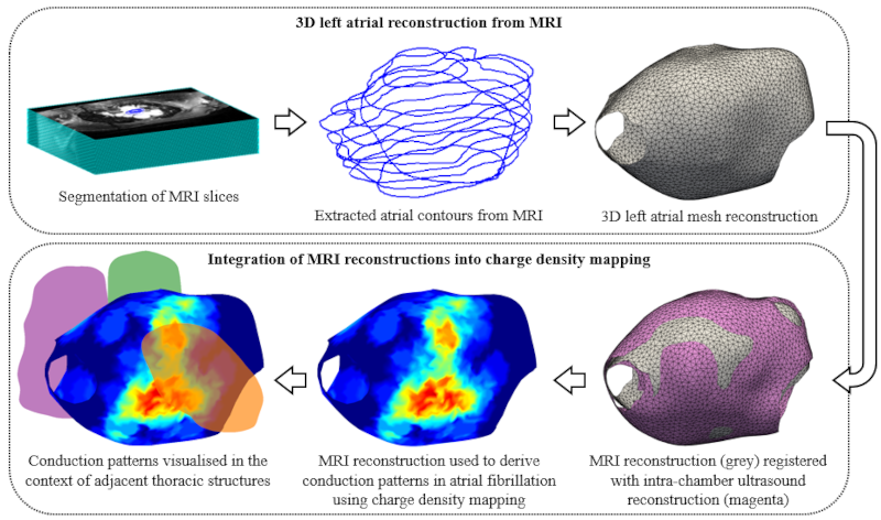
MRI is crucial because it provides high-resolution images of the heart's structure. Dr Banerjee’s MultiMeDIA lab specialises in multimodal medical data integration, and was therefore perfectly poised to develop a new computer tool that creates detailed 3D reconstructions of the heart's left atria from MRI scans. These reconstructions were then integrated with charge density mapping, which shows the patterns of electrical conduction during AF. This combination provides doctors with both structural information from MRI and detailed electrical patterns from charge density mapping - essentially creating a more complete picture of what may be causing AF in a patient.
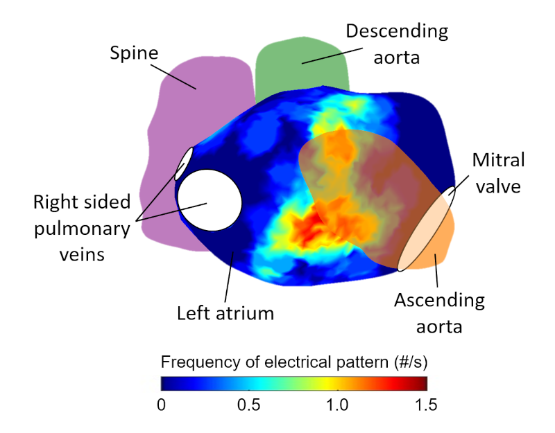
When testing their new approach, the researchers found that problematic electrical patterns were more likely to occur in areas where the heart was close to other structures in the chest, like the aorta and spine. This suggests that pressure from these neighbouring structures might contribute to AF in some patients.
Dr Sharp commented, "Our work provides the foundation for further integrating MRI's ability to characterise heart tissue with electrical mapping, which could lead to better understanding of the unique conditions that lead to AF in a patient and ultimately improve how we target treatment."
“The current study is a significant first step towards demonstrating how the integration of multiple complementary modalities can push our current scientific knowledge and aid clinical guidance”, added Dr Banerjee.
Read the full paper here: Multi-modal integration of MRI and global chamber charge density mapping for the evaluation of atrial fibrillation.
Royal Society Open Science is an open access journal that welcomes the submission of all high-quality science. More information about the submission process can be found on our webpage.
Hero image caption: Integration of Magnetic Resonance Imaging (MRI) with electrical mapping data from charge density mapping can offer new insights into the treatment of atrial fibrillation. This approach allows for a more comprehensive understanding of the structural and electrical characteristics of the heart's atria, which are crucial in planning and improving treatment strategies for this common cardiac arrhythmia.


