Micro-imaging 2018
Shortlisted entries in the Micro-imaging category from the 2018 Royal Society Publishing Photography Competition.
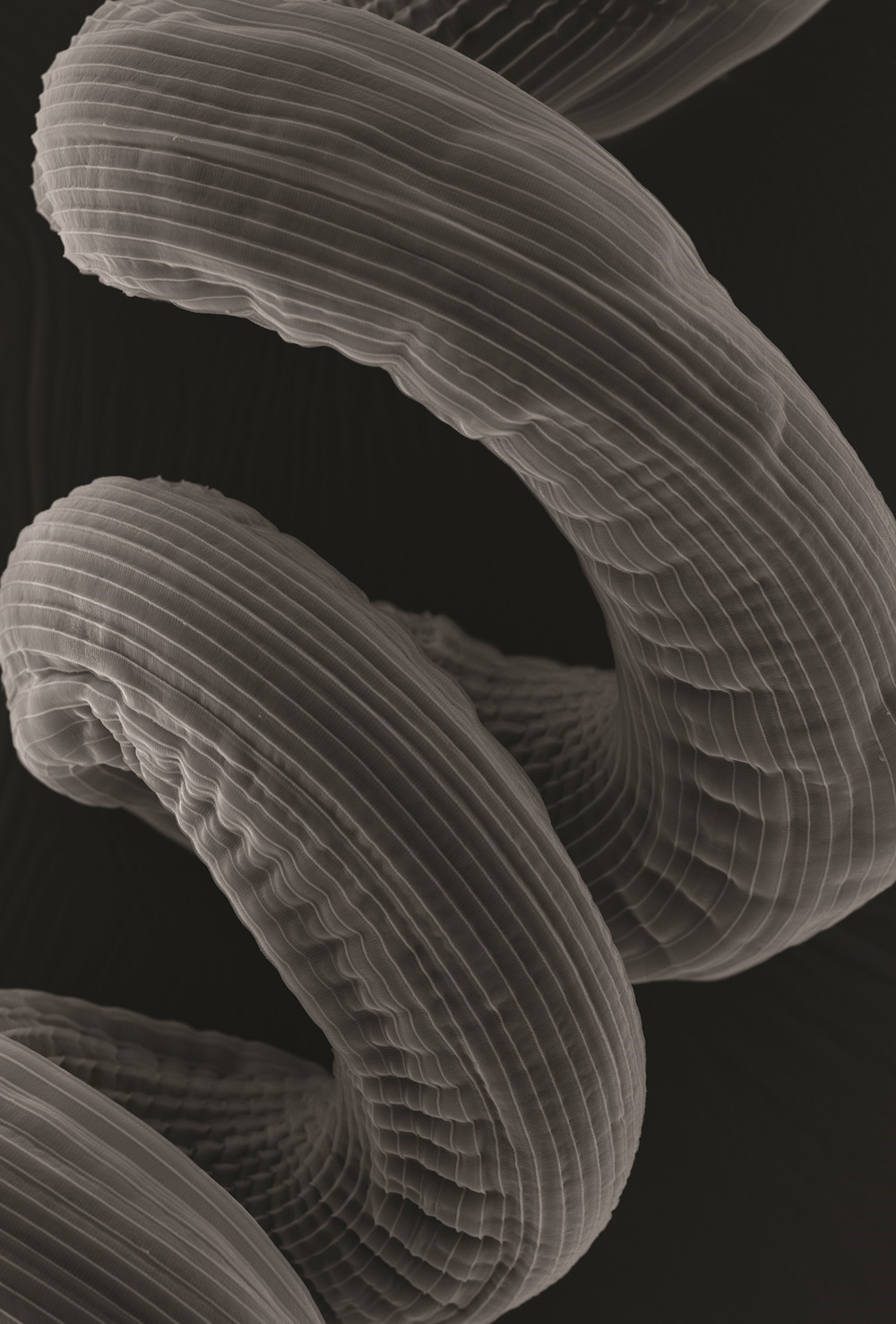
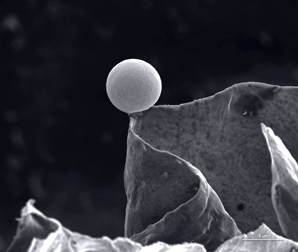
Micro-imaging Winner 'Going round and round' by Leandro Lemgruber. 'Within the Institute of Infection, Immunity and Inflammation, scientists and clinical investigators develop different lines of research, from characterising the immune system to understanding the cell biology of parasites, viruses and bacteria and their relationship with their hosts; aiming to develop new therapies and treatments. One research group (Maizels Laboratory) studies the exploitation of the host's own immune system by helminth parasites and how that could lead to minimising the risk of autoimmunity. The group makes use of a model rodent parasite - Heligmosomoides polygyrus. The adult parasite inhabits the intestinal space of its host and coils closely around the intestinal villi. Here we show the striated external surface of this helminth - the cuticle - observed by scanning electron microscopy.'
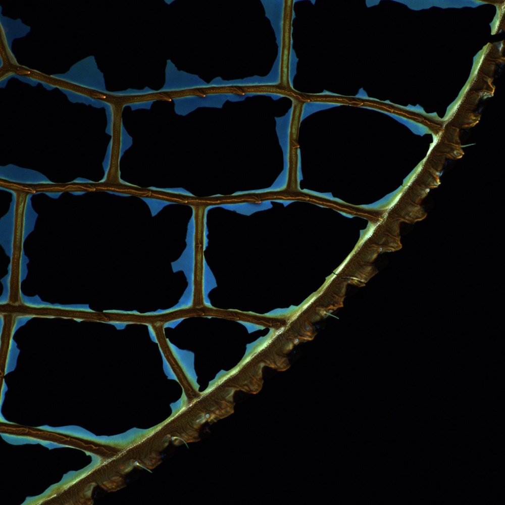
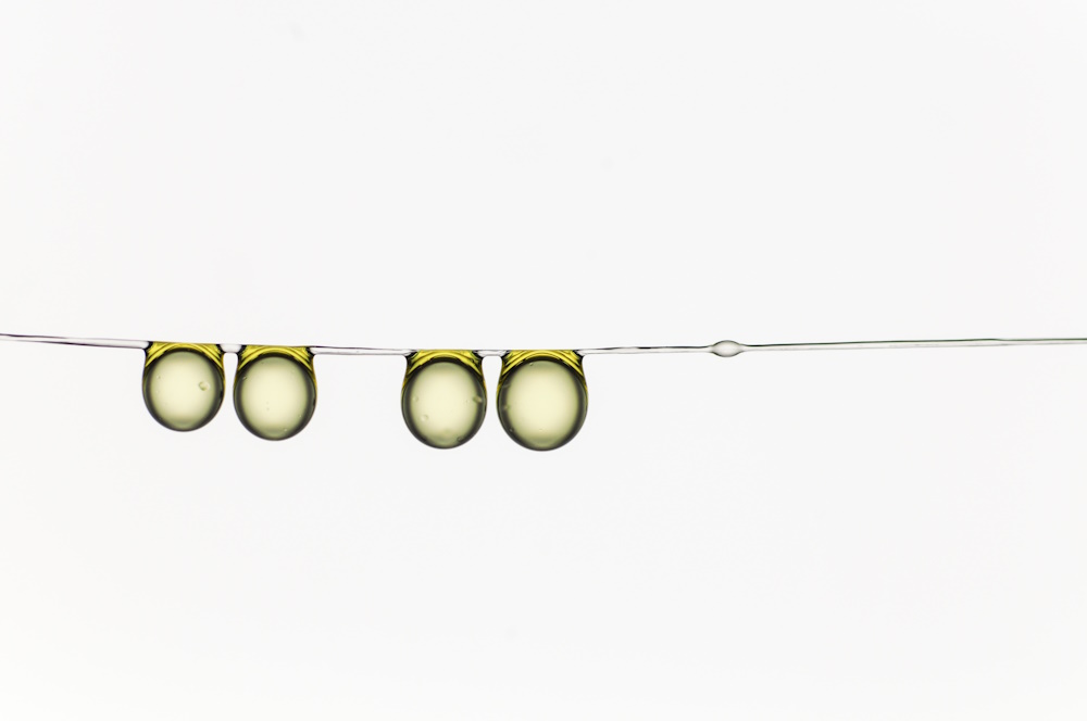
Micro-imaging Runner up 'Broken Window' by Hamed Rajabi. 'Wings experience substantial accidental collisions during the life span of a flying insects. Such collisions often result in irreversible wing damage and, therefore, could significantly influence insect flight ability. In order to investigate the material composition of wings of the dragonfly Acisoma panorpoides, I used confocal laser scanning microscopy (CLSM). During the scan, I observed no autofluorescence from wing membrane. This was very odd, because membranes are known for their blue autofluorescence when subject to laser light. Interestingly, after the scan I found out that wing membranes were all broken.'
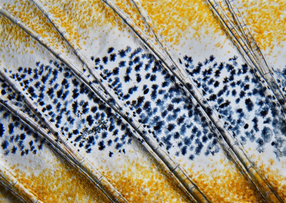
Micro-imaging Honourable mention 'A sunlit bamboo forest – bones and pigment cells in the adult zebrafish fin' by Ivo de Vos. 'While studying zebrafish (Danio rerio) development, we were intrigued by the striking colours and structure of its fins. This image depicts individual pigment cells (black melanocytes and yellow xanthophores), that give the fish its characteristic zebra stripes. Together with the underlying segmented bones, this picture reminded us of a sunlit bamboo forest, fitting with the fishes’ tropical habitat. The image was taken with a Nikon SMZ25 stereomicroscope with SHR Plan Apo 2X lens at 6X magnification and DS-Fi3 colour camera.'
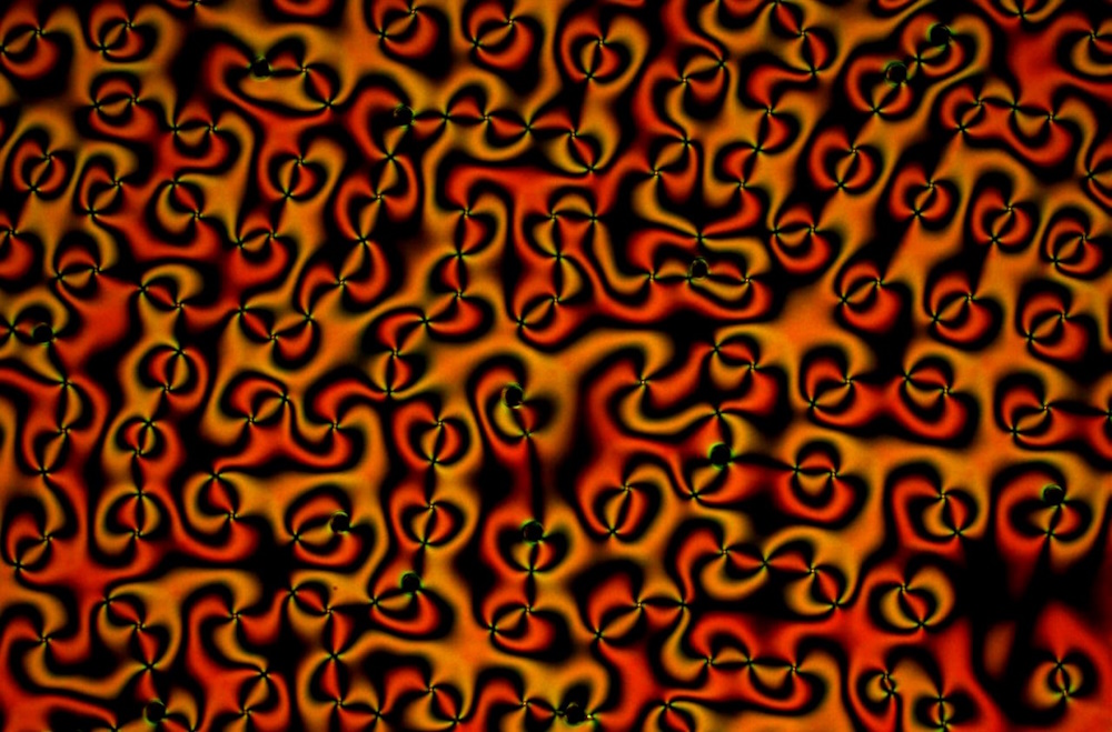
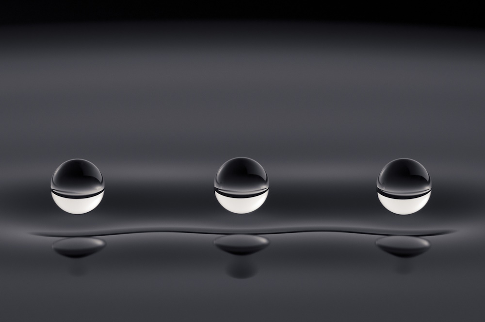
Micro-imaging Honourable mention 'Wings of vortexes pairs' by Valeska Zambra. 'When an oscillatory voltage is applied to a thin film of nematic liquid crystal with negative electric constant and with homeotropic anchoring, the balance between the electric force and the elastic force of the molecules allow the creations of topological defects called vortexes. There are vortexes with positive topological charge and vortexes with negative topological charge. Vortexes with opposite charge are attracted. In this image, we observe many pairs of vortexes that interact with each other. I research vortexes in liquid crystals. When I was taking measures in the laboratory I observed this image, I was impressed and I fell in love with how matter is able to behave. This image was observed through an Olympus BX51 microscope with a 10x objective, with linear cross polarizers and it was captured by a CMOS camera.The lower edge of the image was cut and the contrast was increased.'
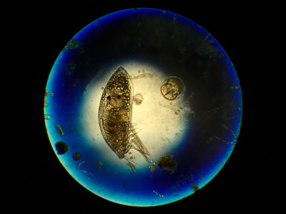
Micro-imaging Honourable mention 'Energy reserves in the Arctic' by Inês Leal. 'Invertebrate larvae face many challenges before settling on hard ground. They must rely on their energy reserves to sustain the dramatic process of metamorphosis. This photograph shows conspicuous lipid droplets (energy storage) within a late larval stage of an Arctic balanid barnacle. Although barnacles are foundation species worldwide, their tiny size means that they are often overlooked. Here, we highlight the fascinating larval ecology of these invertebrates, that still have secrets to yield. Photograph taken at the Greenland Institute of Natural Resources in Nuuk, Greenland (PENTAX Optio WG-1 Camera; Original color).'
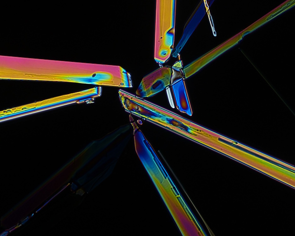
Micro-imaging Honourable mention 'Meeting Place' by Charlotte Johnson. 'Meeting Place is the by-product of a collaboration between myself (Science) and Arts. As a microscopist, I was asked to create crystals and record videos of them forming for an upcoming art installation. The hardest part of this project was creating a solution which would crystallize using a trigger, and at a concentration low enough to produce crystals which formed slowly enough to be recorded in real time. My progress was slow until I realized temperature greatly affected crystal formation. My moving the lab-prepared crystal solution into the much cooler microscope room was causing spontaneous crystal formation! Eventually it all came together and I was able to capture this image of several crystals having reached the same destination. Viewed under polarized light at x20 magnification, the crystals naturally reflected the stunning colors seen here which was made even more striking against the black background. No post-processing needed.'
Related content
Micro-imaging 2019
2019 micro-imaging category shortlist for the Royal Society Publishing Photography Competition
Micro-imaging 2017
2017 micro-imaging category shortlist for the Royal Society Publishing Photography Competition
Micro-imaging 2016
2016 micro-imaging category shortlist up for the Royal Society Publishing Photography Competition