Micro-imaging 2019
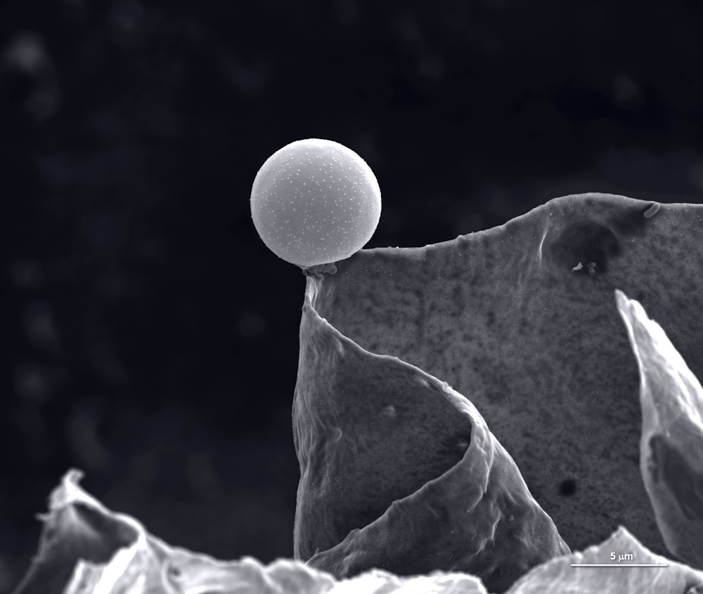
Shortlisted entries in the Micro-imaging category from the 2019 Royal Society Publishing Photography Competition.
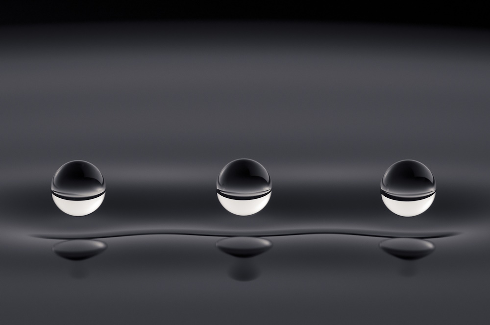
Competition winner 'Quantum droplets' by Aleks Labuda, in collaboration with Jan Belina. 'This photograph represents experimental proof of the theoretical work in the field of hydrodynamic quantum analogs. These silicone oil droplets are bouncing indefinitely above a vibrating pool of silicone oil at 15 Hz. The surface waves generated by the droplets are analogous to quantum mechanical waves that guide the dynamics of quantum particles. While the droplets move like quantum particles, they behave like quantum waves. The droplets' wave fields mediate their interactions with their surroundings, with each other, and even with themselves - similarly to electrons in the double-slit experiment. Such bouncing droplets were discovered in 2005 by Yves Couder (Université Paris Diderot) and mark the first real-world demonstration of the pilot-wave theory postulated by de Broglie in 1927. This behaviour provides measurable and intuitive insight into the mystery of particle-wave duality. Post processing involved simple contrasting and colour balancing. Camera: Nikon D700: 1/200; f/16; ISO200; 105mm.'
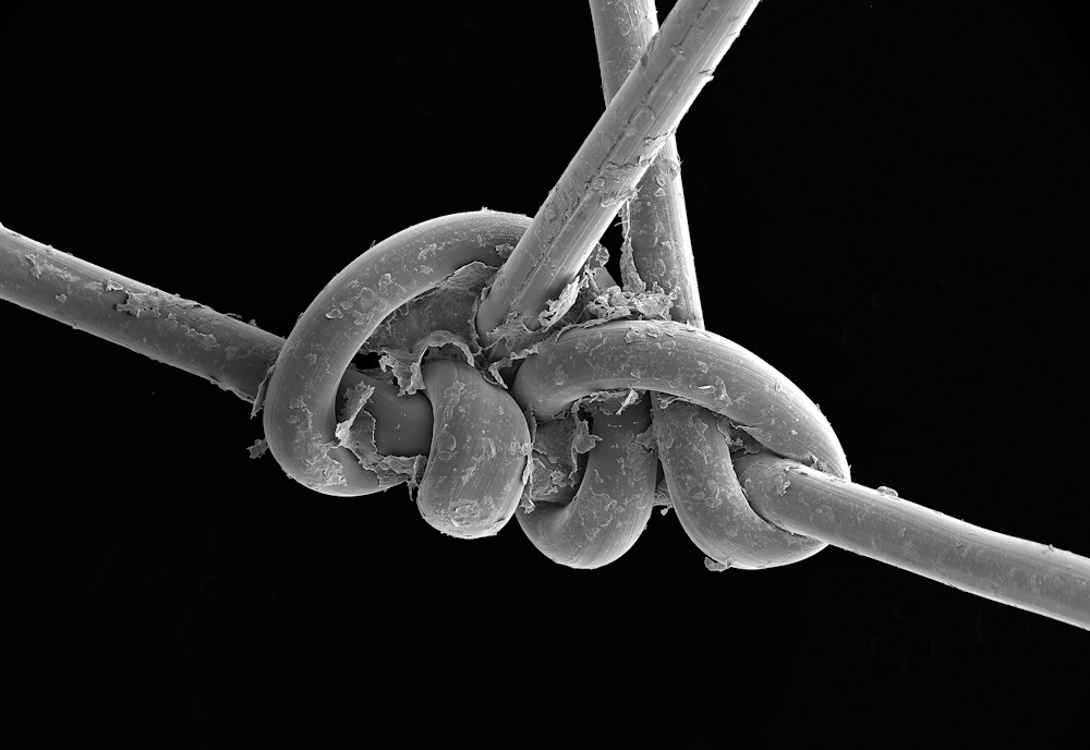
Micro-imaging Honourable mention 'Completely stitched up' by Anne Weston. 'This is a scanning electron micrograph of surgical thread used to stitch a head wound. The surgical thread was removed from the human patient after seven days, and debris from the skin and the area surrounding the wound can be seen still attached to the thread after removal. What this image shows in great detail is the skill required by healthcare professionals in being able to accurately tie such an intricate knot with a piece of thread less than 0.25mm in diameter, in what is a "routine procedure". Brightness and contrast of background altered and image cropped to remove imaging status bar. The image was captured using an FEI Quanta scanning electron microscope.'
Related content

Micro-imaging 2018
2018 micro-imaging category shortlist for the Royal Society Publishing Photography Competition
Micro-imaging 2017
2017 micro-imaging category shortlist for the Royal Society Publishing Photography Competition
Micro-imaging 2016
2016 micro-imaging category shortlist up for the Royal Society Publishing Photography Competition