Micro-imaging 2016
Shortlisted entries in the Micro-imaging category from the 2016 Royal Society Publishing Photography Competition.
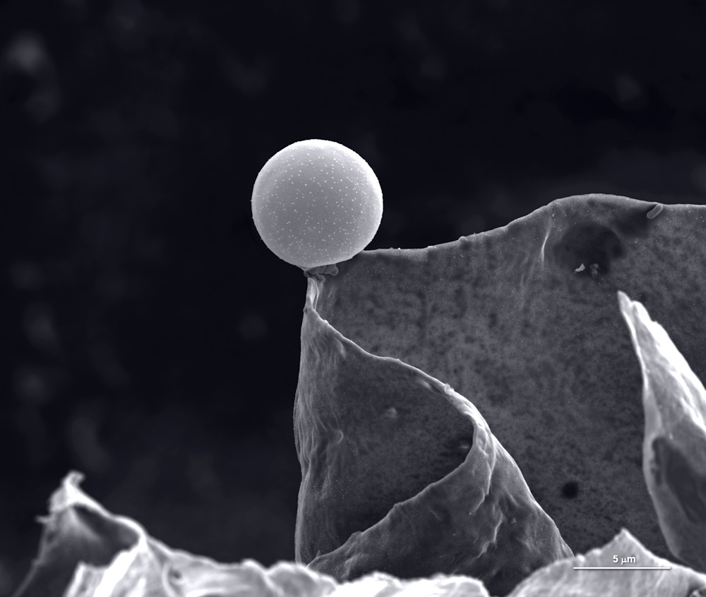
Category winner: Micro-imaging. "In balance" by María Carbajo Sánchez. Electron microscopy reveals the alien landscape of the surface of an activated carbon grain. It’s a lot like photography but instead of light, electron microscopes use a beam of electrons to make an image. This image is magnified 5000 times to see the structures of the surface of an activated carbon grain. Just as carbon is used to filter water it can be used in waste treatment for power plants. Activated carbon is prepared from biomass that is rich in carbon - wood, nutshells, olive pits. This piece of activated carbon was prepared from nutshells by a research team looking at using biomass waste for energy production. It may have been the roughness and variation in the surface of the nutshell that caused the curious and surprising images we obtained using the electron microscope, including this one of a microsphere in perfect balance.
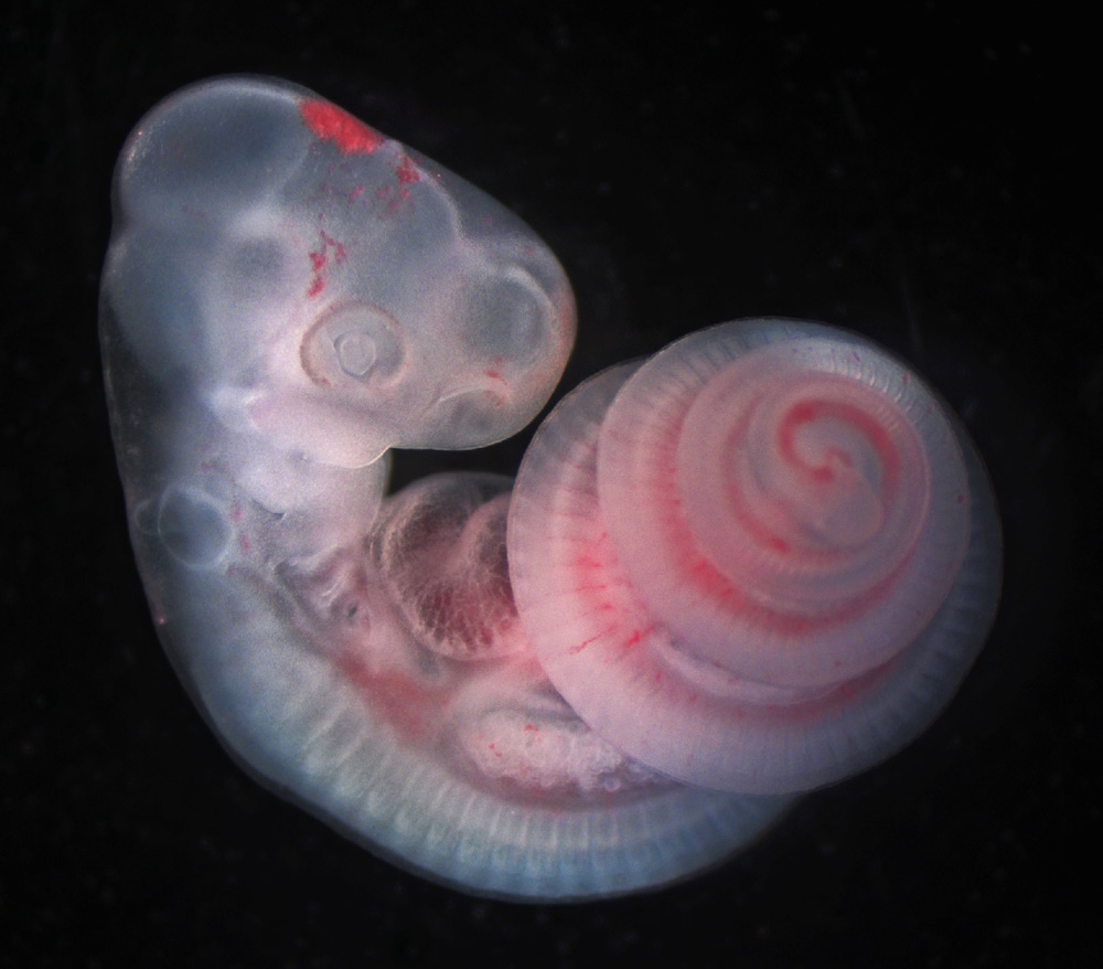
Runner up: Micro-imaging. "The spiralled snake axis" by Tyler Square. During early growth and development, most vertebrate animals look quite similar. Here, in this image of a one-day post-oviposition African house snake (Boaedon fuliginosus), we see many features that tie all vertebrates together: pharyngeal arches surrounding the mouth and throat (with gill slits between them), muscle segments (called somites) which will eventually contribute to the spine and musculature, and a chambered heart connected to a closed circulatory system to move oxygen and nutrients to tissues. During and after these stages, the novelties specific to a species or lineage begin to develop - here the 5mm long elongated body of the snake develops as a spiral to fit inside the egg.
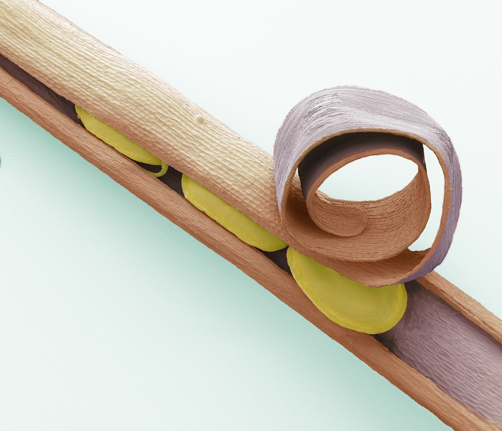
Special commendation. "Carbon nanotube jellyfish" by Clare Collins. Seemingly a swarm of jellyfish, this image was actually created using carbon nanotubes grown in a pillar formation. The metal disks that make up the jellyfish bodies are made by ‘sputtering’ charged aluminium and iron ions onto a surface to deposit a thin film of the metals. The carbon nanotubes grow from this thin disk. The disks are 5 micrometers in diameter- 5 1/1000ths of a millimetre - and are 10 micrometers apart. If you zoomed out of the image, you would be able to see that they make up part of a 2 mm x 2 mm pattern across the surface of a silicon chip. I research carbon nanotubes to study the emission of electrons, called the field emission, from a number of different configurations. The applications for field emission could be for displays or as X-ray sources.
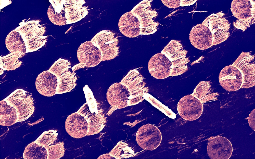
"SEM Brochosomes" by Steve Gschmeissner. This image is a coloured scanning electon micrograph (SEM) showing brochosomes. Brochosomes are intricately structured microscopic granules secreted by leafhoppers. After each molt, leafhoppers release droplets of the brochosome-containing fluid through the anus and actively spread them over the newly formed integument. The resulting coating makes the integument highly repellent to water (superhydrophobic) and to the liquid excreta of the leafhopper itself. Magnification: x5000 when printed 10cm wide.
"SEM Hairy bitercress" by Steve Gschmeissner. Hairy bittercress (Cardamine hirsuta). Coloured scanning electron micrograph (SEM) of hairy bittercress seed pod. Cardamine hirsuta is an annual species, common throughout the British Isles, particularly on bare ground, path-sides, and on walls. Bittercress has an explosive seed mechanism (explosive dehiscence) by which seeds can be dispersed up to 1m (3ft) away or considerably further if carried by the wind. Magnification: x22 when printed 10 centimetres wide.
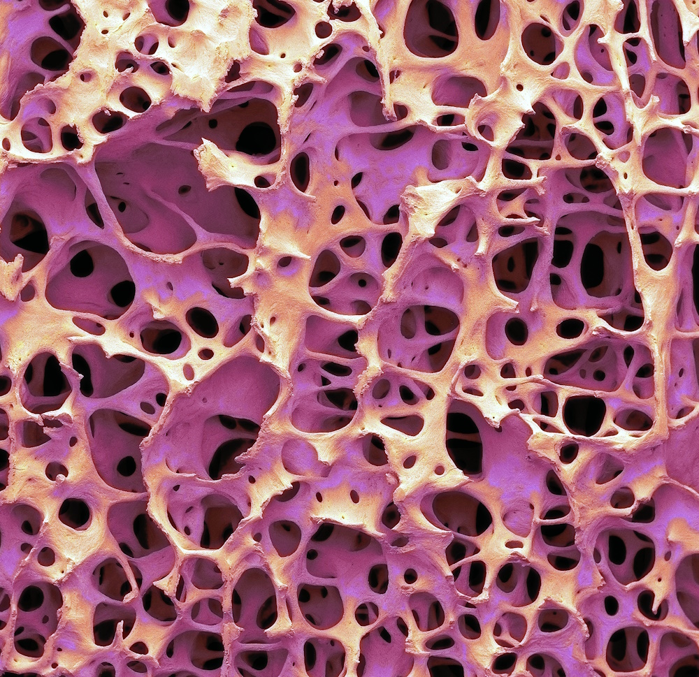
"SEM Bone" by Steve Gschmeissner. Coloured scanning electron micrograph (SEM) of human cancellous (spongy) bone. Cortical bone usually makes up the exterior of the bone, while cancellous bone is found in the interior. Cancellous bone is characterised by a honeycomb arrangement, comprising of a network of trabeculae. These structures provide support and strength to the bone. The spaces within this tissue contain bone marrow (not seen), a blood forming substance. Magnification: x20 when printed 10cm wide.
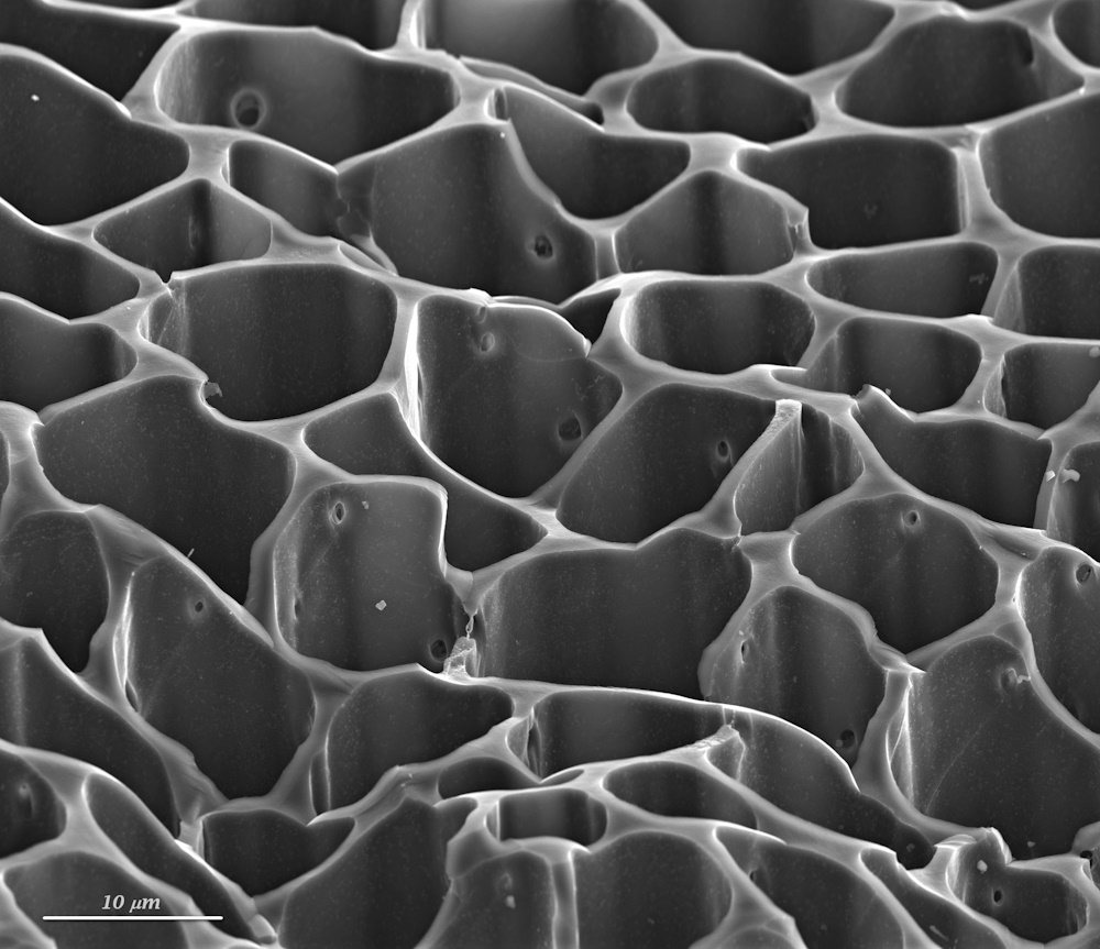
"Labyrinth of pores" by María Carbajo Sánchez. Activated carbon is a porous carbon material that is well known for a high adsorption capacity. This is due to the structure and surface which depends on the starting material and the method followed in the preparation properties. This image shows the porous structure of an activated carbon made from agricultural waste. The pore structure of activated carbons is given by the geometry of the cavities or channels of the particles. They have a high surface area which can reach values over 1000 square meters per gram of carbon.
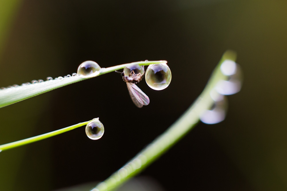
"Hanging on by a drop" by Merlin Fox. Caught in a droplet of dew, the wings of this creature drew the initial attention of the photographer. On closer inspection, it becomes clear that it is hanging by the dew drop rather than the blade of grass. Discovering these details in nature is inspirational for taking close-up photographs. Canon 6D, 50mm with macro rings, f3.2, 1/60, spot removal, luminance detail and smoothing, contrast adjustment.
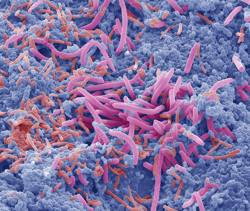
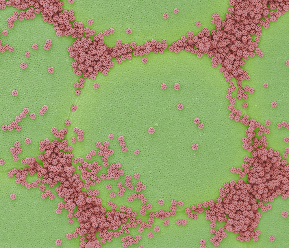
"SEM Tongue bacteria" by Steve Gschmeissner. Coloured scanning electron micrograph (SEM) of bacteria on the surface of a human tongue. Large numbers of bacteria can form a visible layer on the surface of the tongue. The mouth contains a large number of bacteria, most of which are harmless or even beneficial. However, some bacteria can cause throat infections or cause the formation of plaque deposits on the teeth, which may lead to decay and bad breath. Magnification: x6000 at 10cm wide.
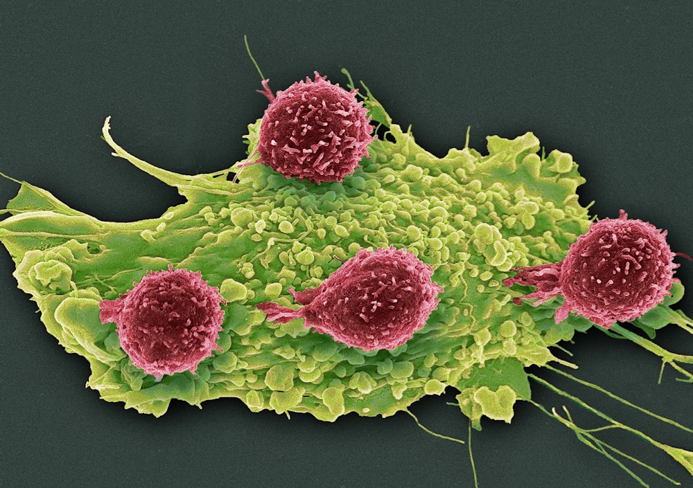
"SEM T lymphocytes and cancer cell" by Steve Gschmeissner. Coloured scanning electron micrograph (SEM) of T lymphocyte cells (red) attached to a cancer cell. T lymphocytes are a type of white blood cell and one of the components of the immune system of the body. T lymphocytes then signal for other immune system cells to eliminate the cell. Cytotoxic T lymphocytes eliminate the cell themselves by releasing a protein that forms pores in the membrane of the cell. Magnification: x1250 at 10 centimetres wide.
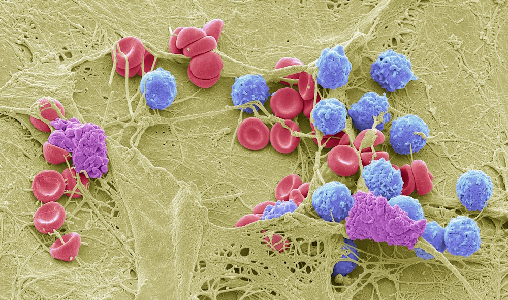
"SEM Skin wound" by Steve Gschmeissner. Coloured scanning electron micrograph (SEM) of a skin wound. A fibrin mesh supports a variety of blood cells at the site of a wound, red and white blood cells as well as platelet clots are visible. Magnification: x650 when printed 10cm wide.
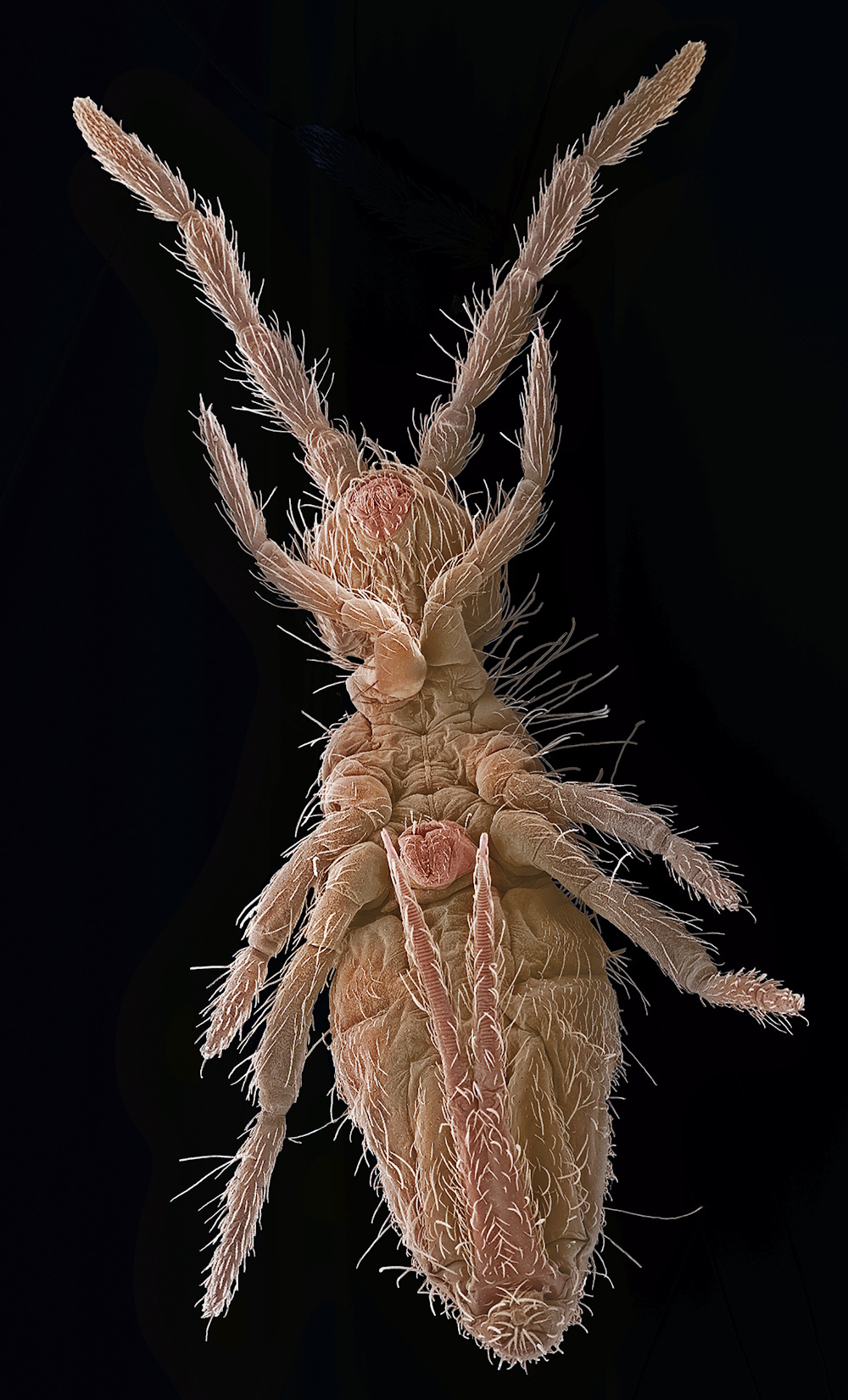
"SEM Springtail" by Steve Gschmeissner. Coloured scanning electron micrograph (SEM) of a springtail. The name "springtail" refers to the specialised jumping appendage (furcula) at the end of the abdomen. This allows the springtail to leap relatively long distances. Springtails have remained anatomically unchanged for millions of years, and are considered living fossils. They are wingless, and their antennae are the main sense organs, as their eyes are small. Magnification: x10 when printed 10cm high.
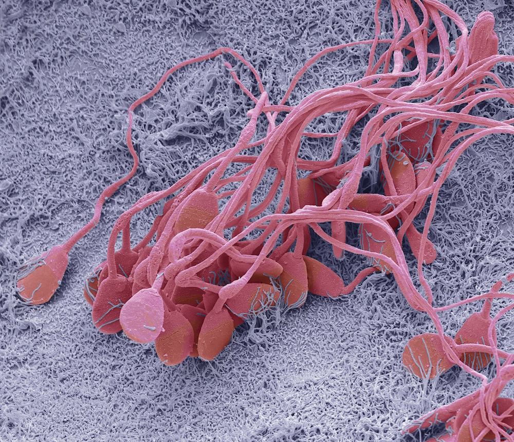
"SEM Sperm in the epididymis" by Steve Gschmeissner. Coloured scanning electron micrograph (SEM) of immature sperm in the epididymis. This immature sperm is characterised by a small amount of unshed residual cytoplasm below the head. The epididymis is a 7-metre- long coiled tube that lies behind each testis, receiving sperm from them. The sperm mature as they pass slowly through this tube. Magnification: x2500 when printed at 10 centimetres wide.
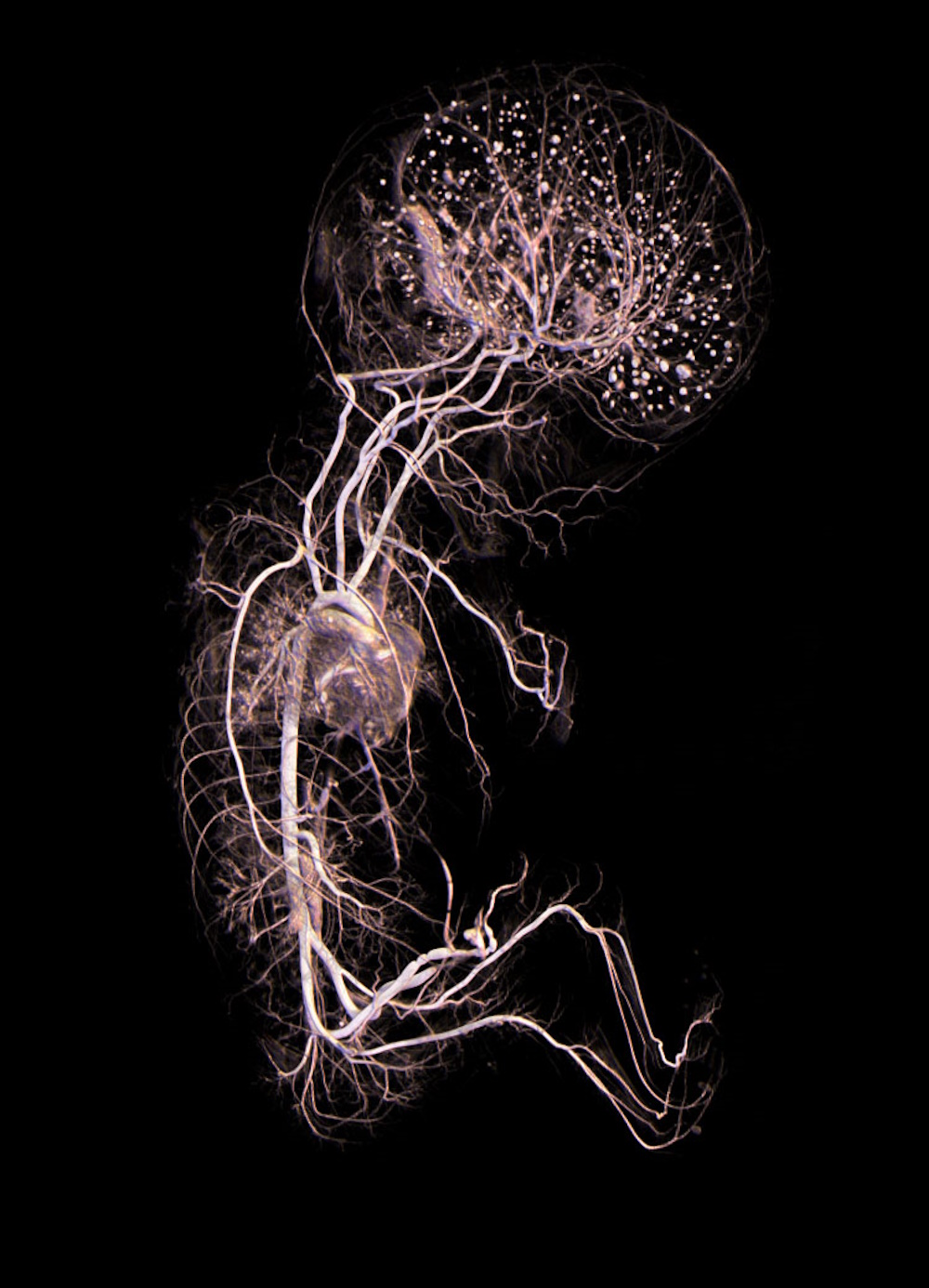
"Vascular tree of the human fetus with Micro-CT" by Guillaume Captier. Visualization 3D of the human fetus at 18 weeks, with a microphone-CT, shows the arterial vascularization of the whole of the body since the umbilical arteries. This arterial tree structure is essential for the fetal growth with a particular density at the cephalic extremity and brain.
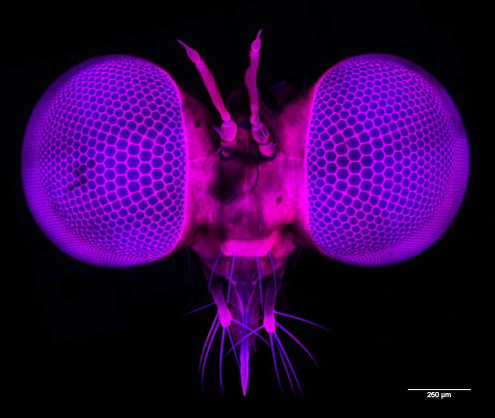
"All the better to see you with: The compound eyes of a tiny predatory fly" by Trevor Wardill. These large forward looking eyes belong to a small predatory robber fly (Holcocephala fusca). Imaging the entire fly head with 2-photon microscopy revealed that the optical and retinal adaptations result in the highest acuity known for a compound eye (0.2 degrees). The image is a maximum intensity projection of a whole head scan (2.9 mm wide x 0.82 mm deep) using an Olympus 25x GMP objective, a Newport Spectra-Physics InSight® DS+â„¢ laser at 810 nm, and a Bruker in vivo microscope using green and red detection of auto-fluorescence signals (shown as blue and red here). Prior to imaging, tissue was fixed, bleached and cleared. This collaborative project between Trevor Wardill and Paloma Gonzalez-Bellido at the University of Cambridge was sponsored by AFOSR (Air Force Office of Scientific Research).
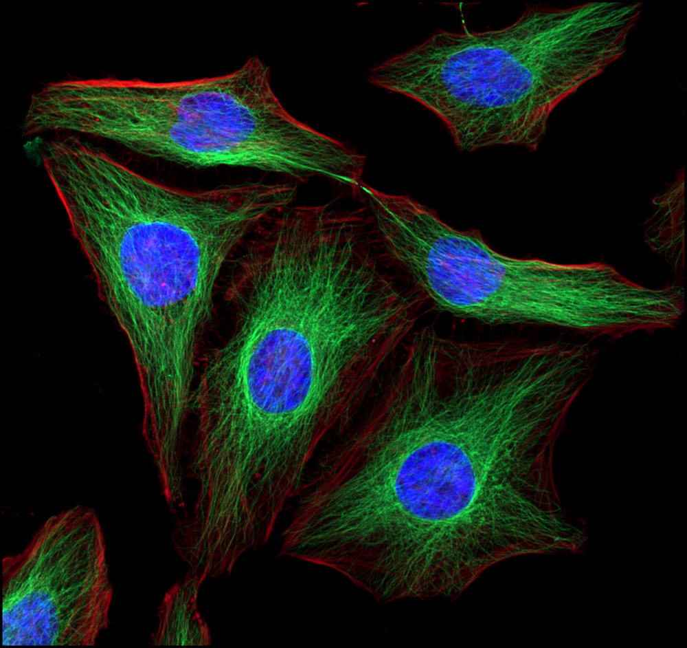
"HeLa cells" by Kevin Mackenzie. HeLa (cancer cell line) cells taken with Zeiss LSM800 confocal with Airyscan. Fluorescently stained to show nuclei in blue, Actin in red and alpha tubulin fibres in green. Taken using the super resolution (Airyscan) mode of the confocal using x63 objective.
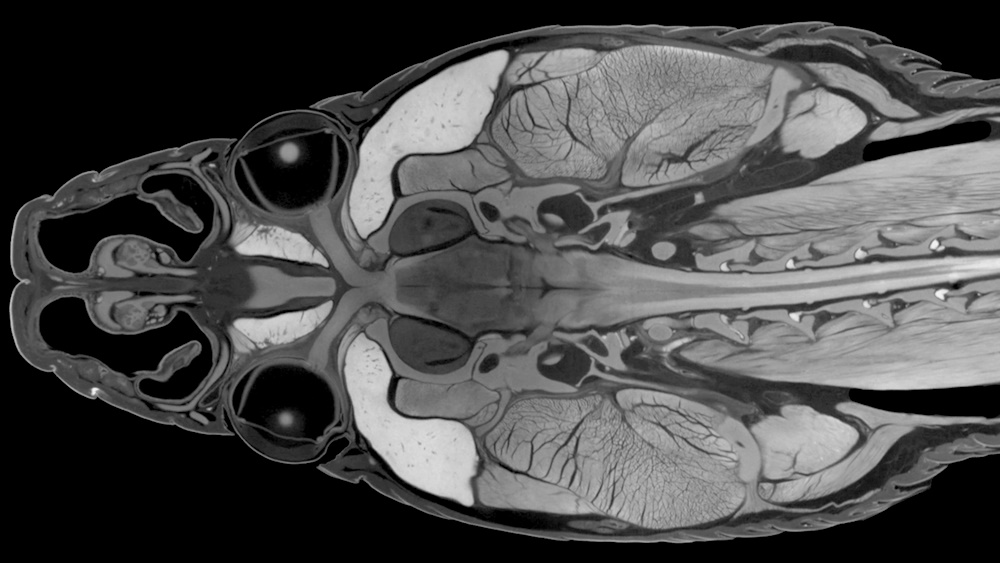
"The cranial anatomy of a diceCT imaged ratsnake in frontal view" by Paul Gignac. Ratsnake (genus Pantherophis) head in frontal view, prepared using diffusible iodine-based contrast enhanced computed tomography (diceCT) imaging, in which an iodine staining solution acts as a contrast agent that non-destructively renders soft tissues visible in X-ray micro-CT scans. The specimen was imaged using a GE Phoenix v | tome | x micro-CT scanner at 29.5 microns. Soft tissues including the brain and its internal structures, spinal cord, optic chiasm, roots and branches of cranial nerves, jaw and neck muscles, glands, nasal epithelia, cranial bones, and integument can all be clearly visualized simultaneously. (Image contrast and brightness were modified to render the background fully black and the neck was cropped to fit the image frame.)
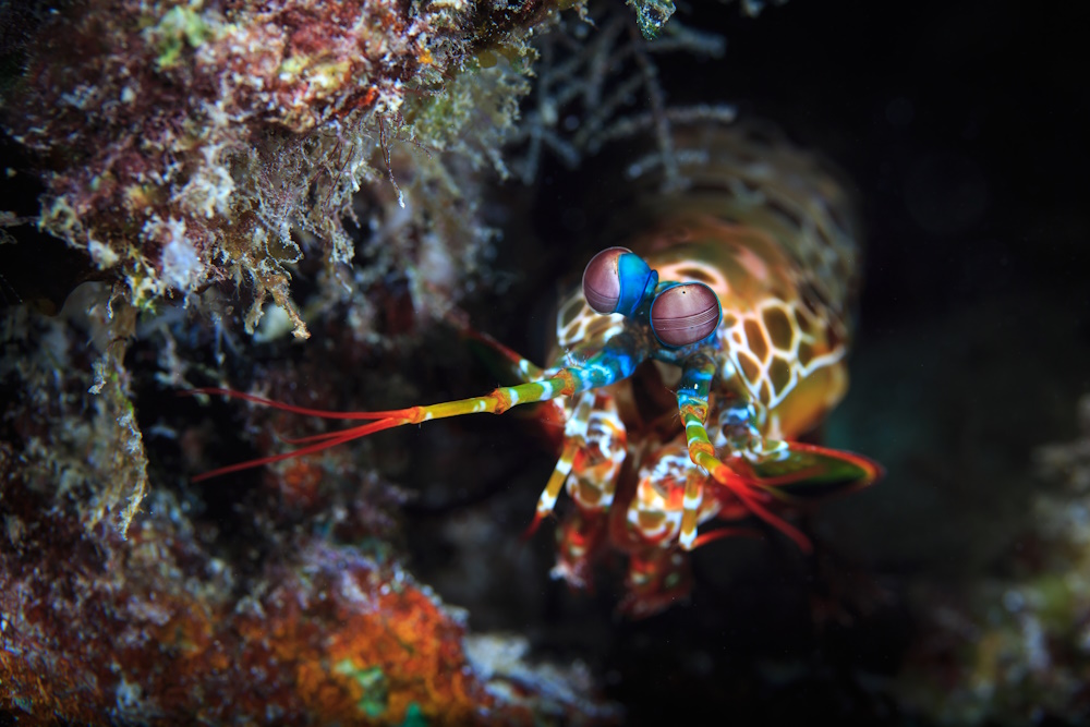
"Finding scientific inspiration in the eyes of nature" by Tane Sinclair-Taylor. A colorful peacock mantis shrimp (Odontodactylus scyllarus) emerges from a crevice in the reef matrix at Magoodhoo Island in the Faafu Atoll, Maldives. With an ability to see circularly polarised light, mantis shrimp visual systems are inspiring the design of new cameras that can detect a variety of cancers and visualise brain activity. Taken using Aquatica A5D3, Macro port, Twin Sea & Sea YS250 Strobes (snooted) 100mm, f7.1, 1/200th, ISO200.
Related content
Micro-imaging 2019
2019 micro-imaging category shortlist for the Royal Society Publishing Photography Competition

Micro-imaging 2018
2018 micro-imaging category shortlist for the Royal Society Publishing Photography Competition
Micro-imaging 2017
2017 micro-imaging category shortlist for the Royal Society Publishing Photography Competition