Micro-imaging 2017
Shortlisted entries in the Micro-imaging category from the 2017 Royal Society Publishing Photography Competition.
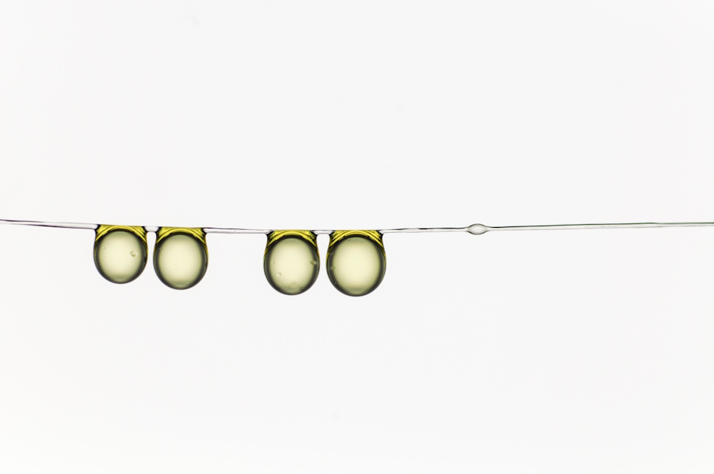
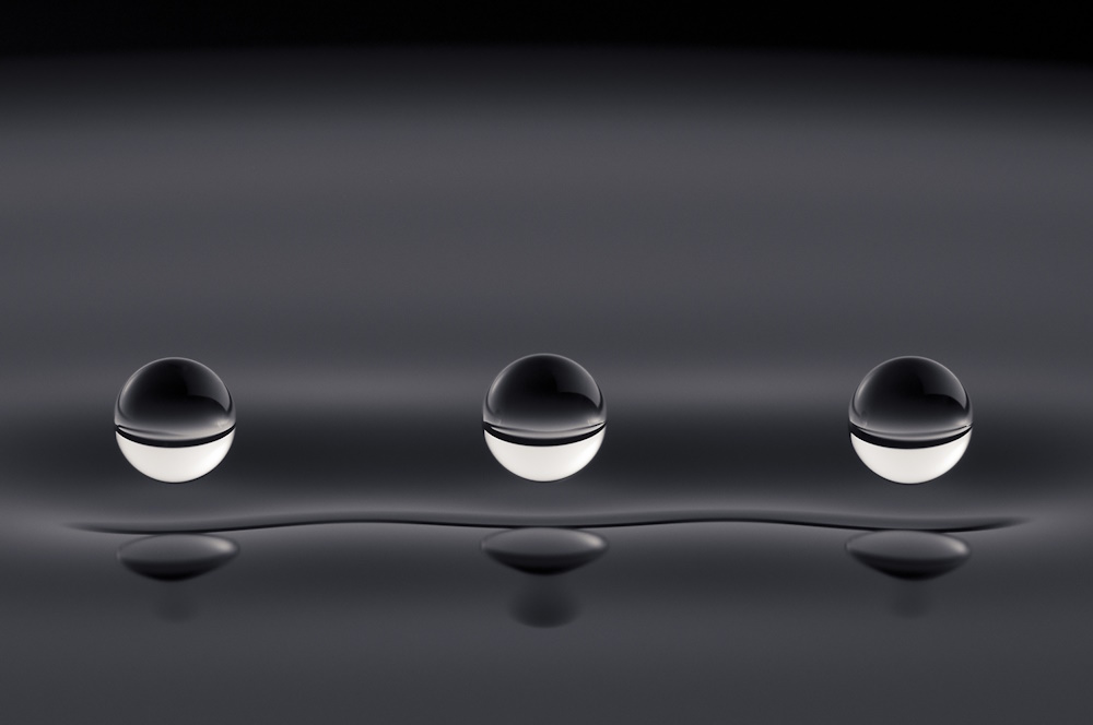
Category winner "Olive oil drop family hanging together." by Hervé Elettro. Inspired by the micro-glue droplets produced by the Nephila Madagascariensis spider to trap its prey, we began thinking to ourselves "What if these droplets could do more than just gluing?". Surface tension, the ability of a fluid to oppose deformation, indeed allows droplets to swallow any fibre made slack under compression, thus tightening the web against natural elements. A first step in the understanding of this mechanism was to use a model system for capture silk: drops on a thin soft fibre. The hanging olive oil drop family was born. Taken with a Nikon D300 camera and Phlox backlight at 10000 lux. Rudimentary Photoshop corrections.
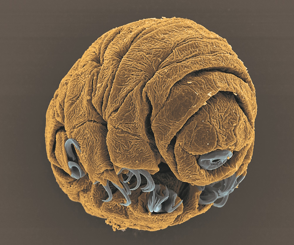
Runner up "Water bear embryo." by Vladimir Gross. Tardigrades (aka water bears) are tiny invertebrate animals that are able to survive extreme environmental conditions. This image depicts a 50-hour-old embryo of the species Hypsibius dujardini, taken with a scanning electron microscope at a magnification of 1800x. The embryo in the image is approximately 1/15 of a millimeter in length.
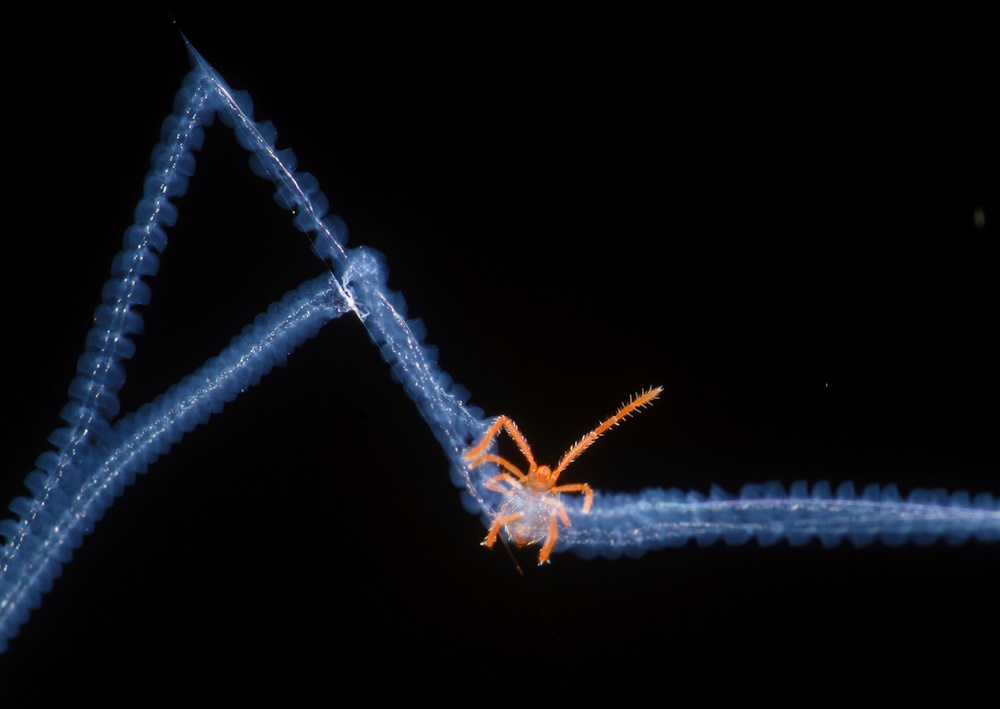
Honourable mention "Acari trapped in spiderweb." by Bernardo Segura. The spiders of the genus Austrochilus build some very conspicuous webs in Chilean temperate forests, and it's impossible to not be amazed by the gigantic horizontal sheet of spiders up to a meter long. After taking some photos of it near Nahuelbuta National Park, I discovered that some threads have some incredible beautiful bluish tones. I also realized that those threads are probably specialized in prey capture, and the spring-like structure that can be seen inside the threads probably has something to do with the elasticity. While taking photos of this amazing structure I saw a small acari hanging from the web, which may have fallen into the web and the spider didn't notice.
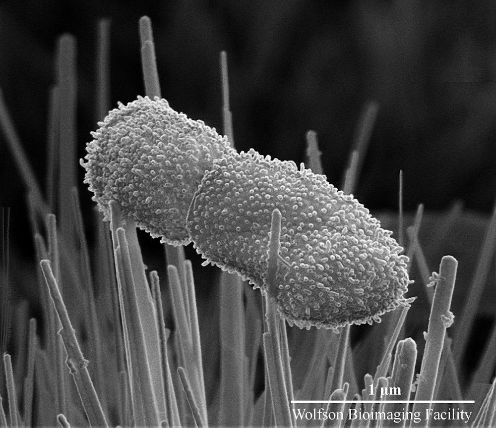
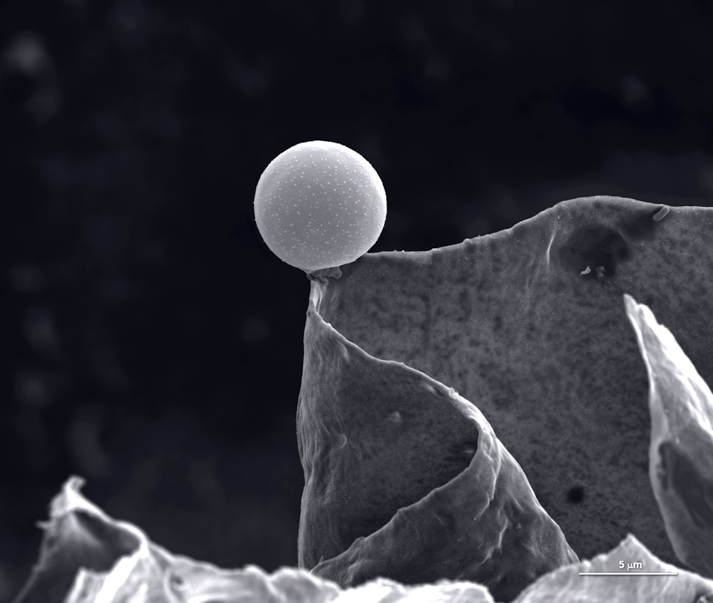
Shortlisted "Lucky escape: bacteria on a bed of nanospikes." by Joshua Jenkins. This image captures two bacterial cells (Klebsiella pneumoniae) in contact with titanium dioxide nanospikes. Titanium surfaces modified with these nano features have the ability to rupture and kill bacterial cells without the use of any chemical agents. In a time when bacteria are rapidly evolving resistance to antibiotics, these surfaces may help to prevent infections associated with titanium medical implants and devices. This image was taken at 100,000 times magnification in a scanning electron microscope to see if the nanospikes had penetrated the bacterial cells. The bacterium on the left has landed on a nanospike whereas the bacterium on the right has escaped by a narrow margin. After taking this image, it became clear that I needed to reduce nanospike spacing to ensure no bacteria can fall inbetween.
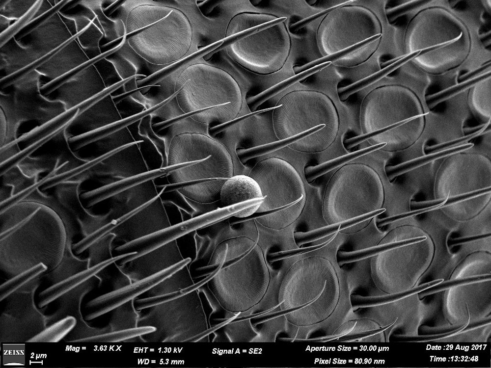
Shortlisted "Alone!" by Geetha Gangasandra Thimmegowda. This image is a non-coated micrograph of an antenna surface of honey bee (Apis dorsata). As we know, the antenna is the sensory gateway for an insect, which have sensors which are used to detect odors, touch, pressure, temperature, humidity and even sound! The body parts of the bee are the reflection of local atmosphere as the tiny hairs end up trapping pollen, dust, pollutants, etc. This honeybee was caught in a relatively clean location shown by the presence of pollen and absence of pollution. The image was taken with Zeiss merlin compact vp microscope at 1.30 kv EHT, 3.63 KX magnification settings. This micrograph is the part of a research working on the effects of air pollution on pollinators from NICE- naturalist-inspired chemical ecology lab, National Centre for Biological Sciences, Bangalore.
Related content
Micro-imaging 2019
2019 micro-imaging category shortlist for the Royal Society Publishing Photography Competition

Micro-imaging 2018
2018 micro-imaging category shortlist for the Royal Society Publishing Photography Competition
Micro-imaging 2016
2016 micro-imaging category shortlist up for the Royal Society Publishing Photography Competition